


7 Steps in phage display
- 2026-05-12
- 131
About the Author: KMD Bioscience is dedicated to becoming a leading provider of therapeutic antibody discovery and related support services. With technology R&D at our core, we deliver high-quality CRO (Contract Research Organization) services to scientists and research institutions worldwide, driving advancement and innovation in medical technology.
Phage display technology has revolutionized the field of molecular biology and drug discovery. By establishing a physical link between the phenotype (the displayed protein or peptide) and the genotype (the DNA sequence inside the phage), this high-throughput screening method allows for the rapid identification of high-affinity ligands against a vast array of targets. Whether you are developing therapeutic antibodies, identifying enzyme inhibitors, or mapping protein-protein interactions, understanding the fundamental steps in phage display is critical for success.
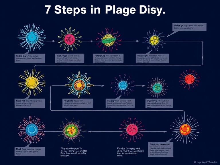
From our experience at KMD Bioscience, the efficiency of a phage display campaign is determined long before the first round of biopanning begins. It relies on the meticulous construction of the library and the strategic design of the selection pressure. In this guide, we provide a deep technical dive into the essential steps in phage display, reflecting the authoritative practices used in our state-of-the-art CRO laboratories.
Table of Contents
1. Phage Display Library Construction
The first and arguably most influential of the steps in phage display is the construction of the phage library. A library is a collection of billions of different phage particles, each displaying a unique peptide or protein variant on its surface. The diversity of the library (often ranging from 10^7 to 10^{11} unique clones) dictates the likelihood of finding a binder with the desired affinity and specificity.
We recommend starting with a high-quality genetic source. For antibody libraries, this often involves isolating mRNA from B cells (immunized or naive), followed by cDNA synthesis. The variable regions (VH and VL) are amplified via PCR and cloned into a phagemid vector. The choice of the display format—such as ScFv (single-chain variable fragment) or Fab (fragment antigen-binding)—is a strategic decision. From our experience, ScFv libraries are often easier to construct and display, while Fab libraries may provide better stability and represent the native antibody structure more accurately.
Once the DNA is cloned into the phagemid, it is transformed into electrocompetent E. coli cells. This leads to the production of phage particles through the use of a helper phage. The resulting library must be validated for its titer (the concentration of infectious phage) and its diversity through Next-Generation Sequencing (NGS).
2. Target Preparation and Immobilization
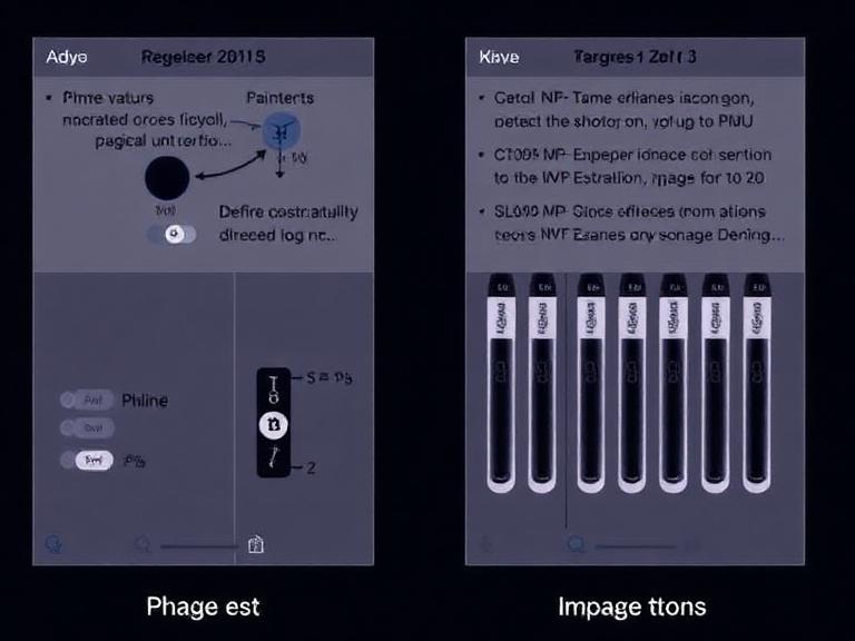
Before initiating the steps in phage display selection, the target molecule must be prepared. The target can be a purified protein, a small molecule, a carbohydrate, or even a whole cell. The success of selection depends heavily on the target remaining in its native, biologically active conformation.
Immobilization is the process of attaching the target to a solid support, such as a 96-well immunoplate or magnetic beads. We recommend using biotin-streptavidin systems for immobilization when possible, as this allows for "solution-phase" panning, which minimizes the risk of exposing neo-epitopes that are not present in the native environment. If you are working with difficult-to-express proteins, utilizing a robust protein expression systems overview is essential to ensure high purity and correct folding before biopanning starts.
3. The Biopanning Cycle: The Heart of Phage Display
Biopanning is the iterative process used to enrich the library for phages that bind to the target. This cycle is performed 3 to 5 times, with each round increasing the selection stringency. The biopanning cycle consists of four distinct sub-steps:
A. Binding
The phage library is incubated with the immobilized target. This allows high-affinity phages to attach to the target epitopes. During the first round, we recommend a longer incubation time and a lower selection pressure to ensure that rare, high-affinity clones are not lost.
B. Washing
This is where stringency is applied. Unbound or weakly bound phages are washed away using buffers containing detergents like Tween-20. From our experience, increasing the number of washes and the concentration of detergent in later rounds is vital to eliminate background noise and non-specific binders.
4. Elution and Phage Amplification
Once the non-specific phages are removed, the specifically bound phages must be recovered. This is the elution phase of the steps in phage display.
C. Elution
Elution is typically achieved by disrupting the protein-protein interaction through pH changes (using acidic glycine buffer) or competitive elution with a known ligand. We recommend neutralizing the pH immediately after acidic elution to preserve the infectivity of the recovered phages.
D. Amplification
The eluted phages are used to infect a logarithmic phase culture of E. coli. Using an E. coli protein expression system approach at this stage allows for the rapid replication of the selected phage DNA. Following infection and the addition of helper phage, a new, enriched sub-library is produced for the next round of selection.
5. Characterization of Positive Clones
After several rounds of panning, the pool of phages is highly enriched for target binders. The next of the steps in phage display is the transition from a "pool" to "individual clones." Individual colonies are picked from agar plates, and phage clones are produced at a small scale.
These clones are then screened using Phage ELISA (Enzyme-Linked Immunosorbent Assay). A positive signal in ELISA indicates that the specific phage clone binds to the target but not to a control protein (such as BSA). The DNA of the positive clones is then sequenced to identify unique sequences and to analyze the consensus motifs.
6. Transitioning to Recombinant Expression
Once the sequence of a high-affinity binder is identified, it must be expressed as a soluble protein for further functional validation. This is a critical transition. The genetic sequence is moved from the phagemid vector into an expression vector.
For antibody fragments or peptides, we frequently utilize our recombinant protein expression service. This allows for the production of mg to gram quantities of the lead candidate. If the goal is a full-length IgG, mammalian expression systems are typically preferred to ensure correct glycosylation. KMD Bioscience offers a comprehensive protein expression platform services suite to handle this entire workflow, from phage sequence to purified recombinant protein products.
7. Summary of Steps in Phage Display
| Step Number | Process Name | Key Objective | Expert Tip |
|---|---|---|---|
| 1 | Library Construction | Generate genetic diversity | Use NGS to verify library quality before starting. |
| 2 | Target Preparation | Maintain native epitope structure | Prefer solution-phase panning with biotinylation. |
| 3 | Incubation/Binding | Allow specific interactions | Use negative selection (pre-clearing) to reduce noise. |
| 4 | Washing | Remove non-specific phage | Increase detergent concentration in later rounds. |
| 5 | Elution | Recover bound phage | Neutralize pH immediately to keep phage alive. |
| 6 | Amplification | Increase clone numbers | Ensure E. coli is in the log phase (OD600 0.4-0.6). |
| 7 | Screening/Sequencing | Identify lead candidates | Verify binding with a secondary assay (e.g., SPR or BLI). |
8. Frequently Asked Questions (FAQs)
How many rounds of biopanning are usually required?
From our experience, 3 to 4 rounds are typically sufficient. More than 5 rounds can lead to the "over-selection" of clones that have a growth advantage in E. coli but might not have the best affinity for the target.
What is the difference between pIII and pVIII display?
pIII is a minor coat protein (3-5 copies), which is ideal for monovalent display and identifying high-affinity binders. pVIII is the major coat protein (thousands of copies), which is used for multivalent display, enhancing the binding of low-affinity clones through the avidity effect.
Why is E. coli used in phage display?
E. coli is the host for phage infection and replication. Its rapid growth rate and well-understood genetics make it the perfect bio-factory for amplifying selected phage clones between rounds.
Can phage display be used for cell-surface targets?
Yes, this is known as "cell panning." It is more complex due to the high background of non-specific proteins on the cell surface, but it is highly effective for discovering antibodies against GPCRs or ion channels in their native state.
9. References
Smith, G. P. (1985). "Filamentous fusion phage: novel expression vectors that display cloned antigens on the virion surface." Science, 228(4705), 1315-1317. (Academic Source)
Bradbury, A. R., et al. (2011). "Beyond natural antibodies: the value of in vitro display technologies." Nature Biotechnology, 29(3), 245-254. (Major News/Academic)
KMD Bioscience Internal Technical Protocols: Antibody Discovery and Phage Display Platforms 2026.
Successfully navigating the steps in phage display requires a blend of precise laboratory technique and strategic experimental design. At KMD Bioscience, we pride ourselves on helping our partners overcome the technical hurdles of library screening to find the next generation of therapeutic candidates. By paying attention to detail in library construction and selection stringency, researchers can harness the full power of this elegant technology to drive medical innovation.

- Protein Platform
- Protein Expression Services
- Protein Modification & Detection Services
- Protein Interaction Services
- Protein Purification Service
- Antibody Discovery Platform
- VHH Antibody Platform
- Phage Display Platform
- Antibody Humanization Platform
- Single B Cell Screening Platform
- Antibody Expression & Validation Platform
- Peptide Library Platform
- Custom Antibody Platform
- Innovative Drug Discovery Platform
- Product Center
- Proteins
- Antibodies
- Raw Materials Reagents
- Small Molecule Reagents
- Peptide Products
- Nanoparticle Products
- Contact Us
- Phone:400-621-6806
- Mobile:13110936064(WeC
- E-mail:[email protected]
- Address:Room 108, Building 10, No. 6 Hechang Road, Wuqing Development Zone, Tianjin City
-

WeChat
-

Service Account
-
 400-621-6806
 -
 3599163699
 -
 Monday to Friday 9:00-18:00
 -

-
 0
0
-
