Home
>>
Antibody Platform
>>
Peptide Library Platform
>>
12-mer Peptide Library Construction & Screening
Download Center
Quick Inquiry & Consultation

12-mer Peptide Library Construction & ScreeningIntroduction

The Phage Display Platform offered by KMD Bioscience is an ultrahigh-throughput ligand screening system. This platform enables presentation of up to 1013 peptide or antibody clones per milliliter on phages. Leveraging this platform, we construct 7-mer, 12-mer, and cyclic 7-mer peptide libraries for clients. Successful screening of phage display libraries depends critically on target properties, library size and diversity, and library quality. Library quality is reflected in diversity, which estimates the conservation or variability of amino acid sequences relative to peptide copy numbers using statistical methods.

Exogenous peptides are fused to structural proteins of filamentous M13 phage through insertion of corresponding nucleotide sequences into protein-encoding genes. This insertion facilitates protein/peptide display on the phage surface, provided the insert does not disrupt protein function. When sufficiently exposed on the phage surface, the peptide acts as a ligand, enzyme, immunogen, or participates actively in biochemical processes. Target-specific peptides can be conjugated to various carrier systems (nanoparticles, liposomes, phage virions), offering advantages over antibodies: lower immunogenicity, simpler production, reduced costs, higher surface density (increased peptide copies per unit area enhancing avidity), minimized particle size augmentation, and superior tissue penetration.


Construction of Phage Display 12-mer Peptide Library

Peptide and Antibody Fragment Display is achieved through conjugation to the minor coat protein pIII of filamentous phage M13, enabling presentation of desired molecules. Located at one end of the phage capsid, pIII comprises three functionally autonomous domains (D1, D2, and D3) connected by glycine-rich linkers. During Gram-negative bacterial infection, the N-terminal D1 domain mediates viral DNA translocation into the host cytoplasm. The D2 domain binds the bacterial F-pilus and executes essential functions in infection. The C-terminal D3 domain is indispensable for stable capsid assembly and serves as a prerequisite for phage production. In most phage display vectors, deletion of D1 and D2 domains in pIII yields phage particles with reduced infectivity.

A randomly synthesized 12-mer peptide-encoding oligonucleotide fragment is chemically synthesized and inserted into the gene encoding the phage coat protein. The engineered gene is then transformed into E. coli using recombinant DNA technology. A random 12-mer peptide is fused to the M13 bacteriophage minor coat protein (pIII), creating a combinatorial library. The amplified phage particles display the foreign peptides on their surface, with each phage expressing one unique foreign peptide. This collection of phage constitutes a pre-made random peptide library. The phage vector contains both phage and bacterial origins of replication, a phage packaging signal, a selectable marker gene, and the gene for the chosen coat protein fused to the insert. This design facilitates easy vector preparation and maintenance, high yields of double-stranded DNA (dsDNA), and improved transformation efficiency. 

To assemble functional phage particles, co-infection with a helper phage (e.g., M13K07) is required. The physical link between the phage-encoded genotype and the displayed phenotype is maintained because the helper phage has an origin of replication and a packaging signal, but its gene III protein function is compromised. Consequently, the phage vector is preferentially packaged into new viral particles over the helper phage genome. For multivalent display purposes, alternative helper phages, such as hyperphage, are used.

Using client-provided target molecules, high-affinity binding phage clones are isolated from pre-fabricated randomized 12-mer peptide libraries through screening, amplification, and panning. Determination of phage DNA sequences reveals the amino acid sequences of displayed peptides.

Screening of Phage Display 12-mer Peptide Library

The overarching principle of Phage Peptide Library Screening exploits the varying affinities existing between certain biomolecules. Using a biomolecule as the target, specific binding peptides with desired affinity can be isolated from the peptide library through single or multiple rounds of screening. The Phage Display Screening method typically employs a biological selection technique termed biopanning. This process utilizes biological manipulation to isolate the desired phage-displayed peptides from a random phage peptide library. The specific screening methodology may differ based on the nature of the target, the scale of the screening, and operator preferences.

Classical screening methods primarily consist of solid-phase screening and solution-phase screening. 

In Solid-Phase Screening, the antigen is coated onto a solid support (e.g., microtiter plates, nitrocellulose membranes, or immunotubes). The antigen is then incubated with the peptide library, allowing specific phage-displayed peptides to bind. Its advantage is the capacity to adsorb more target protein per unit area of the solid support. However, a limitation of solid-phase screening is that the target molecule—particularly conformationally labile targets—may undergo conformational changes during the immobilization process. 

Solution-phase screening was developed to address this limitation. In this method, the target molecule is typically biotinylated, mixed with the peptide library in solution to allow interaction, and the mixture is then added to immobilized avidin. High-affinity phage-displayed peptides are enriched through the binding of biotin to avidin.

Phage Display Peptide Library: Construction & Screening Workflow

ProjectService ContentTimeline
12-mer Peptide Gene Library Synthesis

Trimer codon mutagenesis for saturated codon mutation synthesis of 12-amino acid peptide libraries

3-5

weeks

12-mer Peptide Library Construction Service

Phagemid Construction and Transformation: Homologous recombination assembly of target genes with pMECS vector, transformation into E. coli TG1 host strain for M13 phage display 12-mer library construction

4-6

weeks

12-mer Peptide Library Screening Service

Three rounds of biopanning (additional rounds at KMD Bioscience discretion at no extra cost); Sequence analysis of 50-80 blue clones

4-5

weeks

Common Target Molecules for Peptide Library Screening

Molecular Type

Introduction

Membrane Receptor Extracellular DomainUtilizing isolated full-length membrane receptors as targets in in vitro library screening presents challenges due to their marked hydrophobicity and dependence on lipid bilayers for native folding. Membrane receptors typically feature large multi-domain extracellular regions capable of interacting with diverse peptide sequences. Isolated ligand-binding domains serve as excellent surrogate targets for selecting peptides mimicking endogenous ligands. Targeting shorter receptor fragments can also enhance specificity toward specific members of highly conserved receptor families. Conversely, synthetic peptides corresponding to specific receptor domains may adopt conformations divergent from native proteins, potentially compromising receptor recognition in physiological contexts. It is therefore advisable to first screen phage libraries against synthetic peptides, followed by affinity selection of enriched libraries against native proteins.
Neutralizing Antibodies Targeting Endogenous Binding PartnersNeutralizing antibodies targeting endogenous ligands can serve as surrogate targets for identifying bioactive peptides. The antigen-binding region of the antibody mimics the ligand-binding site of the receptor, thereby providing a "template" for retrieving peptides that cross-react with the receptor. Neutralizing antibodies do not necessarily recognize the exact receptor-binding epitope located on the ligand. Instead, they exert their antagonistic effect by sterically blocking the ligand-receptor interaction, potentially through binding to sites adjacent to these regions.
Whole-Cell ScreeningPhage Display Libraries can also be screened against live cells expressing the membrane receptor of interest. This eliminates concerns about misfolding of the target protein and allows selection against the extracellular region of the receptor in its native conformation. Target cells can be in suspension or adherent. The primary goal is to identify peptides binding to tumor-specific receptors, which can be used for targeted delivery of drugs or diagnostic agents.

Future Prospects of Phage Display Technology

Two drugs developed by screening phage-displayed random peptide libraries are the pegylated erythropoietin receptor agonist peginesatide (Hematide, Affymax) and the Fc-fused thrombopoietin receptor agonist romiplostim (Nplate, Amgen). Additionally, two other drugs—the anti-TNF-α antibody adalimumab and the small protein inhibitor of plasma protease kallikrein, ecallantide—have been developed using phage display technology, establishing this technique alongside other established methods in the drug discovery field.

KMD Bioscience Service Advantages

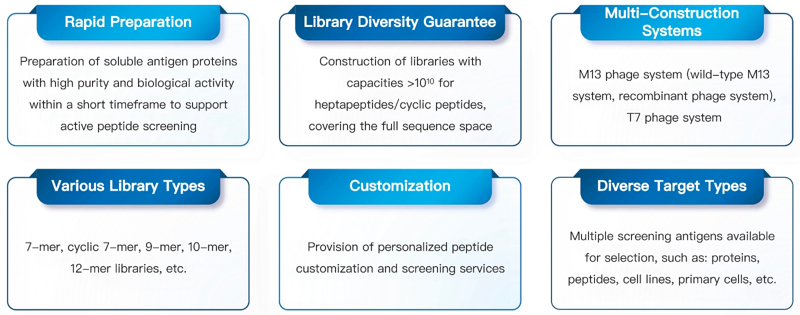
71-KMDBioscience-12peptide-library-construction-and-screening.jpg

Login

Don’t have an account?Sign Up Now

Register

Already have an account?Log In Now
