


Troubleshooting Common Issues in SDS-PAGE
- 2026-03-27
- 251
I. SDS-Polyacrylamide Gel Electrophoresis
SDS-Polyacrylamide Gel Electrophoresis (SDS-PAGE) is one of the most commonly used and essential techniques in protein analysis. The principle of SDS-PAGE relies on three key components: SDS, polyacrylamide gel, and reducing agents (β-mercaptoethanol or DTT).
1. SDS
SDS is an anionic detergent that binds to the hydrophobic regions of proteins at a ratio of approximately one SDS molecule per two amino acids, coating the proteins with a large number of negative charges. This negative charge vastly exceeds the intrinsic charge of the proteins, effectively masking differences in their isoelectric points and resulting in all proteins carrying a uniform negative charge density. Additionally, SDS disrupts hydrogen bonds and hydrophobic interactions within proteins, causing them to unfold from their folded conformations into linear rod-like structures.
2. Polyacrylamide Gel
Polyacrylamide gel is a synthetic polymer formed by the polymerization of acrylamide and the crosslinker N,N'-methylenebisacrylamide (Bis-acrylamide). By adjusting the concentration of acrylamide, the pore size of the gel can be controlled. When proteins migrate toward the anode (positive electrode) in an electric field, they must navigate through the gel pores. Smaller proteins, due to their smaller size, move faster through the pores; larger proteins are more likely to be retarded by the gel matrix and migrate more slowly. This differential migration enables the separation of proteins based on their molecular weight during electrophoresis.
3. Reducing Agents (β-Mercaptoethanol or DTT)
Reducing agents such as β-mercaptoethanol or DTT are typically included in the sample loading buffer. During sample preparation, these agents reduce disulfide bonds within proteins, completely dissociating multi-subunit proteins into individual linear polypeptide chains. Consequently, the band positions observed on the gel reflect the molecular weight of individual subunits. If reducing agents are omitted from the sample buffer, proteins may retain secondary structures that result in a more compact conformation, leading to faster migration during electrophoresis and bands appearing at relatively lower positions on the gel.
II. Experimental Protocols for SDS-Polyacrylamide Gel Electrophoresis
1. Choice Between Denaturing and Non-Denaturing Gels
•Denaturing Gels (SDS-PAGE)
(1) Purity Assessment (most common): For example, after nickel column purification (His-tag purification), denaturing gels can clearly display the bands of elution fractions to determine the presence of the target protein and any contaminating proteins.
(2) Molecular Weight Determination: Confirming whether the expressed protein is of the correct size (e.g., predicted size 50 kDa and observed band near 50 kDa). Since proteins are fully linearized, molecular weight estimation is relatively accurate.
(3) Expression Analysis: Comparing expression levels before and after induction, or between different clones.
•Non-Denaturing Gel (Native-PAGE)
(1) Protein Activity Detection: If downstream applications such as in-gel activity staining (e.g., enzyme substrate degradation producing color) are planned, native gels must be used, as only proteins in their native conformation retain activity.
(2) Analysis of Protein Complex Composition: To determine whether a protein exists as a monomer, dimer, multimer, or in complex with other subunits under native conditions, native-PAGE is appropriate.
(3) Analysis of Charge Isoforms: Some proteins exhibit different post-translational modifications (e.g., phosphorylation) that alter their net charge. In native-PAGE, even proteins of the same molecular weight may migrate to different positions due to charge differences.
(4) Lipoprotein or Membrane Protein Complex Analysis: Certain protein complexes that require lipid environments for stability can only be maintained intact under native conditions.
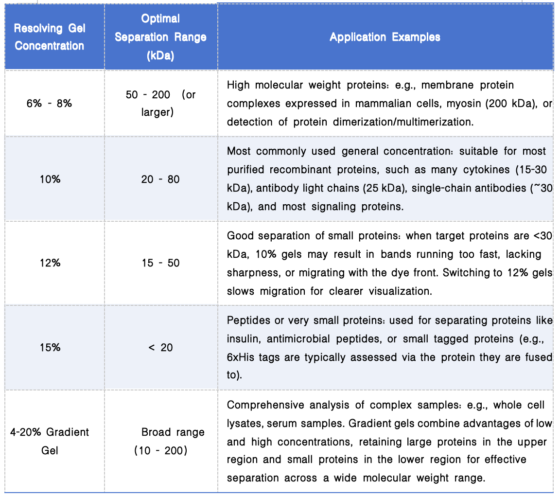
2. Selection of Resolving Gel Concentration
III. SDS-Polyacrylamide Gel Electrophoresis Procedure
1. Preparation of SDS-Polyacrylamide Gels
(1) Assemble glass plates and casting stand, and perform a leak test with water. If leakage occurs, adjust plate assembly until sealing is achieved before proceeding.
(2) Prepare the resolving gel (lower gel): Mix gel solution according to the desired concentration protocol, add polymerization initiators (APS and TEMED), mix well, and carefully dispense the solution into the assembled plate cavity using a pipette. Gently overlay with distilled water or ethanol to create a flat interface.
(3) Prepare the stacking gel (upper gel): Discard the overlay liquid (water or ethanol) and absorb residual liquid with filter paper. Prepare the stacking gel solution, pour it onto the polymerized resolving gel, and gently insert the comb. Avoid disturbing the casting stand during gel polymerization to prevent bubble formation.
2. Sample Preparation
Mix protein samples with an appropriate volume of loading buffer (containing SDS/reducing agent/glycerol/bromophenol blue). Heat at 100°C in a metal bath or water bath for 5-10 minutes. For native-PAGE, this heating step is omitted, and the loading buffer must not contain SDS or reducing agents.
3. Loading and Electrophoresis
(1) Mount the prepared gel cassette onto the electrode assembly and place it into the electrophoresis tank.
(2) Fill the inner chamber with fresh SDS running buffer (containing Tris, glycine, SDS). The outer chamber may be partially filled with recirculated buffer.
(3) Carefully remove the comb and inspect the wells for straightness and integrity. Rinse wells with a syringe or pipette to remove any unpolymerized acrylamide fragments.
(4) Using a fine pipette tip or gel-loading tip, slowly load the prepared samples into the wells. Load steadily to prevent sample from flushing out of the wells.
(5) Close the lid, connect the power supply (red to red, black to black), and run at constant voltage (typically 80 V) during the stacking phase, allowing samples to concentrate into a sharp line. When the bromophenol blue front reaches the resolving gel and compresses into a straight line, increase the voltage to 120-150 V (constant). Continue running until the bromophenol blue dye front reaches the bottom edge of the gel, then turn off the power supply.
4. Staining and Destaining
(1) Gel removal: Remove the glass plates from the apparatus and gently pry apart the short plate using a spatula. Cut away the stacking gel and transfer the resolving gel to a staining container.
(2) Staining: Add Coomassie Brilliant Blue R-250 staining solution (typically dissolved in methanol/acetic acid) ensuring complete coverage of the gel. Incubate with gentle shaking on an orbital shaker for 30-60 minutes (or heat in a microwave for 10-20 seconds to accelerate, taking care to avoid boiling).
(3) Destaining: Discard the staining solution (may be saved and reused multiple times) and add destaining solution (typically a methanol/acetic acid/water mixture). Shake gently on an orbital shaker, changing the destaining solution several times until the background becomes transparent and blue protein bands are clearly visible.
KMD Bioscience provides fundamental molecular biology services including SDS-PAGE validation, Western Blot detection, and ELISA assays. Additionally, we offer advanced services built upon these techniques, such as protein expression and functional validation, and protein-protein interaction analysis, supporting the research endeavors of numerous scientists.
引用:
[1] Matsumoto H, Haniu H, Komori N. Determination of Protein Molecular Weights on SDS-PAGE. Methods Mol Biol. 2019;1855:101-105.
[2] Brunelle JL, Green R. One-dimensional SDS-polyacrylamide gel electrophoresis (1D SDS-PAGE). Methods Enzymol. 2014;541:151-9.
[3] Kielkopf CL, Bauer W, Urbatsch IL. Sodium Dodecyl Sulfate-Polyacrylamide Gel Electrophoresis of Proteins. Cold Spring Harb Protoc. 2021 Dec 1;2021(12).
- SDS-PAGE
- SDS-Polyacrylamide Gel Electrophoresis

- Protein Platform
- Protein Expression Services
- Protein Modification & Detection Services
- Protein Interaction Services
- Protein Purification Service
- Antibody Discovery Platform
- VHH Antibody Platform
- Phage Display Platform
- Antibody Humanization Platform
- Single B Cell Screening Platform
- Antibody Expression & Validation Platform
- Peptide Library Platform
- Custom Antibody Platform
- Innovative Drug Discovery Platform
- Product Center
- Proteins
- Antibodies
- Raw Materials Reagents
- Small Molecule Reagents
- Peptide Products
- Nanoparticle Products
- Contact Us
- Phone:400-621-6806
- Mobile:13110936064(WeC
- E-mail:[email protected]
- Address:Room 108, Building 10, No. 6 Hechang Road, Wuqing Development Zone, Tianjin City
-

WeChat
-

Service Account
-
 400-621-6806
 -
 3599163699
 -
 Monday to Friday 9:00-18:00
 -

-
 0
0
-
