


Exploration of Isoquinoline Alkaloids as Potential Inhibitors against Human Islet Amyloid Polypeptide
- 2026-05-14
- 97
I. Literature Background
1. Type-2 Diabetes Mellitus (T2DM)
Type-2 diabetes mellitus has a high incidence, numerous complications, and is difficult to treat, making it one of the most concerning public health issues. Human islet amyloid polypeptide (hIAPP) is closely associated with T2DM because its abnormal self-assembly leads to membrane damage and cellular dysfunction. Developing potential inhibitors to prevent hIAPP fibrillation is a promising strategy for intervention and treatment of diabetes.
2. Human Islet Amyloid Polypeptide (hIAPP)
hIAPP is a hormone secreted by pancreatic β cells, consisting of 37 amino acids. It can cross the blood-brain barrier, activate specific receptors in the brain, inhibit glucagon release, and play a key role in maintaining glucose homeostasis. However, under abnormal conditions, misfolded hIAPP forms toxic oligomers and amyloid fibrils, thereby disrupting the membrane barrier and leading to β-cell death. Meanwhile, amylin can accumulate in vital organs such as the heart, brain, and kidney, causing more severe complications. Many recent studies have focused on hIAPP to inhibit its aggregation and find effective drug candidates for preventing and treating T2DM.
3. Research Direction
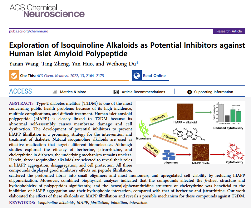
Natural isoquinoline alkaloids have been used as potent drugs against different biomolecules. Although studies have explored the efficacy of berberine, jatrorrhizine, and chelerythrine in diabetes, the underlying mechanisms remain unclear.
Based on the above, a research paper titled "Exploration of Isoquinoline Alkaloids as Potential Inhibitors against Human Islet Amyloid Polypeptide" was published in ACS Chemical Neuroscience (IF=5.78), with Professor Weihong Du from the Department of Chemistry, Renmin University of China as the corresponding author. In this study, three isoquinoline alkaloids were selected to reveal their roles in hIAPP aggregation, disaggregation, and cytoprotection. All three compounds effectively inhibited peptide fibrillation, dispersed fibrils into small oligomers and mostly monomers, and upregulated cell viability by inhibiting hIAPP oligomerization.
4. Differences Among the Three Isoquinoline Alkaloids
▶Berberine(BBR)
Berberine has attracted widespread attention due to its broad distribution and pharmacological effects. Recent research on BBR has focused on its multi-target potential in neurodegenerative diseases. BBR affects the biological functions of cholinesterase and monoamine oxidase, as well as the aggregation of β-amyloid (Aβ) associated with Alzheimer's disease. In addition, BBR is the main hypoglycemic component of Coptis alkaloids, promoting the repair of pancreatic β cells and enhancing glucose utilization in hepatocytes. Its glucose-lowering effect has been confirmed in diabetic mice and patients.
▶Jatrorrhizine(JAT)
Jatrorrhizine (JAT) is a metabolite of BBR, sharing the same tetracyclic skeleton, and can be obtained by opening the oxygen-containing five-membered ring at the terminal of BBR. The terminal hydroxyl modification increases the polarity of JAT, which may be related to the physiological activity of BBR in vivo.JAT is also a major hypoglycemic component of Coptis, specifically binding to α-glucosidase.
▶Chelerythrine (CHE)
Chelerythrine (CHE) is an active component of the traditional natural medicinal plant Chelidonium majus, possessing significant anti-tumor, antibacterial, antifungal, antipyretic, and detoxifying activities. CHE differs from BBR in that it has a benzo[c]phenanthridine structure. The N atom on ring 2 is methylated, thus reducing the polarity of this compound. CHE has been used as a novel bifunctional scaffold targeting cholinesterase and Aβ aggregation. Due to its potential binding to PPARγ, it may improve insulin sensitivity.
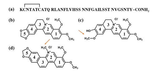
Figure 1. Differences among the three isoquinoline alkaloids.
II. Research Findings
1. Inhibitory Effect of Isoquinoline Alkaloids on hIAPP Aggregation
ThT assay was used to study the aggregation behavior of hIAPP in the absence or presence of different molar ratios of the alkaloids. The results showed that CHE exhibited a more pronounced inhibitory effect on hIAPP aggregation than the other two alkaloids.
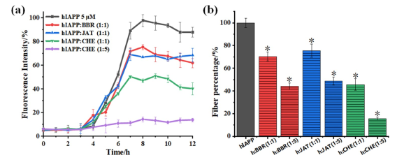
Figure 2. Inhibitory effect of isoquinoline alkaloids on hIAPP aggregation.
2. Analysis of Particle Size and Morphological Changes of hIAPP
Dynamic light scattering (DLS) provided information on particle size distribution in solution. The results indicated that all three alkaloids, especially CHE, effectively dispersed peptide aggregates, confirming the inhibitory effect of alkaloids on hIAPP aggregation.
Atomic force microscopy (AFM) further confirmed the inhibitory effect of the three isoquinoline alkaloids on hIAPP fibrillation. After 72 h of incubation at 310 K, the peptide alone exhibited unique long, thick, and unbranched fibrillar structures, consistent with the ThT and DLS data.
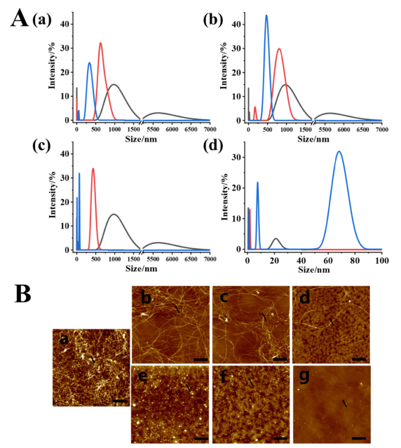
Figure 3. Particle size and morphological analysis of hIAPP.
3. Remodeling of Preformed hIAPP Fibrils
Many inhibitors can remodel mature fibrils, holding great promise in drug development. In this study, the remodeling of hIAPP fibrils was assessed by ThT kinetic analysis and AFM imaging. The results demonstrated that all three alkaloids effectively remodeled mature fibrils; the peptide disaggregation process was essentially completed within 9 hours, with CHE showing relatively stronger disaggregation capacity. The inhibitory effects of the three alkaloids were similar to their disaggregation capacities at the same molar ratios.
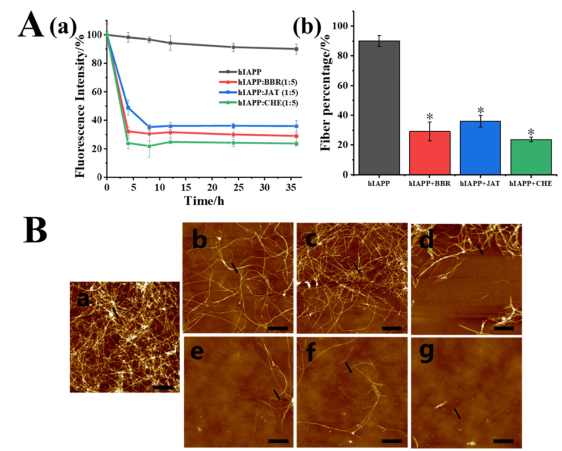
Figure 4. Remodeling of preformed hIAPP fibrils.
4. Reduction of Peptide Oligomerization
Enzyme-linked immunosorbent assay (ELISA) was used to detect the reduction of peptide oligomerization by the alkaloids. The results indicated that all three alkaloids reduced peptide oligomerization, and this method could distinguish spherical oligomers, revealing differences between CHE and the other two oligomeric species.
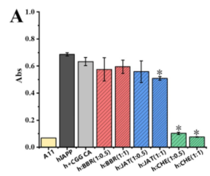
Figure 5A. Reduction of peptide oligomerization.
5. Modulation of hIAPP-Induced Cytotoxicity
Oligomers and fibrils formed by hIAPP aggregation can lead to cytotoxicity and β-cell dysfunction. The (4,5-dimethylthiazol-2-yl)-2,5-diphenyltetrazolium bromide (MTT) assay was performed using rat insulinoma (INS-1) cells to evaluate the protective effect of the alkaloids on pancreatic cells. The data indicated that the self-toxicity of these three alkaloids was relatively higher than that of some well-known inhibitors such as EGCG and curcumin, which may limit the application of these alkaloids in T2DM. However, at low concentrations, the upregulatory effects of the three small-molecule compounds were superior to curcumin. Therefore, they still effectively inhibit hIAPP fibrillation, and their intrinsic mechanisms related to T2DM require further study.
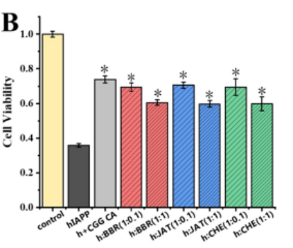
Figure 5B. Modulation of hIAPP-induced cytotoxicity.
6. Alkaloid-Induced Conformational Changes of hIAPP
Circular dichroism (CD) spectroscopy was used to determine the secondary structure of hIAPP in the presence of the alkaloids. The results showed that to some extent, all three alkaloids prevented the structural transition of hIAPP from its initial structure to β‑sheet components, thereby inhibiting amyloid fibril formation. When the molar ratio increased, CHE caused greater conformational changes. The CD results were largely consistent with ThT, AFM, and ELISA analyses, indicating that the compounds induced secondary structure changes and inhibited hIAPP aggregation.
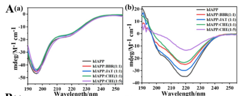
Figure 6A. Alkaloid-induced conformational changes of hIAPP.
7. Interaction Between Isoquinoline Alkaloids and hIAPP
Nile red quenching, isothermal titration calorimetry (ITC), and intrinsic fluorescence quenching were used to analyze and demonstrate the binding behavior of the alkaloids to hIAPP, and possible interaction mechanisms were deduced. The results indicated that all three alkaloids bound to unaggregated hIAPP, thereby stabilizing the monomeric structure and reducing the formation of hydrophobic regions on the hIAPP surface. CHE exhibited the greatest effect on hIAPP hydrophobicity.
Figure 6B. Interaction between isoquinoline alkaloids and hIAPP.
ITC experiments performed at 310 K showed that the ΔG values for all titrations were negative, meaning that the binding of the alkaloids to hIAPP was spontaneous. Moreover, at this temperature, all reactions were endothermic, with positive ΔH values.
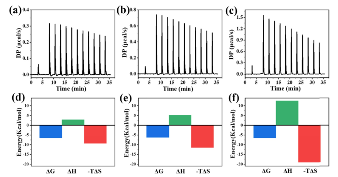
Figure 7. ITC thermograms of 50 μM hIAPP titrated with BBR, JAT, and CHE (310 K).
For all three alkaloids, both ΔH and ΔS were greater than zero, indicating that hydrophobic interactions dominated the binding between hIAPP and the three compounds. In addition, the dissociation constants and the number of binding sites (N) are shown in Table 1. The N values suggest non‑specific binding interactions between the small molecules and the peptide, as found for other inhibitors of Aβ and hIAPP aggregation.
Table 1. Thermodynamic parameters for the binding of compounds to hIAPP at 310 K from ITC experiments.

The A-11 antibody (KMD Bioscience) used for ELISA experiments at 277 K was supplied by KMD Bioscience and was a critical experimental material in this study. Research on peptides continues to be a hot topic. KMD Bioscience offers a variety of peptide‑related services, including but not limited to peptide library screening services, peptide synthesis, custom peptide libraries, etc., meeting diverse customer experimental needs.
[1] https://doi.org/10.1021/acschemneuro.2c00206.
Summary of Common FAQs
Q1:How to design a library?
A1:
Sequence length: Typically 7-15 amino acids. Too short results in insufficient diversity; too long may reduce quality due to synthesis errors.
Randomized codon usage: The most commonly used degenerate codons are NNK or NNG (N = A/T/C/G, K = G/T). These encode all 20 amino acids while reducing stop codons to only one (TAG).
Library size: The total number of independent clones should be no less than 10⁹. A larger library size covers a broader sequence space.
Q2:How to evaluate library quality?
A2:
Library size: The total number of independent clones in the library. A larger library size covers a broader sequence space, increasing the chance of finding high‑affinity ligands.
Diversity: Assessed by randomly picking a small number of clones for sequencing. Sequences should be evenly distributed without obvious bias. A high‑quality library should possess both large library size and high diversity.
Q3:What should be done about false‑positive / non‑specific binding in screening results?
A3:False‑positive rates can be effectively reduced by the following strategies:
Use blocking agents: Block non‑specific sites on the target and container surfaces with BSA, non‑fat milk, etc., before screening.
Increase washing stringency: Increase the number of washes or add mild detergents (e.g., Tween‑20) before the elution step.
Perform “negative selection”: Pre‑incubate the library with a matrix lacking the target (e.g., uncoated beads or plates) to absorb clones that bind to the matrix itself, thereby improving screening specificity.
Q4:Why sometimes high‑affinity peptides cannot be isolated?
A4:
Possible reasons include:
Target issues: Loss of target protein activity, incorrect conformation, or masking of the epitope due to immobilization.
Library issues: Library size too small or insufficient diversity to cover potential binding sequences.
Overly stringent screening conditions: Washing or elution conditions too harsh, causing loss of all binding clones.
The target itself is “undruggable”: The surface of certain targets may lack a suitable “pocket” for small molecule or peptide binding.
Q5:What are the application areas of peptide library screening?
A5:
Drug discovery: Identification of peptide‑based lead compounds and development of novel therapies for diseases such as cancer and autoimmune disorders. For example, peptides with nanomolar affinity for transferrin receptor 1 (TfR1) have been successfully isolated from libraries, offering potential solutions for targeted drug delivery.
Epitope mapping: Localization of linear or conformational epitopes on target proteins, providing critical sequence information for vaccine design and antibody development.
Protein‑protein interactions: Investigation of protein interaction networks and identification of key domains or motifs to help understand complex cell signaling pathways.
GPCR ligand screening: G protein‑coupled receptors (GPCRs) are important drug targets; peptide libraries can be used to screen for their agonists or antagonists.
Enzyme substrate or inhibitor screening: Discovery of substrates for specific enzymes or potent inhibitors for the development of metabolic or antiviral drugs.
Diagnostics and biomarker discovery: Screening of specific peptides as diagnostic probes or biomarkers for in vitro diagnostics and early cancer detection.

- Protein Platform
- Protein Expression Services
- Protein Modification & Detection Services
- Protein Interaction Services
- Protein Purification Service
- Antibody Discovery Platform
- VHH Antibody Platform
- Phage Display Platform
- Antibody Humanization Platform
- Single B Cell Screening Platform
- Antibody Expression & Validation Platform
- Peptide Library Platform
- Custom Antibody Platform
- Innovative Drug Discovery Platform
- Product Center
- Proteins
- Antibodies
- Raw Materials Reagents
- Small Molecule Reagents
- Peptide Products
- Nanoparticle Products
- Contact Us
- Phone:400-621-6806
- Mobile:13110936064(WeC
- E-mail:[email protected]
- Address:Room 108, Building 10, No. 6 Hechang Road, Wuqing Development Zone, Tianjin City
-

WeChat
-

Service Account
-
 400-621-6806
 -
 3599163699
 -
 Monday to Friday 9:00-18:00
 -

-
 0
0
-
