Home
>>
Technical Platform
>>
Molecular Platform
>>
Fluorescence In Situ Hybridization (FISH)
Download Center
Quick Inquiry & Consultation
Fluorescence In Situ Hybridization (FISH)Introduction

KMD Bioscience has been dedicated to cell biology technologies for many years. With an experienced team of scientific experts, highly skilled laboratory personnel, a well-established Cell Culture Platform, and comprehensive laboratory equipment and facilities, we have accumulated extensive expertise in providing Fluorescence In Situ Hybridization (FISH) Services. Over time, we have built a robust FISH Technology Platform that enables us to deliver one-stop services—from probe design, sample preparation, hybridization, and gene expression studies to data analysis. Our standardized procedures and technical systems ensure improved accuracy, minimized false positives and negatives, and consistently high-quality results. Our fluorescent reagents and probes are cost-effective and safe, enabling rapid completion of experiments. This approach significantly reduces the complexity and time required for laboratory-based FISH operations, lowers experimental costs, conserves valuable samples, and provides reliable support for customers’ research projects.

FISH is a non-radioactive molecular cytogenetic technique. It utilizes fluorescently labeled probes that specifically bind to target chromosomal sequences, revealing highly complementary regions. These hybridized regions are typically visualized using a fluorescence microscope. FISH serves as a powerful tool for karyotyping, cell genotyping, cancer diagnostics, species identification, and gene expression analysis, allowing researchers to visualize DNA or locate RNA within cells. By providing a means to map or visualize genetic material, such as specific genes or gene fragments,  FISH is indispensable for understanding chromosomal abnormalities and other genetic mutations at the single-cell level. Its flexibility, safety, and high specificity make FISH broadly applicable across various species (e.g., plant or animal tissues) and highly versatile in genomic structural studies, fine-scale chromosomal variation analysis, prenatal diagnostics, tumor genetics, and microbial analyses.
Principles of Fluorescence In Situ Hybridization (FISH)

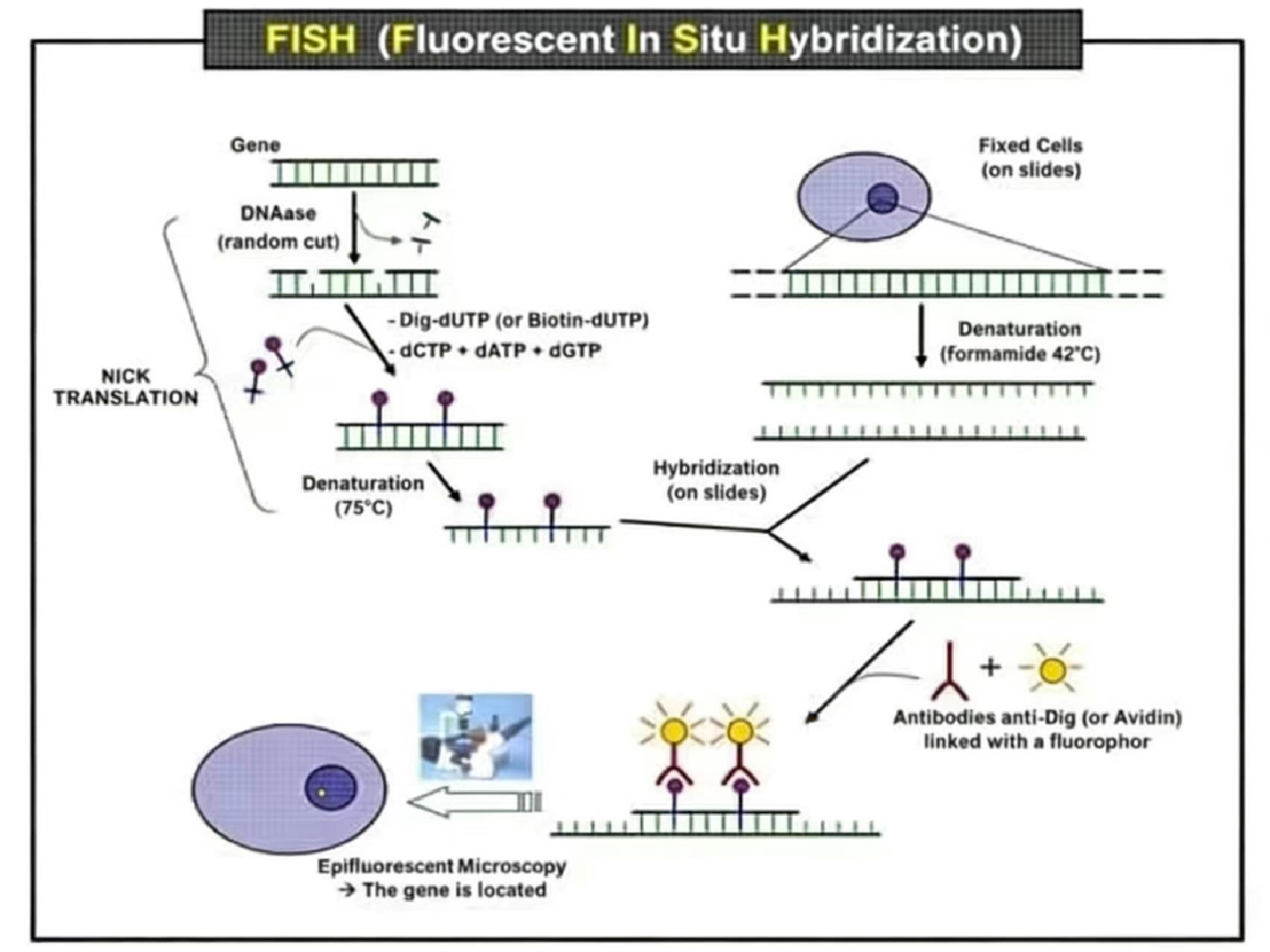
In Situ Hybridization (ISH) refers to the process in which a labeled nucleic acid probe of known sequence hybridizes with target nucleic acids within cells or tissue sections, enabling precise localization and quantification of specific nucleic acid sequences. The basic principle relies on the ability of two single-stranded nucleic acid fragments to form double-stranded molecules, such as DNA-DNA, DNA-RNA, or RNA-RNA, through hydrogen bonding under appropriate conditions. Labeled DNA or RNA probes (radioisotopes, fluorophores, biotin, digoxigenin, or other non-radioactive labels) are hybridized with nucleic acids (RNA or DNA) in cells or tissue sections. Detection is performed using autoradiography or other visualization methods to observe and localize the target mRNA or DNA under light or electron microscopy.

Fluorescence in situ hybridization replaces isotopic labeling with fluorescent labeling, making the method safer and more economical. Additionally, probes labeled with different fluorophores can be used simultaneously to detect multiple sequences within the same sample.

Fluorescent labeling of FISH probes can be performed using direct or indirect methods. Indirect labeling involves biotin-labeled DNA probes detected via fluorescently tagged avidin or streptavidin, often amplified using an avidin–biotin–fluorophore complex. Direct labeling covalently attaches fluorophores to nucleotides or the sugar-phosphate backbone of the probe, or incorporates fluorescent nucleotide triphosphates during nick translation labeling. While direct labeling simplifies the detection process, it is less sensitive than indirect labeling due to the absence of signal amplification.

FISH is characterized by rapidity, high sensitivity, and specificity. It allows simultaneous quantitative analysis of multiple cells in both metaphase and interphase; detection of cryptic or microstructural chromosomal abnormalities and complex karyotypes; and visualization of DNA fragments and genes, including their relative positions and orientations with precise spatial resolution.

图片22.png

Figure 1 Schematic diagram illustrating the principle of gene localization in the nucleus using FISH (Fluorescence In Situ Hybridization).

Service scope

We offer FISH Services covering a wide range of research areas, including human, animal, plant, and microbial studies. Based on specific customer requirements, our technical experts provide professional one-on-one customized solutions and detection services for samples from different species, delivering strong support for research projects. For example, Microbial FISH Detection Services involve detecting and identifying microbial rRNA, enabling in situ detection, identification, and selection of specific bacterial and archaeal groups.

image.png

Service process

图片23.png

Provided by the customer

-- Relevant information of the samples to be tested.

-- Relevant information or sequence of the target gene to be tested.

-- Sample quantity information.

-- Detection requirements.

Our technical experts provide a comprehensive customized Fluorescence In Situ Hybridization (FISH) service, ranging from probe design and chromosome/cell/bacteria preparation/culture to final result presentation. For details, please fill out our inquiry form or contact our staff directly.

The delivery content of KMD Technology

-- Remaining probes, slides, and samples.

-- Original hybridization and staining images.

-- A detailed experimental report (including complete workflow and result analysis).

Service advantages

-- High-specificity probe design services.

-- Short service timeline (1–2 weeks) with precise localization, delivering results promptly.

-- Broad detection range covering human, animal, plant, and microbial samples.

-- Imaging tailored to customer needs.

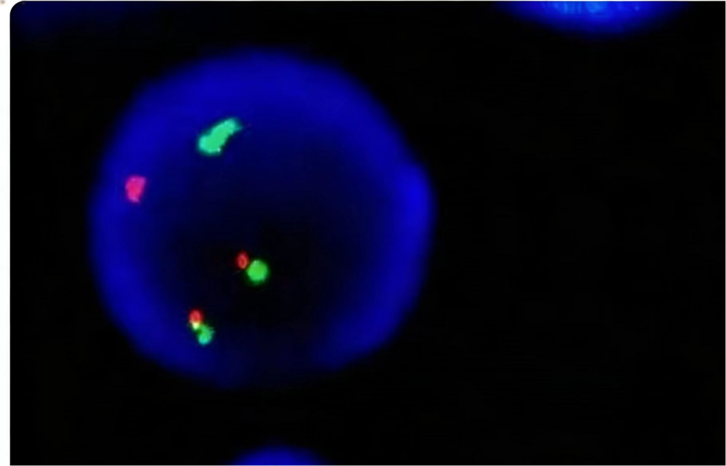
-- Capability to visualize up to three colors (red, green, and blue) simultaneously in a single nucleus for multiplex sequence detection.

Login

Don’t have an account?Sign Up Now

Register

Already have an account?Log In Now