


A Guide to Avoiding Pitfalls in Peptide Library Screening: A Practical Analysis of Nonspecific Binding and Misinterpretation of Data in Phage Display
- 2026-08-21
- 211
Peptide library screening---particularly phage display technology---has served as the gold standard for peptide screening and peptide drug discovery since its invention by George P. Smith in 1985[1]. However, anyone who has conducted peptide library screening knows that while the workflow appears straightforward—loading, binding, elution, amplification, and sequencing—the actual process is fraught with mysteries, and nonspecific binding hangs like the Sword of Damocles over every researcher’s head.Whether using pre-made peptide libraries or custom peptide libraries, the technical capabilities of the peptide service provider directly impact the success or failure of the screening results.
The most significant drawback of phage display is that phages displaying peptides with no binding affinity for the target are nonspecifically enriched and selected. In this article, I'll discuss common pitfalls related to nonspecific binding that researchers often encounter during peptide library screening and, drawing on practical experience in peptide services, provide a guide to help you avoid them.
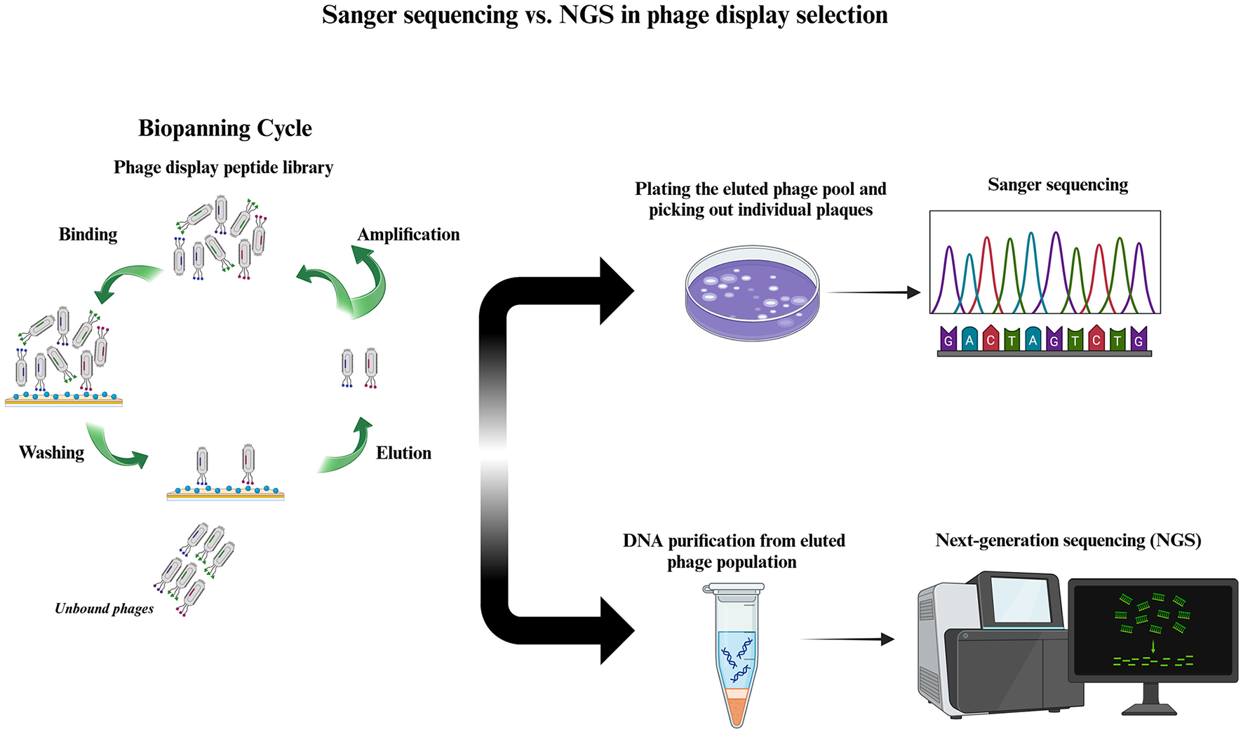
Figure 1: Schematic Diagram of the Entire Phage Display Screening Process[1]
I. Insufficient Purity of the Target Protein
(i) Phenomenon
After the first round of peptide library screening, the phage titer eluted appears promising, and the second round of amplification went smoothly, but when the samples were sent for sequencing after the third round, the resulting sequences were highly diverse, with an extremely low repetition rate—let alone any shared motifs. After repeated verification, it was found that these sequences had absolutely no affinity for the target protein.
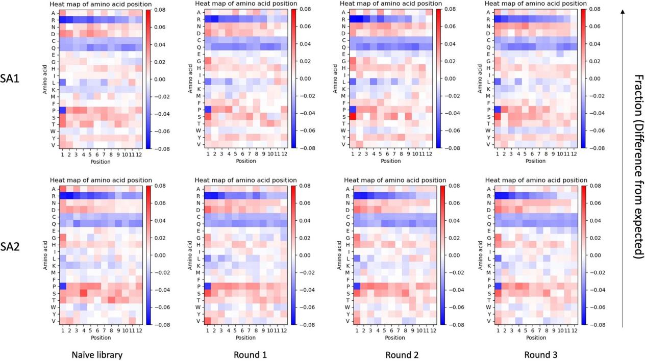
Figure 2: Changes in Sequence Abundance Before and After Library Amplification[2]
(ii) Origin
The origin is insufficient purity of the target protein. Screening of a phage library follows the principle that the most abundant protein dominates; if your target protein is contaminated with 5% of a foreign protein, the clones in the phage library that happen to bind to that foreign protein will be disproportionately enriched during the selection process, resulting in sequences that lack affinity for the target protein.
(iii) Our Solutions
Ø Target Quality Control Prerequisites: The target protein must undergo SDS-PAGE electrophoresis, and its purity must be >95% before proceeding to the next step of library screening. This is the most critical quality control step before peptide screening..
Ø Set up a non-target control: In each round of screening, include blank wells without the target protein and perform the screening in parallel[1].
Ø Screening and Purification: First pass the phage library through wells coated with contaminating proteins to remove phages that bind nonspecifically, then use the supernatant to bind the actual target.
II. Phage Amplification Issues
(i) Phenomenon
After the first round of selection, the eluted phages were used for titer determination, and the plates were covered with dense plaques. After amplification, they were introduced into the second round, and everything proceeded normally. However, after the third round, a high-frequency sequence appeared in the sequencing results; when the peptide corresponding to this sequence was synthesized and tested via ELISA, the signal was extremely weak.
(ii) Origin
The origin of this phenomenon is competitive inhibition during the phage amplification phase. Amplification is an essential step in the phage screening process, but it is also a double-edged sword, as it can lead to the enrichment of targets through nonspecific binding.
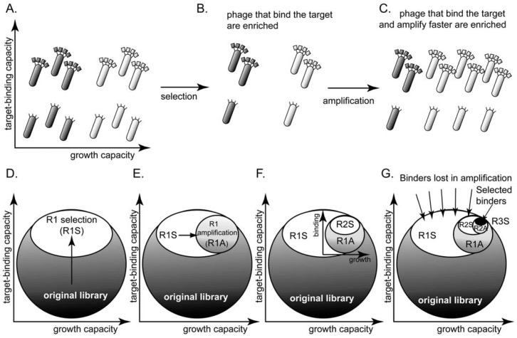
Figure 3: Flowchart of the Phage Display Library Construction Process[3]
(iii) Our Solutions
Ø Control the number of screening rounds: More peptide library screening rounds are not necessarily better. We typically limit the process to 3–4 rounds; more than 4 rounds only marginally improve binding affinity, reduce sequence diversity and lead to an increase in nonspecific binding[3].
Ø Control the amplification factor: After each round of amplification, do not strive for an excessively high titer —this is a common operational pitfall in peptide library screening; proceed to the next round once the titer reaches the level typically required for input to reduce the likelihood of competitive accumulation.
Ø Monitoring Library Composition Before and After Amplification via NGS: Perform NGS sequencing on the eluted products before amplification and the amplified library, and compare changes in their sequence distributions.
III. A High Sequencing Frequency Does not Necessarily Indicate High Affinity
(i) Phenomenon
Analysis of the NGS output data revealed that the top-ranked clone accounted for more than 30% of the total. However, SPR affinity assays conducted on the synthesized version of this sequence showed that its KD value was only in the micromolar range, indicating limited affinity. In contrast, sequences that were not as highly ranked exhibited very strong affinity.
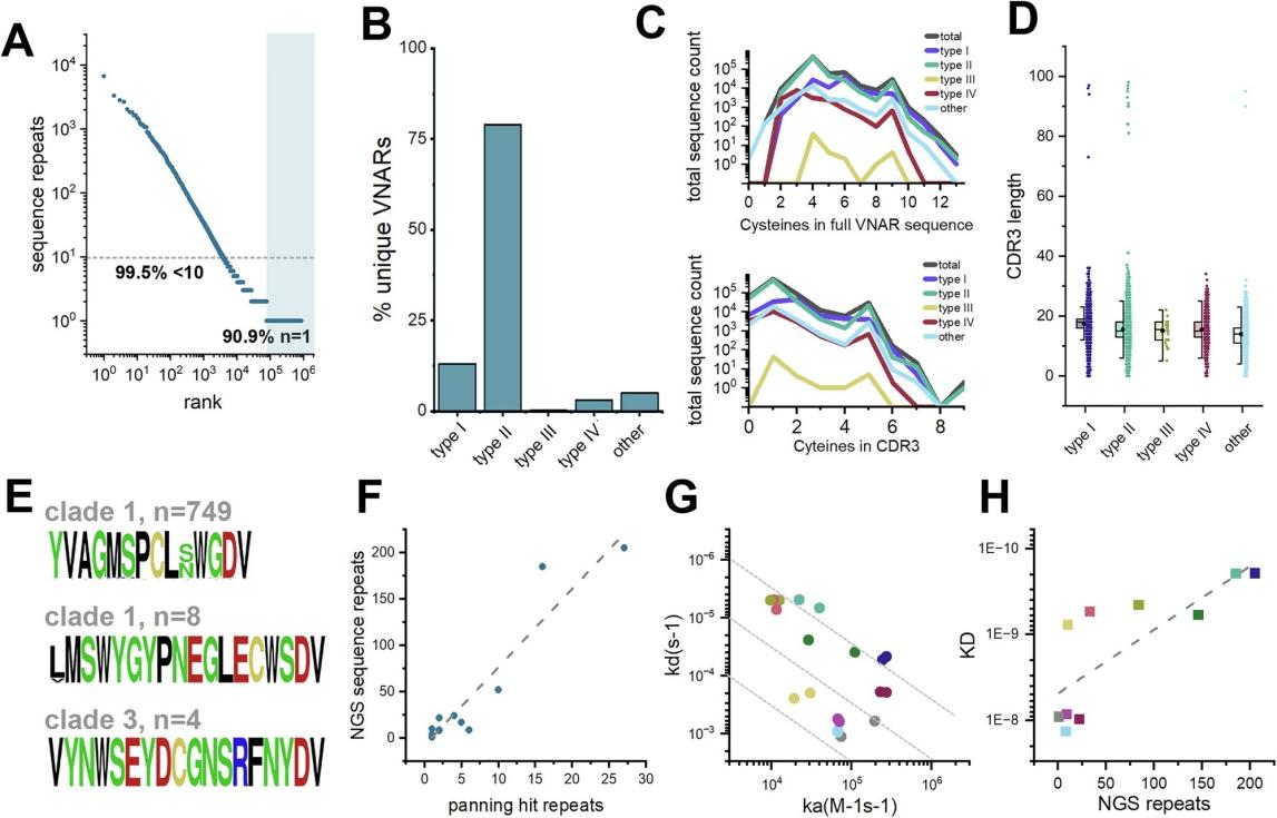
Figure 4: Scatter Plot Showing the Correlation Between NGS Sequence Repeat Counts and Affinity[4]
(ii) Origin
This is the issue of decoupling sequencing frequency from binding affinity. NGS can tell you which sequences have been enriched, but it cannot directly tell you which ones bind most strongly[1].
(iii) Our Solutions
Synthesis validation is not based solely on the highest scores: For the top 20 NGS sequences, sort them by enrichment fold and select those with high enrichment folds—but not necessarily the highest absolute frequencies—for simultaneous synthesis and validation.
Incorporating AI-assisted screening: Deep sequencing data can be used to train machine learning models to identify sequence features in weakly enriched regions that may have functional potential.
Looking back at the three pitfalls mentioned above, their root causes can all be traced back to the same core issue: peptide library screening is not a simple, standardized assembly line, but rather a systematic engineering process that requires peptide service providers to possess deep technical expertise and meticulous quality control at every stage. Insufficient purity of the target protein causes the screening to veer off course; competitive bias during amplification distorts the true composition of the library; and the decoupling of NGS frequency from affinity serves as a warning that we cannot simply equate high-throughput data with functional validation results.
Selecting a professional CRO service provider is the key to effectively mitigating these risks. Leveraging its proven phage display platform, KMD Bioscience has developed a systematic solution to address the challenges mentioned above. In addition, KMD Bioscience offers a one-stop, closed-loop service ranging from peptide synthesis and activity validation to animal immunization and monoclonal antibody production, ensuring full control over the entire process from candidate peptide screening to functional validation.
KMD Bioscience’s phage display platform can generate up to 1013 peptide or antibody clones per mL, offering a variety of pre-built libraries—including random 7-peptide libraries, 12-peptide libraries, and cyclic 7-peptide libraries—as well as customized services using the T7 and M13 phage systems. The essence of peptide library screening is finding that “needle” in a haystack of 109 possibilities, and a professional platform combined with rigorous quality control is the key to making this “search for a needle in a haystack” both efficient and reliable.
[1]Bakhshinejad B, Kjaer A. NGS and the design of an optimized phage display workflow for peptide discovery. Amino Acids. 2025 Dec 26;58(1):5. doi: 10.1007/s00726-025-03492-z. PMID: 41452380; PMCID: PMC12775084.
[2]Sinkjaer AW, Sloth AB, Andersen AO, Jensen M, Bakhshinejad B, Kjaer A. A comparative analysis of sequence composition in different lots of a phage display peptide library during amplification. Virol J. 2025 Feb 1;22(1):24. doi: 10.1186/s12985-024-02600-x. PMID: 39893369; PMCID: PMC11786364.
[3]Derda R, Tang SK, Li SC, Ng S, Matochko W, Jafari MR. Diversity of phage-displayed libraries of peptides during panning and amplification. Molecules. 2011 Feb 21;16(2):1776-803. doi: 10.3390/molecules16021776. PMID: 21339712; PMCID: PMC6259649.
[4]Gunaratne GS, Gallant JP, Ott KL, Broome PL, Celada S, West JL, Mixdorf JC, Aluicio-Sarduy E, Engle JW, Boros E, Meimetis L, Lang JM, Zhao SG, Hernandez R, Kosoff D, LeBeau AM. The characterization of variable new antigen receptors targeting FAP isolated from a novel immunized library. Commun Biol. 2025 Aug 13;8(1):1210. doi: 10.1038/s42003-025-08610-x. PMID: 40804296; PMCID: PMC12350670.
FAQs
Q1: “Setting up a non-target control” sounds simple, but how exactly is it done? How should the results be analyzed to effectively distinguish true binding from background?
The specific procedure for a non-target control is as follows: Coat parallel wells with the same blocking solution as the experimental group (e.g., BSA or skim milk), but do not add the target protein; all other steps (blocking, library incubation, washing, elution, and amplification) are identical. The key lies in the cross-comparison during the NGS data analysis stage—excluding high-frequency clones that appear in both the experimental group and the no-target control group, and retaining only those sequences that are significantly enriched in the experimental group.
Specific procedure for elimination screening: First, incubate the phage library with wells or magnetic beads immobilized with “contaminating proteins” (such as purified tagged proteins or host cell lysates), collect the supernatant containing unbound phages, and then transfer this supernatant to wells coated with the target protein for forward screening. Since all clones bound to contaminating proteins are removed in the first step, the target specificity of the subsequent forward screening is significantly improved.
Q3: The article says to “limit the screening to 3–4 rounds,” but if the proportion of positive clones in the sequencing results is very low after 3 rounds, should we proceed to a 5th round? How do we determine when to stop?
To determine whether additional rounds are needed, it is recommended to conduct a comprehensive assessment based on two indicators:
(i) Changes in the Input-to-Output Ratio—If the increase in the recovery rate (output pfu/input pfu) from Round 3 to Round 4 narrows significantly, it indicates that enrichment is approaching saturation, and continuing to increase the number of rounds will only further reduce diversity;
(ii) Monitoring NGS Sequence Diversity—If the number of unique sequences in the library has dropped significantly by the third round and clear common motifs have emerged, the process can be stopped.
Q4: How exactly is the enrichment fold mentioned in NGS analysis calculated? What threshold is typically set when using this metric to screen candidate peptides?
The enrichment factor is typically calculated as log₂(Output/Input), which is the ratio of the frequency of a specific sequence in the post-selection library (Output) to its frequency in the pre-selection library (Input). This metric reflects the degree to which the sequence is enriched under selection pressure, rather than its absolute abundance.
Q5: The article states that “the sequences ranked 5th and 8th actually have better affinity.” Is it possible that sequences ranked 50th or even 500th might have better affinity? How many sequences should be screened during NGS analysis to be considered reasonable?
Theoretically, this possibility exists, but the probability decreases significantly as the rank drops. The recommended strategy is to reorder the top 20–50 NGS sequences by enrichment fold and select the 15–30 candidate sequences with the highest enrichment folds for synthetic validation; this is an empirical threshold that strikes a balance between validation costs and hit rates. For projects with high screening pressure (where target concentration decreases with each round and washing conditions are stringent), sequences with high enrichment multiples show a stronger correlation with measured affinity.
- Phage display technology
- peptide library screening
- peptide drug discovery
- non-specific binding

- Protein Platform
- Protein Expression Services
- Protein Modification & Detection Services
- Protein Interaction Services
- Protein Purification Service
- Antibody Discovery Platform
- VHH Antibody Platform
- Phage Display Platform
- Antibody Humanization Platform
- Single B Cell Screening Platform
- Antibody Expression & Validation Platform
- Peptide Library Platform
- Custom Antibody Platform
- Innovative Drug Discovery Platform
- Product Center
- Proteins
- Antibodies
- Raw Materials Reagents
- Small Molecule Reagents
- Peptide Products
- Nanoparticle Products
- Contact Us
- Phone:400-621-6806
- Mobile:13110936064(WeC
- E-mail:[email protected]
- Address:Room 108, Building 10, No. 6 Hechang Road, Wuqing Development Zone, Tianjin City
-

WeChat
-

Service Account
-
 400-621-6806
 -
 3599163699
 -
 Monday to Friday 9:00-18:00
 -

-
 0
0
-
