


9 Common Causes of Poor Phage Display Enrichment: An Expert Diagnostic Guide
- 2026-09-15
- 46
In the high-stakes arena of biopharmaceutical drug discovery and therapeutic antibody engineering, failure is expensive. Researchers routinely invest months constructing libraries and executing biopanning protocols, only to discover that their output titers have stagnated. When you execute three to four rounds of panning and your output-to-input ratio remains flat, you are experiencing a catastrophic failure in your selection process. Understanding the root Causes of Poor Phage Display Enrichment is not just an academic exercise; it is a critical commercial necessity required to salvage your research budget and project timeline.

From our experience engineering advanced biotherapeutics at KMD Bioscience, we observe a consistent pattern of failure among research teams attempting in-house panning. They often blame the target antigen, when in reality, the failure stems from fundamental errors in library construction, fluid dynamics during the washing phase, or profound biological toxicity within the E. coli host. In this uncompromising diagnostic guide, we will strip away the academic theory and provide hard, practical judgment. We will detail the 9 definitive Causes of Poor Phage Display Enrichment, explain exactly how to troubleshoot your protocol, and help you determine whether your project requires intervention from a professional custom antibody development service.
Quick Answer: Why Is Your Panning Failing?
If you are experiencing stagnant or negative enrichment ratios across your biopanning rounds, the primary Causes of Poor Phage Display Enrichment typically fall into these immediate categories:
Garbage In, Garbage Out: Your initial library lacks functional diversity or is dominated by truncated, non-expressing clones.
Antigen Denaturation: The target protein has lost its native conformation during the immobilization process on the immunotube or magnetic bead.
Matrix Binders Taking Over: Your library is actively selecting for streptavidin, BSA, or the plastic surface itself rather than your target.
Amplification Toxicity: High-affinity clones are biologically toxic to the E. coli host and are being outcompeted by fast-growing, non-binding junk clones during the rescue phase.
In most professional situations, we recommend aborting a failed panning campaign after round three. Do not waste capital sequencing a flat output. Instead, verify your library diversity or consult an antibody customization solutions provider to restructure your selection strategy.
Table of Contents
What is Phage Display Enrichment?
Phage display enrichment is the mathematical and biological verification that your Darwinian selection process is working. It is calculated by taking the output titer (the number of phages recovered after washing and elution) and dividing it by the input titer (the number of phages initially applied to the antigen).
In a healthy, successful panning campaign, this ratio should increase logarithmically across sequential rounds. For example, round one might yield an enrichment ratio of 10-6, round two 10-4, and round three 10-2. This progressive increase proves that phages displaying high-affinity binders are surviving the wash steps and multiplying. When this ratio stalls or drops, you are witnessing the Causes of Poor Phage Display Enrichment in real-time.
How Biopanning Should Work
The mechanics of the system are unforgiving. A library of bacteriophages—often a nanobody library construction service output—is incubated with an immobilized target antigen. Phages that lack affinity are washed away. The bound phages are then subjected to extreme pH (usually acidic elution with Glycine-HCl) or enzymatic cleavage to release them from the target. These surviving phages are then used to infect a fresh culture of E. coli (like TG1 or XL1-Blue) to amplify the population for the next round. If any variable in this delicate mechanical and biological chain is compromised, enrichment halts.
The 9 Causes of Poor Phage Display Enrichment
To successfully troubleshoot your campaign, you must ruthlessly evaluate your protocol against these nine definitive failure points.
1. Insufficient Initial Library Diversity
You cannot fish a trophy catch out of a barren lake. If your library claims a diversity of 109, but 90% of those clones contain frameshift mutations, stop codons, or severe folding defects, your functional diversity is actually 108 or less. Poor library construction is the leading factor among the Causes of Poor Phage Display Enrichment. Always verify your library via Next-Generation Sequencing (NGS) before initiating panning.
2. Antigen Denaturation During Immobilization
Proteins are highly fragile. When you coat a polystyrene immunotube with your target antigen using carbonate buffer at pH 9.6, you risk unfolding the protein. If the protein denatures, you will enrich for antibodies that bind to a linear, unfolded epitope. When you test these clones later in a functional assay against the natively folded protein, they will fail completely.
3. Proliferation of Matrix Binders (Background Noise)
In most professional situations, we utilize biotinylated antigens captured on streptavidin magnetic beads. However, streptavidin and the blocking agent (like BSA or milk) are massive, highly immunogenic targets. If you do not perform rigorous "depletion" or "negative selection" steps prior to exposing the library to your actual target, your output will be entirely dominated by useless anti-streptavidin or anti-BSA clones.
4. Amplification Bias and Host Toxicity
Not all phages grow at the same rate. Some antibody fragments are biologically toxic to the E. coli host. During the overnight rescue and amplification phase, clones that do not bind the target but happen to grow 10% faster will rapidly outcompete your toxic, high-affinity binders. This biological reality collapses library diversity and halts enrichment.
5. Inadequate Washing Stringency
Washing is where the actual selection occurs. If your wash buffer (typically PBS with Tween-20) lacks sufficient detergent, or if you do not increase the number of wash cycles in rounds two and three, you will carry over thousands of low-affinity, non-specific background phages. These background phages will dilute your high-affinity binders in the output.
6. Suboptimal Elution Conditions
If you have an incredibly high-affinity binder, a standard pH 2.2 Glycine-HCl elution might not be strong enough to break the bond. You will literally leave your best clones stuck to the plastic tube while recovering only moderate-affinity binders. In our testing, switching to Triethylamine (TEA) alkaline elution or utilizing competitive elution with soluble antigen often rescues seemingly failed campaigns.
7. Insufficient Antigen Concentration Control
To drive selection pressure, you must decrease the antigen concentration in subsequent rounds. If you use 10 µg/mL of antigen in round one, round two, and round three, you are applying zero evolutionary pressure. Without dropping the concentration, low-affinity binders will continue to survive, leading to flat enrichment curves.
8. Helper Phage Contamination
If your helper phage (e.g., M13KO7) is degraded or added at the incorrect Multiplicity of Infection (MOI), the packaging of your phagemid DNA will fail. The E. coli will simply produce wild-type helper phages that display no antibody fragments, flooding your output titer with useless viral particles.
9. Valency Artifacts (Avidity vs. Affinity)
If you are utilizing a multivalent display system rather than a true monovalent display, weak binders can latch onto the target using multiple arms. This "avidity" effect allows garbage clones to survive stringent washing. You must ensure your system promotes monovalent display to select strictly for high intrinsic affinity.
Benefits of Optimizing Your Protocol
Correcting the Causes of Poor Phage Display Enrichment yields immediate commercial benefits. A properly executed campaign compresses the discovery timeline from months to weeks. It allows you to isolate sub-nanomolar binders against highly toxic, non-immunogenic, or highly conserved human targets that traditional animal immunization simply cannot handle. Review the latest protein research insights to see how optimized libraries are accelerating oncology and autoimmune pipelines.
Limitations and Hard Truths of the Technology
We must present practical judgment: phage display is a robust in vitro tool, but it is not a perfect mirror of mammalian biology. The primary limitation is that bacteria do not perform post-translational modifications (PTMs) like glycosylation. A scFv or VHH that folds perfectly on the tip of an M13 phage might aggregate or lose affinity when reformatted into a full-length IgG and expressed in mammalian CHO cells.
Who Should Use Phage Display (And Who Shouldn't)
Who Should Use It: For commercial users, biotech startups, and diagnostic developers requiring rapid access to vast panels of recombinant antibodies, phage display is unparalleled. It is the definitive choice for generating nanobody antibody products where single-domain stability is required.
Who Does Not Need It: If your project strictly demands preserving the native heavy and light chain pairing of a natural immune response—such as isolating antibodies directly from a convalescent patient—phage display is the wrong tool. The random combinatorial nature of the library destroys native pairing. In these heavy-duty applications, you must transition to a single B cell screening service, which interrogates intact, naturally matured B cells. For more details on this alternative, consult the single B cell antibody discovery guidelines.
Common Mistakes During Execution
Expert Insight: The most frequent operational mistake we observe is researchers "over-panning" their libraries.
Executing five or six rounds of panning does not guarantee better antibodies; it guarantees diversity collapse. By round five, amplification bias has entirely taken over. You will typically be left with one or two fast-growing clones that dominate 99% of the output, wiping out the rare, high-affinity binders you were actually trying to isolate. Halt your panning at round three or four and proceed immediately to ELISA screening.
Expert Buying Considerations
If your internal team has spent months fighting flat enrichment ratios, it is time to perform a cost-benefit analysis. The reagent costs, sequencing fees, and labor hours wasted on failed panning vastly exceed the cost of outsourcing. When selecting a vendor to rescue your project, demand transparency regarding their library diversity metrics and their negative selection protocols. Staying updated with the antibody development news will help you identify partners utilizing the latest sequencing technologies to validate their libraries.
Summary and Troubleshooting Tables
Quick Summary Table: Diagnosing Flat Enrichment
| Symptom | Probable Cause | Immediate Corrective Action |
|---|---|---|
| High output titer in Round 1, flat in Round 2. | Matrix Binders / Background | Implement rigorous negative selection against blocked beads/tubes before panning. |
| Titer drops to near zero in Round 3. | Excessive Wash Stringency | Reduce Tween-20 concentration or decrease wash cycle time. |
| ELISA shows high binding, but fails in flow cytometry. | Antigen Denaturation | Switch from direct plastic coating to biotin-streptavidin capture to preserve native conformation. |
| Output clones are identical (low diversity). | Amplification Bias / Toxicity | Reduce overnight amplification time; harvest early in the log phase. |
Comparison Table: Phage Display vs. Single B Cell Screening
| Platform Feature | Phage Display Library | Single B Cell Screening |
|---|---|---|
| Native Chain Pairing | Destroyed (Combinatorial matching) | Preserved (Direct from host B cell) |
| Speed of Discovery | Very Fast (In vitro selection) | Fast, but requires animal immunization first |
| Target Constraints | Can target toxic/self-antigens | Limited by animal immune tolerance |
| Best Use Case | Synthetic/Naive libraries, VHH discovery | High-affinity therapeutic IgGs, infectious disease |
Pros and Cons of In-House vs. Outsourced Panning
| Strategy | Pros (Advantages) | Cons (Limitations) |
|---|---|---|
| In-House Panning | Absolute control over proprietary antigens; builds internal team expertise. | High risk of failure; requires massive capital expenditure for infrastructure. |
| Outsourced Services | Guaranteed deliverables; access to verified, pre-built 10^10 diversity libraries. | Requires upfront commercial investment and IP negotiation. |
Expert Recommendation: Partnering with KMD Bioscience
In most professional situations, fighting the Causes of Poor Phage Display Enrichment internally is a drain on your pipeline. If your goal is to advance a molecule to clinical trials, your core competency should be functional validation, not troubleshooting E. coli growth curves. We highly recommend leveraging established, high-diversity platforms.
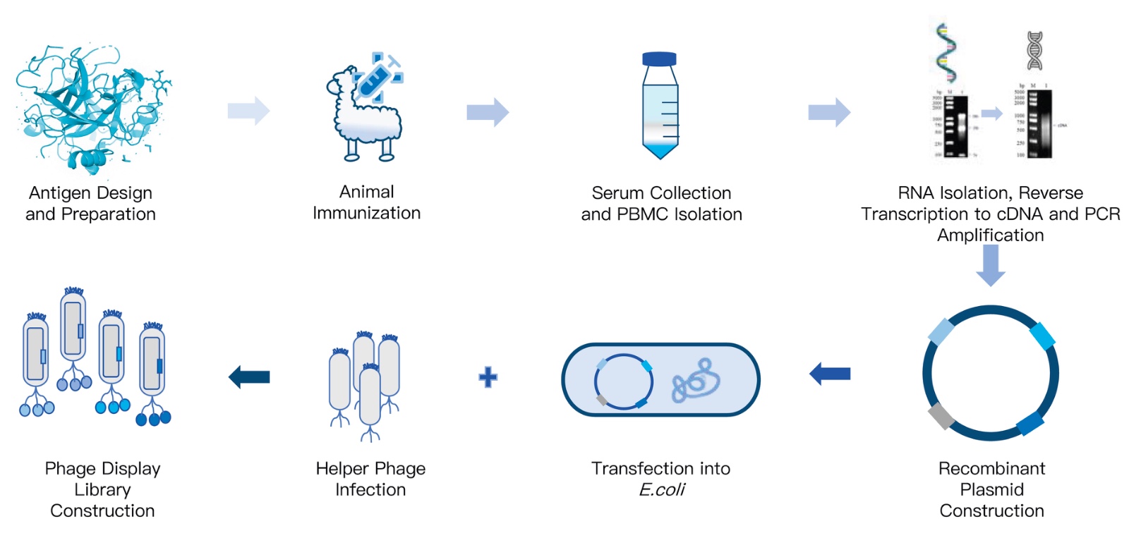
Phage Display Library Construction Services
KMD Bioscience possesses extensive project experience in Antibody Library Construction, dedicated to providing efficient Research Services to universities, research institutes, and enterprises globally.
We offer diverse options to meet most research needs, expertly constructing Naive, Immune, Synthetic, and Semi-synthetic libraries from humans, mice, rabbits, chickens, alpacas, sheep, sharks, and non-human primates. Tissue sources include PBMC, bone marrow, lymph nodes, or spleen. The antibody gene formats we construct are highly varied, encompassing scFv, Fab, VHH, bispecific antibody genes, and bispecific ligand-antibody conjugates.
Explore Custom Library Services
By partnering with a facility that actively monitors the biotech industry trends blog to implement the latest panning algorithms, you safeguard your project from failure.
Frequently Asked Questions (FAQ)
What is an acceptable output titer after the first round of panning?
In most professional situations, an acceptable output titer after round one is between 10^4 and 10^6 colony-forming units (cfu). If your output is vastly higher (e.g., 10^8), you likely have insufficient washing stringency or severe non-specific background binding.
Why do all my selected clones bind to the blocking agent instead of the antigen?
This is a classic manifestation of the Causes of Poor Phage Display Enrichment. It occurs when you fail to perform negative selection. Before exposing the library to your antigen, you must incubate the library with the blocking agent (like Milk or BSA) alone. You discard the bound phages and only proceed with the unbound supernatant.
How do I know if my target antigen is denatured?
If your panning campaign successfully enriches clones that show strong signals in an ELISA (where the antigen is coated on plastic) but show zero binding in flow cytometry or cell-based assays (where the antigen is in its native conformation), your antigen was denatured during the immobilization process.
Authoritative Industry References
To ensure our diagnostic guidelines align with rigorous biopharmaceutical standards, we reference data from the following authoritative bodies:
National Center for Biotechnology Information (NCBI / NIH): Providing peer-reviewed literature and methodological optimization protocols for resolving amplification bias in recombinant antibody libraries. Visit NCBI
U.S. Food and Drug Administration (FDA): Regulatory guidelines concerning the characterization, stability, and IND requirements for therapeutic monoclonal antibodies derived from in vitro display technologies. Visit FDA Guidance
Nature Protocols / mAbs Journal: The premier scientific publications tracking the clinical success rates and structural biology constraints of biotherapeutics generated via phage display. Visit Nature

- Protein Platform
- Protein Expression Services
- Protein Modification & Detection Services
- Protein Interaction Services
- Protein Purification Service
- Antibody Discovery Platform
- VHH Antibody Platform
- Phage Display Platform
- Antibody Humanization Platform
- Single B Cell Screening Platform
- Antibody Expression & Validation Platform
- Peptide Library Platform
- Custom Antibody Platform
- Innovative Drug Discovery Platform
- Product Center
- Proteins
- Antibodies
- Raw Materials Reagents
- Small Molecule Reagents
- Peptide Products
- Nanoparticle Products
- Contact Us
- Phone:400-621-6806
- Mobile:13110936064(WeC
- E-mail:[email protected]
- Address:Room 108, Building 10, No. 6 Hechang Road, Wuqing Development Zone, Tianjin City
-

WeChat
-

Service Account
-
 400-621-6806
 -
 3599163699
 -
 Monday to Friday 9:00-18:00
 -

-
 0
0
-
