


The Definitive Protocol for Phage Display Library Screening: An Expert Guide
- 2026-04-21
- 6394
Authored by KMD Bioscience, a leading provider of therapeutic antibody discovery and related support services. With technology R&D at our core, we deliver high-quality CRO services to scientists and research institutions worldwide, driving advancement and innovation in medical technology.
In the rapidly evolving landscape of therapeutic antibody discovery, the ability to isolate high-affinity, target-specific binders from vast libraries of variants is paramount. Phage display technology remains one of the most robust, versatile, and widely utilized in vitro selection techniques in modern molecular biology. However, the success of any biopanning campaign hinges entirely on the precision and optimization of the methodology employed. Understanding the exact protocol for phage display library screening is the critical differentiator between a successful therapeutic lead discovery and a failed experiment plagued by false positives and high background noise.
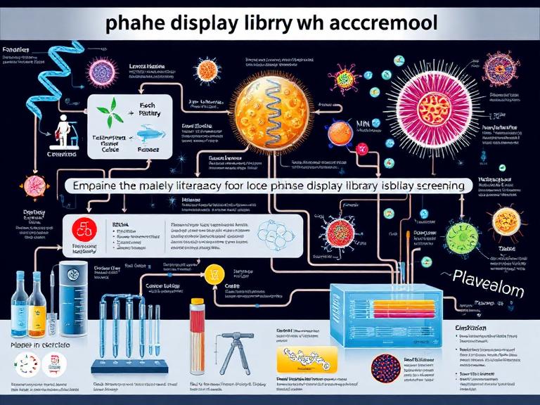
At KMD Bioscience, our scientists have spent years optimizing the protocol for phage display library screening across diverse targets, ranging from complex membrane proteins to small peptides. From our experience, while the overarching concept of biopanning is straightforward, the nuances in buffer selection, washing stringency, and elution strategies dictate the outcome. In this comprehensive guide, we provide an authoritative, step-by-step breakdown of the protocol for phage display library screening, designed to empower researchers and biopharmaceutical developers to achieve superior selection results.
Table of Contents
2. Preparation: The Foundation of the Protocol for Phage Display Library Screening
3. The Biopanning Cycle: Step-by-Step Protocol for Phage Display Library Screening
5. KMD Bioscience's Integrated Platforms for Antibody Discovery
1. Core Principles of Phage Display Screening
Phage display relies on the fundamental principle of genotype-phenotype linkage. A gene sequence encoding a peptide or antibody fragment (such as an scFv or VHH) is fused to a bacteriophage coat protein gene (commonly pIII or pVIII of M13 filamentous phage). When the phage replicates, it "displays" the expressed protein on its surface while encapsulating the corresponding genetic material inside its core.
The protocol for phage display library screening, often referred to as biopanning, is an iterative process of affinity selection. A vast library—often containing upwards of 10^10 unique variants—is exposed to an immobilized target antigen. Non-binding phages are washed away, while specific binders are eluted and amplified in bacterial hosts (such as E. coli TG1 or ER2738). This enriched pool is then subjected to subsequent rounds of panning under increasingly stringent conditions to isolate the highest-affinity binders.
2. Preparation: The Foundation of the Protocol for Phage Display Library Screening
Before initiating the physical steps of the protocol for phage display library screening, meticulous preparation of reagents and libraries is mandatory. We recommend stringent quality control at this stage, as compromised antigens or degraded libraries will inevitably lead to assay failure.
Target Antigen Quality Control
The target antigen must be of the highest possible purity, ideally exceeding 95%. Contaminating proteins can become dominant targets during the screening process, resulting in the isolation of off-target binders. We recommend utilizing our Antibody Expression & Validation Platform to ensure the recombinant proteins utilized for screening are correctly folded and possess the appropriate post-translational modifications.
Library Selection
The choice of the initial library dictates the potential ceiling of your discovery campaign. KMD Bioscience offers access to a premier Phage Display Platform and a highly diverse Peptide Library Platform. Whether you are screening a naive library, an immune library, or a synthetic repertoire, ensuring the library titer is at least 100-fold higher than the library diversity is a critical prerequisite in the protocol for phage display library screening.
3. The Biopanning Cycle: Step-by-Step Protocol for Phage Display Library Screening
The standard protocol for phage display library screening typically involves three to four rounds of biopanning. The following steps outline a standard solid-phase screening methodology using immunotubes or microtiter plates.
Step 1: Coating the Target Antigen
The protocol for phage display library screening begins with antigen immobilization. Coat a Maxisorp immunotube or a 96-well plate with the target antigen. We recommend using a concentration of 10 to 50 micrograms per milliliter diluted in a coating buffer (such as 0.1 M NaHCO3, pH 8.6, or standard PBS, pH 7.4). Incubate the coated surface overnight at 4 degrees Celsius to ensure uniform and stable adsorption of the protein.
Step 2: Blocking Non-Specific Sites
Following antigen coating, the vessel must be blocked to prevent the non-specific binding of phages to the plastic surface. Discard the coating solution and wash the tube once with PBS. Fill the tube completely with a blocking buffer—typically 3% to 5% Bovine Serum Albumin (BSA) or 5% non-fat skim milk dissolved in PBS or PBST (PBS containing 0.05% Tween-20). Incubate at room temperature for 1 to 2 hours, or overnight at 4 degrees Celsius. In parallel, it is crucial to block the phage library itself in a separate tube using the same blocking buffer to reduce background binding.
Step 3: Phage Binding (Incubation)
This is the interaction phase of the protocol for phage display library screening. Add the blocked phage library (typically 10^11 to 10^12 colony-forming units, cfu) to the antigen-coated and blocked well. Seal the vessel and allow it to incubate at room temperature for 1 to 2 hours. We recommend gentle rotation during the first 30 minutes to facilitate mass transfer, followed by static incubation to allow low off-rate binders to stabilize their interaction with the antigen.
Step 4: Stringent Washing
Washing is the most critical parameter to optimize within the protocol for phage display library screening. The goal is to remove weak and non-specific binders while retaining high-affinity phages. Discard the unbound phage solution. Wash the tube with PBST (PBS + 0.05% Tween-20). For the first round of panning, 5 to 10 washes are generally sufficient. In subsequent rounds (Rounds 2, 3, and 4), we recommend increasing both the number of washes (up to 20 times) and the concentration of the detergent (up to 0.1% or 0.2% Tween-20) to exert high selection pressure.
Step 5: Elution of Bound Phages
Once non-specific binders are removed, the target-specific phages must be recovered. The most common method in the protocol for phage display library screening is acidic elution. Add 1 mL of 0.2 M Glycine-HCl (pH 2.2) to the tube and incubate for exactly 10 minutes at room temperature with gentle shaking. Prolonged exposure to low pH can permanently damage the phage infectivity. Immediately neutralize the eluate by transferring it into a fresh tube containing 150 microliters of 1 M Tris-HCl (pH 9.1). Alternatively, competitive elution utilizing an excess of soluble target antigen can be employed to isolate highly specific conformer-dependent binders.
Step 6: Amplification of Eluted Phages
The neutralized eluate represents the enriched phage pool. To proceed to the next round of the protocol for phage display library screening, these phages must be amplified. Infect an exponentially growing culture of E. coli TG1 cells (OD600 = 0.4 to 0.6) with the eluted phages for 30 minutes at 37 degrees Celsius without shaking. Plate a small serial dilution to determine the output titer. Centrifuge the remaining infected cells and plate them on large 2xTY agar plates supplemented with the appropriate antibiotic (e.g., Ampicillin) and 2% glucose. Incubate overnight at 30 degrees Celsius. The following day, scrape the bacterial lawn, rescue the phages using a helper phage (such as M13KO7 or VCSM13), precipitate the amplified phages utilizing PEG/NaCl, and resuspension for the next round of panning.
4. Screening and Validation of Positive Clones
After three to four rounds of executing the protocol for phage display library screening, the output pool should be highly enriched for target-specific binders. The next phase involves isolating individual colonies to identify monoclonal binders.
Monoclonal Phage ELISA
Randomly pick 96 to 192 individual colonies from the final titration plates. Grow these in a 96-well deep-well plate, superinfect with helper phage, and produce monoclonal phage supernatants. Perform an Enzyme-Linked Immunosorbent Assay (ELISA) by coating plates with the target antigen and using an anti-M13 antibody conjugated to HRP to detect binding. Clones exhibiting an absorbance signal significantly higher than the negative control are considered positive hits.
Sequencing and Bioinformatics
Positive clones identified via ELISA must be subjected to Sanger sequencing to determine the DNA sequence of the displayed antibody fragment or peptide. From our experience, analyzing the sequence diversity at this stage is crucial to identifying unique clonal families. KMD Bioscience offers robust bioinformatics support to cluster these sequences and identify the most promising candidates for downstream application.
5. KMD Bioscience's Integrated Platforms for Antibody Discovery
Executing the protocol for phage display library screening is just one facet of the drug discovery pipeline. KMD Bioscience functions as a comprehensive CRO, offering a seamless transition from initial screening to preclinical validation.
VHH Antibody Platform: Capitalizing on the unique properties of camelid single-domain antibodies, we construct and screen highly diverse nanobody libraries, ideal for targeting cryptic epitopes and penetrating dense tissue environments.
Antibody Humanization Platform: Once murine or camelid leads are identified through phage display, our proprietary algorithms and structural modeling techniques humanize the sequences to reduce immunogenicity while retaining picomolar affinity.
Innovative Drug Discovery Platform: We provide end-to-end solutions, transforming raw hits from the protocol for phage display library screening into developable therapeutic candidates with optimized biophysical properties.
Single B Cell Screening Platform: For projects requiring the preservation of natural heavy and light chain pairing, we complement our phage display services with advanced microfluidic single B cell interrogation to isolate rare, high-affinity antibodies directly from immunized hosts.
Custom Antibody Platform: Whether you require traditional hybridoma generation or recombinant antibody production, our custom services are tailored to meet exact specifications for diagnostic, therapeutic, or research reagents.
6. Troubleshooting: From Our Experience
Even with strict adherence to the protocol for phage display library screening, researchers frequently encounter challenges. Drawing from decades of collective laboratory experience, KMD Bioscience recommends the following troubleshooting strategies:
Low Output Titer After Round 1: This is a common occurrence and not necessarily indicative of failure. A highly diverse naive library will inherently have very few specific binders. However, if the output titer drops below 10^3 cfu, consider assessing the structural integrity of your coated antigen or utilizing a more benign elution strategy, such as enzymatic cleavage (if a protease site is engineered into the linker) rather than acidic elution.
High Background (False Positives): If the number of phages binding to the blocking agent (e.g., BSA) rivals those binding to the target, the selection pressure is too low. We recommend alternating blocking agents between rounds (e.g., use BSA in Round 1, Skim Milk in Round 2) to eliminate phages that are specifically binding to the blocking protein. Additionally, implementing a "depletion" or "negative panning" step—where the library is incubated in an empty, blocked tube prior to antigen exposure—drastically improves the signal-to-noise ratio in the protocol for phage display library screening.
7. Summary Table: Biopanning Protocol Overview
| Biopanning Stage | Recommended Buffer / Reagent | Incubation Time / Conditions | Expert Optimization Tip |
|---|---|---|---|
| Antigen Coating | 0.1 M NaHCO3 (pH 8.6) or PBS (pH 7.4) | Overnight at 4°C | Ensure antigen purity >95% to avoid selecting off-target binders. |
| Blocking | 3-5% BSA or 5% Skim Milk in PBST | 1-2 Hours at Room Temperature | Alternate blocking agents in each round to reduce background. |
| Phage Binding | Blocked Phage Library (10^11 - 10^12 cfu) | 1-2 Hours at Room Temperature | Use negative depletion panning prior to target incubation. |
| Washing | PBST (0.05% to 0.2% Tween-20) | 5 to 20 washes (increasing per round) | Increase Tween-20 concentration in later rounds to select for high affinity. |
| Elution | 0.2 M Glycine-HCl (pH 2.2) | 10 Minutes exactly (Neutralize with Tris-HCl) | Never exceed 10 minutes to prevent permanent phage degradation. |
| Amplification | E. coli TG1, Helper Phage (M13KO7) | Overnight at 30°C on 2xTY-Amp-Glu plates | Maintain cultures in exponential growth phase (OD600 ~0.5) for optimal infection. |
8. Frequently Asked Questions (FAQs)
How many rounds are required in the protocol for phage display library screening?
From our experience, 3 to 4 rounds of biopanning are optimal. Stopping at round 2 often yields too many non-specific binders, while exceeding 5 rounds can lead to the over-amplification of fast-growing phages (parasitic clones) that outcompete the high-affinity, slow-growing binders.
Can I screen against whole cells instead of purified proteins?
Yes. Cell-based biopanning is a highly effective modification of the protocol for phage display library screening, particularly for multi-pass transmembrane proteins that lose their native conformation when purified. It requires specialized centrifugation steps and extensive negative depletion against non-target cells to reduce background.
What is the difference between a naive library and an immune library?
A naive library is constructed from non-immunized donors and can be screened against any antigen, making it highly versatile. An immune library is constructed from an animal (or human) that has been specifically exposed to the target antigen, resulting in a pre-enriched pool of naturally affinity-matured binders. KMD Bioscience offers comprehensive services for the construction and screening of both library types.
9. Academic References
- Protocol for Phage Display Library Screening

- Protein Platform
- Protein Expression Services
- Protein Modification & Detection Services
- Protein Interaction Services
- Protein Purification Service
- Antibody Discovery Platform
- VHH Antibody Platform
- Phage Display Platform
- Antibody Humanization Platform
- Single B Cell Screening Platform
- Antibody Expression & Validation Platform
- Peptide Library Platform
- Custom Antibody Platform
- Innovative Drug Discovery Platform
- Product Center
- Proteins
- Antibodies
- Raw Materials Reagents
- Small Molecule Reagents
- Peptide Products
- Nanoparticle Products
- Contact Us
- Phone:400-621-6806
- Mobile:13110936064(WeC
- E-mail:[email protected]
- Address:Room 108, Building 10, No. 6 Hechang Road, Wuqing Development Zone, Tianjin City
-

WeChat
-

Service Account
-
 400-621-6806
 -
 3599163699
 -
 Monday to Friday 9:00-18:00
 -

-
 0
0
-
