


Is phage the same as bacteriophage?
- 2026-08-03
- 116
In the rapidly evolving landscape of molecular biology and antibody engineering, precise terminology is non-negotiable. Whether you are drafting a grant proposal, communicating with a contract research organization (CRO), or designing a complex screening protocol, using the correct nomenclature ensures your project parameters are understood. One of the most common semantic questions we encounter from junior researchers and procurement managers alike is: is phage the same as bacteriophage?
From our experience operating at the forefront of biotechnology, linguistic confusion can lead to operational delays. The short answer is yes, but the implications of how these biological entities are utilized in modern laboratories are vast and highly complex. In this comprehensive guide, we will definitively answer the nomenclature question, break down the mechanics of these viral vectors, and explain why mastering this technology is absolutely critical for commercial antibody development.
Quick Answer
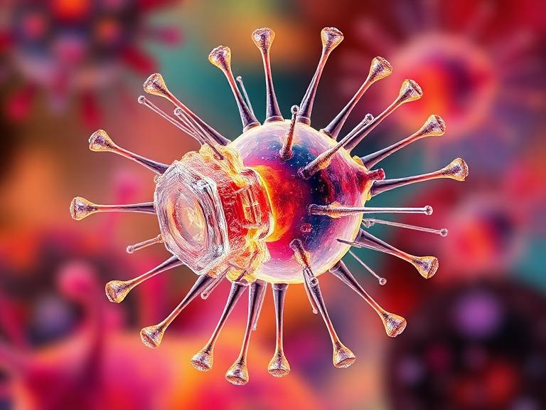
Yes, is phage the same as bacteriophage? Absolutely. The term "phage" is simply the universally accepted scientific abbreviation for "bacteriophage." Both terms refer to the exact same biological entity: a virus that specifically infects and replicates within bacteria. In most professional situations and daily laboratory vernacular, scientists use the shortened term "phage" to save time, particularly when discussing complex industrial applications such as phage display technology.
Table of Contents
What It Is: Phage vs. Bacteriophage
To understand the terminology, we must look at the etymology. The word "bacteriophage" is derived from "bacteria" and the Greek word "phagein," which translates to "to devour." Therefore, a bacteriophage is literally a "bacteria eater." They are the most abundant biological entities on the planet, found wherever bacteria exist.
In the commercial biotechnology sector, nobody uses the full term in daily operations. We refer to "phage display," "phage libraries," and "phage titers." Make no mistake: when a CRO offers a Phage Display Platform, they are engineering bacteriophages. The most commonly utilized phage in laboratory environments is the M13 filamentous phage, primarily because it infects Escherichia coli (E. coli) without killing the host cell outright, allowing for the continuous secretion of assembled phage particles.
How It Works in Commercial Biotechnology
Understanding that phage and bacteriophage are the same is only the first step. You must understand how we exploit their biological mechanisms for commercial gain. In the wild, a phage attaches to a specific bacterial host, injects its genetic material, and hijacks the host's cellular machinery to replicate itself.
In our laboratories, we genetically modify the phage genome. By inserting a specific DNA sequence (coding for a foreign peptide or antibody fragment) into the gene that codes for the phage's coat protein, the phage will physically "display" this new protein on its outer surface while encapsulating the corresponding DNA inside. This creates a direct, physical link between genotype (the DNA inside) and phenotype (the protein outside).
When we construct libraries containing billions of these unique phages, we can screen them against a target antigen in a process called "panning." Phages that bind strongly to the target are captured, while non-binders are washed away. The binders are then eluted and amplified by infecting E. coli, providing researchers with highly specific antibody candidates in a matter of weeks.
Benefits of Phage Technology in Antibody Discovery
From our experience, the commercial advantages of utilizing phages for drug discovery are undeniable. For heavy-duty applications requiring rapid turnaround, this technology is unmatched.
In Vitro Selection: Unlike animal immunization, phage display occurs entirely in test tubes. This allows for the selection of antibodies against toxic antigens, highly conserved proteins, or non-immunogenic targets that an animal's immune system would reject.
Massive Diversity: A high-quality synthetic or naive phage library can contain up to 10^11 unique clones. This vast repertoire significantly increases the probability of finding a high-affinity binder compared to traditional hybridoma methods.
Speed: In most professional situations, a standard panning protocol can isolate target-specific binders in 3 to 4 weeks. This rapid timeline drastically accelerates the R&D pipeline.
Nanobody Discovery: Phage technology is particularly well-suited for discovering single-domain antibodies. Sourcing a nanobody library construction service relies almost entirely on phage display to isolate these incredibly stable and compact molecules.
Limitations to Consider
We believe in commercial transparency. While phages are incredibly powerful, they are not flawless. The primary limitation of phage display is the lack of mammalian post-translational modifications (PTMs). Because the phages replicate inside E. coli (a prokaryote), the displayed proteins are not glycosylated. If your target antibody relies on specific glycosylation patterns for its biological function, the phage-derived candidate will require significant downstream engineering.
Furthermore, certain eukaryotic proteins are toxic to bacterial hosts or misfold when expressed on a phage coat. In these scenarios, the diversity of your library can be unintentionally truncated as the bacteria clear the toxic clones from the pool.
Who Should Use Phage Technologies
We recommend phage display for pharmaceutical companies, academic research institutions, and biotech startups operating in the early stages of biologics discovery. If you are developing diagnostics, therapeutics, or research reagents, the ability to rapidly screen billions of candidates is vital. It is especially critical for teams exploring camelid antibodies and seeking nanobody antibody products, as phage display is the gold standard for panning VHH domains.
Who Does Not Need This Technology
For commercial users who already possess a fully validated, high-producing hybridoma cell line, transitioning backward to phage display is generally an unnecessary expenditure. Similarly, if your project strictly requires the immediate isolation of fully glycosylated, full-length IgGs directly from a human or animal host with zero downstream formatting, you should bypass phage display and instead utilize a Single B Cell Screening Platform.
Common Mistakes in Phage Applications
In our testing and client consultations, the most frequent mistake is poor library selection. Clients often assume that a generic, off-the-shelf naive library will solve every problem. If you are targeting a highly complex, multi-pass membrane protein, a standard library will likely fail. You must invest in customized immune libraries or specialized synthetic libraries tailored to your antigen's structural biology.
Another profound mistake is mishandling the panning wash stringency. If the wash buffer is too weak, you will isolate thousands of false-positive background binders. If the wash is too harsh, you will strip away your rare, high-affinity therapeutic candidates. Panning is an art form that requires deep industry expertise.
Buying Considerations for Phage Services
When you are preparing to outsource your discovery phase, you must evaluate CROs with intense scrutiny. Do not accept vague promises.
Library Size and Diversity: Demand proof of library size. A library smaller than 10^9 clones is rarely sufficient for demanding commercial applications.
QC Metrics: Ask for insertion rates and open reading frame (ORF) percentages. A massive library is useless if 50% of the clones contain stop codons or frame shifts.
Downstream Capabilities: Isolating a phage binder is only step one. Ensure your partner possesses an Antibody Expression & Validation Platform to convert your phage-derived fragment (like an scFv) into a functional, full-length IgG for functional testing.
Pros vs Cons Table: Phage Display Technology
| Pros of Phage Display | Cons of Phage Display |
|---|---|
| Massive library capacity (up to 10^11 clones). | Lacks mammalian post-translational modifications (no glycosylation). |
| Does not require animal immunization (in vitro selection). | Some mammalian proteins may misfold on the phage coat. |
| Can generate antibodies against highly toxic or conserved antigens. | Requires highly specialized downstream reformatting to create full IgGs. |
| Rapid panning timelines (weeks instead of months). | High background noise if panning protocols are poorly optimized. |
Comparison Table: Phage Display vs Hybridoma vs Single B Cell
| Feature / Metric | Phage Display | Hybridoma Technology | Single B Cell Screening |
|---|---|---|---|
| Discovery Speed | Very Fast (3-6 weeks) | Slow (3-6 months) | Fast (1-2 months) |
| In Vivo Immunization | Not Required (Naive/Synthetic) | Strictly Required | Strictly Required |
| Antibody Format Discovered | Fragments (scFv, Fab, VHH) | Full-length IgG | Full-length IgG |
| Target Limitations | Virtually None (Can target toxic proteins) | Cannot target toxic/lethal antigens | Cannot target toxic/lethal antigens |
Expert Recommendation
Why You Should Partner with KMD Bioscience
If you have definitively decided that phage-based discovery aligns with your R&D objectives, selecting the right contract manufacturer is your most critical commercial decision. Building libraries in-house requires immense capital expenditure, highly specialized personnel, and months of protocol optimization.
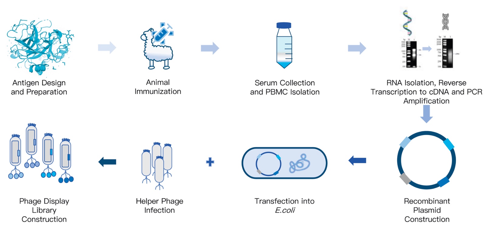
KMD Bioscience possesses an advanced phage display technology platform, offering customized high-quality phage display library construction services. We can create diverse natural libraries (mouse, rabbit, human, etc.), immune libraries (such as VHH libraries, scFv libraries, Fab libraries), synthetic libraries (e.g., peptide libraries), and semi-synthetic libraries for our clients, along with professional library panning and screening services. Through this platform, clients can rapidly identify antibodies or ligands with high affinity and specificity for their target of interest.
In our professional judgment, partnering with an established provider that integrates discovery with downstream production is the safest strategy. For clients operating in specialized domains, our VHH Antibody Platform is specifically optimized to leverage phage display for camelid single-domain antibodies. Furthermore, if you require a tailored approach from inception to commercial scale, exploring our custom antibody development service ensures your project moves seamlessly from a viral vector in a petri dish to a validated therapeutic candidate.
Frequently Asked Questions (FAQ)
Is phage the same as bacteriophage?
Yes. As established throughout this guide, phage is simply the abbreviated, commonly used scientific term for bacteriophage. They both refer to the exact same entity: a virus that exclusively infects and replicates within bacteria.
Can bacteriophages infect human cells?
No. Bacteriophages are highly specific to their bacterial hosts. They completely lack the biological mechanisms, surface proteins, and receptors required to attach to, infect, or replicate within human or other mammalian cells. This biological specificity makes them incredibly safe for laboratory researchers to handle and is the foundational reason they are currently being researched for phage therapy to combat antibiotic-resistant bacterial infections in humans.
What is the primary use of phages in antibody discovery?
In commercial and academic antibody discovery, phages are primarily used in a technique called "phage display." By genetically modifying the phage genome, researchers force the phage to display billions of different antibody fragments (like scFvs or VHH nanobodies) on their outer coat. This massive library is then washed over a target disease antigen. The phages that stick are isolated, giving researchers a fast, highly specific antibody candidate without the need to immunize animals. For more updates on this evolving field, we recommend checking the antibody research news and updates.
Authoritative References & Industry Standards
To ensure our operational recommendations are aligned with global scientific consensus, we rely on data from the following authoritative institutions:
National Center for Biotechnology Information (NCBI): Foundational literature and peer-reviewed studies detailing the molecular biology of M13 filamentous bacteriophages and their application in phage display. Access NCBI Databases.
Nature Portfolio: High-impact scientific protocols and advancements regarding in vitro selection methodologies and synthetic antibody library construction. Review Nature Scientific Journals.
American Society for Microbiology (ASM): Comprehensive guidelines and historical context on bacteriophage biology, viral taxonomy, and emerging phage therapies. Explore ASM Resources.

- Protein Platform
- Protein Expression Services
- Protein Modification & Detection Services
- Protein Interaction Services
- Protein Purification Service
- Antibody Discovery Platform
- VHH Antibody Platform
- Phage Display Platform
- Antibody Humanization Platform
- Single B Cell Screening Platform
- Antibody Expression & Validation Platform
- Peptide Library Platform
- Custom Antibody Platform
- Innovative Drug Discovery Platform
- Product Center
- Proteins
- Antibodies
- Raw Materials Reagents
- Small Molecule Reagents
- Peptide Products
- Nanoparticle Products
- Contact Us
- Phone:400-621-6806
- Mobile:13110936064(WeC
- E-mail:[email protected]
- Address:Room 108, Building 10, No. 6 Hechang Road, Wuqing Development Zone, Tianjin City
-

WeChat
-

Service Account
-
 400-621-6806
 -
 3599163699
 -
 Monday to Friday 9:00-18:00
 -

-
 0
0
-
