


6 Key Components and Applications of High Content Screening in Drug Discovery
- 2026-04-16
- 216
At KMD Bioscience Co., Ltd., we are dedicated to becoming a leading provider of therapeutic antibody discovery and related support services. With technology R&D at our core, we deliver high-quality CRO (Contract Research Organization) services to scientists and research institutions worldwide, driving advancement and innovation in medical technology. Through decades of collaborative research, we have witnessed a paradigm shift in how therapeutic candidates are evaluated. Central to this transformation is the integration of high content screening in drug discovery.
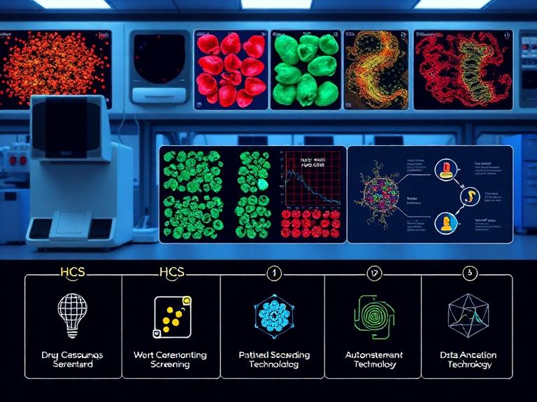
High content screening in drug discovery merges automated microscopy with quantitative image analysis, allowing researchers to extract multiparametric data from cellular populations. Unlike traditional biochemical assays that measure a single endpoint, high content screening in drug discovery assesses complex cellular phenotypes, morphological changes, and subcellular protein localizations simultaneously. From our experience, adopting this multidimensional approach significantly reduces false-positive rates and accelerates the identification of viable therapeutic compounds. We recommend researchers and pharmaceutical developers fully understand the underlying architecture and practical applications of these systems to maximize their return on investment and shorten the clinical development timeline.
Table of Contents
Summary Table: High Content Screening Architecture
To provide a clear overview of how these sophisticated systems operate, we have compiled a summary table detailing the foundational elements and strategic applications of high content screening in drug discovery.
| Component / Application | Functional Description | Impact on Drug Discovery |
|---|---|---|
| Automated Imaging Systems | High-speed robotic microscopes capturing multidimensional cellular images. | Enables rapid scanning of thousands of compounds without manual intervention. |
| Reagents & Probes | Fluorescent markers (Antibodies, Small Molecule Reagents) binding to specific targets. | Provides targeted visibility of subcellular structures and protein dynamics. |
| Cellular Models | 2D cultures, 3D spheroids, and patient-derived organoids. | Ensures physiological relevance to accurately predict human responses. |
| Liquid Handling | Robotic dispensing of cells, compounds, and Raw Materials Reagents. | Maintains precise assay reproducibility across microtiter plates. |
| Target Validation (App) | Confirming that a drug modulates the intended biological target. | Prevents late-stage clinical failures by verifying mechanistic efficacy early. |
| Phenotypic Profiling (App) | Analyzing overall cellular changes rather than a single target. | Identifies novel therapeutic compounds with unknown mechanisms of action. |
The Evolution of Cellular Assays
The pharmaceutical industry historically relied on target-based biochemical assays. While these methods offer high throughput, they inherently lack biological context. A molecule that binds tightly to an isolated target in a test tube may fail to cross a cell membrane, suffer from rapid efflux, or induce severe cytotoxicity. High content screening in drug discovery bridges the gap between high-throughput capacity and deep physiological relevance.
By capturing the spatial and temporal dynamics of cells exposed to vast libraries of compounds, high content screening in drug discovery allows scientists to evaluate efficacy and toxicity simultaneously. We strongly recommend transitioning from single-parameter assays to multiparametric high content approaches, as this minimizes attrition rates in downstream in vivo studies. Implementing this technology requires a synchronized assembly of hardware, software, and highly specific biological consumables.
The 6 Key Components of High Content Screening in Drug Discovery
1. Automated Epifluorescence and Confocal Microscopy
The hardware backbone of high content screening in drug discovery is the automated imaging platform. Modern systems utilize either widefield epifluorescence or spinning disk confocal microscopy. Confocal systems are particularly critical when imaging thick 3D cellular structures, as they reject out-of-focus light to produce crisp optical sections. From our experience, the integration of high-numerical-aperture objectives and rapid laser autofocus mechanisms dictates the speed and clarity of the screening campaign. Hardware stability directly translates to data reproducibility, which is paramount when analyzing subtle morphological shifts.
2. Biological Reagents and Fluorescent Probes
Hardware is entirely dependent on the quality of biological labeling. High content screening in drug discovery relies on multiple fluorescent channels to delineate different cellular compartments—such as the nucleus, cytoskeleton, and specific target organelles. At KMD Bioscience, our extensive catalog perfectly supports these complex labeling requirements. We supply high-affinity Antibodies that ensure specific binding to cellular targets with minimal background noise. Furthermore, our highly purified Proteins, Small Molecule Reagents, and Peptide Products serve as essential controls and modulators in assay development, guaranteeing robust signal-to-background ratios.
3. Physiologically Relevant Cellular Models
The predictive power of high content screening in drug discovery is only as strong as the cellular model deployed. While immortalized 2D cell lines remain a staple for initial high-throughput sweeps, the industry is rapidly moving toward more complex models. We recommend the utilization of primary cells, stem cell-derived models, and 3D organoids for secondary screening phases. These advanced models preserve the native tissue architecture and metabolic profiles, providing a much higher correlation to clinical outcomes.
4. High-Throughput Liquid Handling Systems
In a standard screening campaign, tens of thousands of compounds are tested across 384-well or 1536-well microplates. The precision of high content screening in drug discovery relies on automated liquid handling robotics. These systems must dispense nano-liter volumes of test compounds, wash buffers, and Raw Materials Reagents with absolute uniformity. Any deviation in dispensing volume can create edge effects or concentration gradients across the plate, leading to severe data artifacts and false negatives.
5. Quantitative Image Analysis Algorithms
Generating terabytes of image data is futile without the means to interpret it. The software component of high content screening in drug discovery transforms raw pixels into actionable numeric data. Advanced algorithms automatically segment cells, identify boundaries, and quantify parameters such as intensity, texture, area, and shape. From our experience, the implementation of machine learning and artificial intelligence has revolutionized this step, allowing software to identify complex, non-linear phenotypic changes that a human observer might miss.
6. Multivariate Data Management Informatics
The final component of high content screening in drug discovery involves data informatics. A single well can yield dozens of different parameters, creating a massive multivariate dataset. Effective informatics platforms aggregate this data, perform quality control (such as calculating Z-prime factors), and utilize clustering algorithms to group compounds with similar phenotypic profiles. This level of data management allows researchers to establish structure-activity relationships (SAR) efficiently.
Critical Applications in the Pharmaceutical Pipeline
The applications of high content screening in drug discovery span the entire preclinical continuum. By capturing comprehensive cellular portraits, researchers can answer multifaceted biological questions within a single assay.
Target Identification and Validation
Before a compound library is screened, the biological target must be validated. Using RNA interference (RNAi) or CRISPR-Cas9 technologies, researchers can knock down specific genes and use high content screening in drug discovery to observe the resulting cellular phenotype. If knocking down a gene replicates the therapeutic effect desired, the target is validated. KMD Bioscience supports this phase by providing custom Proteins and Antibodies to verify target expression and localization.
Phenotypic Drug Discovery
Unlike target-based drug discovery, phenotypic screening does not require prior knowledge of a specific molecular target. Instead, compounds are evaluated based on their ability to alter a disease phenotype back to a healthy state. High content screening in drug discovery is the premier tool for this approach. By monitoring complex behaviors like neurite outgrowth in neurodegenerative disease models, or angiogenesis in oncology models, researchers can discover novel first-in-class drugs.
Predictive Toxicology and Nanoparticle Delivery
Drug-induced toxicity is a major cause of clinical trial failure. High content screening in drug discovery allows for early hepatotoxicity, cardiotoxicity, and neurotoxicity profiling by measuring mitochondrial membrane potential, reactive oxygen species (ROS) production, and apoptosis. Additionally, evaluating drug delivery mechanisms is crucial. We supply cutting-edge Nanoparticle Products that can be tracked via high content imaging to study cellular uptake, endosomal escape, and the targeted delivery of active pharmaceutical ingredients.
The KMD Bioscience Advantage
Implementing high content screening in drug discovery requires an ecosystem of reliable reagents. We deliver high-quality CRO services and premium consumables to ensure your assays are reproducible and biologically relevant. Whether your protocol demands specific Small Molecule Reagents for pathway inhibition, highly pure Proteins for assay calibration, or customized Peptide Products for targeted binding studies, KMD Bioscience provides the critical raw materials necessary for screening success. We recommend partnering with a dedicated CRO that understands the nuances of multiparametric imaging from the ground up.
Frequently Asked Questions (FAQs)
What is the main difference between high throughput screening (HTS) and high content screening in drug discovery?
While both methods process large libraries of compounds rapidly, HTS typically measures a single biochemical endpoint (like enzyme activity). High content screening in drug discovery utilizes automated microscopy to extract multiple spatial and temporal parameters from intact cells, providing a much richer, physiologically relevant dataset.
Why are 3D cell models becoming important in high content screening in drug discovery?
2D cell cultures do not accurately represent the complex architecture of human tissues. 3D models, such as spheroids and organoids, better mimic in vivo cell-cell and cell-matrix interactions. From our experience, screening compounds against 3D models significantly improves the accuracy of efficacy and toxicity predictions.
How does KMD Bioscience support high content screening in drug discovery?
We supply the foundational biological materials required for assay development. This includes high-specificity Antibodies for fluorescent labeling, purified Proteins and Peptide Products for target modulation, and reliable Raw Materials Reagents to ensure assay stability across large-scale screening campaigns.
Can high content screening in drug discovery be used for toxicology?
Yes, it is highly effective for predictive toxicology. Multiparametric imaging allows researchers to simultaneously assess cell viability, nuclear morphology, mitochondrial health, and membrane permeability, identifying subtle toxic effects that standard viability assays might miss.
References
To further explore the standards, methodologies, and peer-reviewed advancements regarding high content screening in drug discovery, we recommend consulting the following authoritative sources:
- High Content Screening in Drug Discovery

- Protein Platform
- Protein Expression Services
- Protein Modification & Detection Services
- Protein Interaction Services
- Protein Purification Service
- Antibody Discovery Platform
- VHH Antibody Platform
- Phage Display Platform
- Antibody Humanization Platform
- Single B Cell Screening Platform
- Antibody Expression & Validation Platform
- Peptide Library Platform
- Custom Antibody Platform
- Innovative Drug Discovery Platform
- Product Center
- Proteins
- Antibodies
- Raw Materials Reagents
- Small Molecule Reagents
- Peptide Products
- Nanoparticle Products
- Contact Us
- Phone:400-621-6806
- Mobile:13110936064(WeC
- E-mail:[email protected]
- Address:Room 108, Building 10, No. 6 Hechang Road, Wuqing Development Zone, Tianjin City
-

WeChat
-

Service Account
-
 400-621-6806
 -
 3599163699
 -
 Monday to Friday 9:00-18:00
 -

-
 0
0
-
