


5 Protocols for Phage Display: An Expert Laboratory Guide
- 2026-06-21
- 7091
When it comes to high-throughput screening for monoclonal antibodies, peptides, and engineered proteins, laboratory professionals face a critical choice: rely on outdated, low-yield hybridoma techniques, or transition to advanced in vitro selection methods. From our experience at KMD Bioscience, standardizing your laboratory's protocols for phage display is the single most effective way to accelerate therapeutic antibody discovery. However, navigating the intricate steps of library construction, biopanning, and screening can quickly overwhelm even seasoned molecular biologists if the methodology is not optimized.
In most professional situations, a failed phage display campaign is rarely due to the underlying technology itself; rather, it is the result of poorly executed protocols. If you are struggling with low-affinity binders, high background noise, or lost clones during amplification, your workflow requires an immediate audit. In this comprehensive guide, we will break down the exact protocols for phage display that industry leaders use, explain the commercial realities of setting up these workflows, and provide practical judgment on whether you should build these libraries in-house or outsource them entirely.
Quick Answer: What are the Core Protocols for Phage Display?
To successfully execute a screening campaign, researchers must master five essential protocols for phage display: 1. Library Construction (inserting diverse genetic variants into phagemid vectors); 2. Solid-Phase Biopanning (immobilizing antigens on plastic surfaces); 3. Solution-Phase Biopanning (using biotinylated antigens in liquid suspension); 4. Phage Amplification and Rescue (using helper phages in bacterial cultures); and 5. Phage ELISA (validating the affinity of the selected clones). We recommend Solution-Phase Biopanning for the highest quality conformation-specific antibodies.
Table of Contents
What is Phage Display and How Does It Work?
Phage display is a laboratory technique used to study protein-protein, protein-peptide, and protein-DNA interactions. It works by splicing a gene encoding a protein of interest into a phage coat protein gene (usually the pIII or pVIII protein of the filamentous bacteriophage M13). This genetic engineering causes the phage to "display" the protein on its outside while containing the gene for the protein on its inside. This creates a direct physical linkage between genotype and phenotype.
By creating a library of millions or billions of unique phages, researchers can expose this library to an immobilized target (an antigen). The phages that do not bind are washed away, while the ones that do bind are eluted and amplified in bacterial hosts. To support this highly technical workflow, a robust protein expression systems overview is essential, as you need continuous access to high-quality target antigens and host bacteria to facilitate the selection rounds.
Protocol 1: Library Construction and Preparation
The foundation of all protocols for phage display is the library. If your library lacks diversity or contains a high percentage of non-functional clones, your entire screening campaign is doomed from day one. Construction involves extracting mRNA from B-cells (for immune libraries) or using synthetic oligonucleotides (for naïve or synthetic libraries), performing RT-PCR, and cloning the resulting repertoires (such as scFv or VHH fragments) into a phagemid vector.
From our experience, library construction is the most bottlenecked step for academic and commercial labs. It requires massive electroporation efficiency to achieve library sizes of 10^9 to 10^11 transformants. We recommend that unless your lab specializes strictly in library generation, you should source your target antigens via a reliable recombinant protein expression service to ensure your baits are perfectly folded before you even attempt to screen a library.
Protocol 2: Solid-Phase Biopanning
Solid-phase biopanning is the traditional and most straightforward protocol. The target antigen is directly coated onto a hydrophobic plastic surface, typically an immunotube or a 96-well polystyrene microtiter plate, and incubated overnight. After blocking the remaining binding sites on the plastic with non-fat milk or BSA, the phage library is added.
While inexpensive and easy to set up, solid-phase biopanning has a severe limitation. Immobilizing proteins directly onto plastic often denatures them, exposing hydrophobic core residues that are not present in the native protein structure. In our testing, this frequently leads to the selection of antibodies that bind the denatured antigen beautifully in an ELISA, but fail completely when tested against the native protein on a living cell surface.
Protocol 3: Solution-Phase Biopanning
In most professional situations targeting complex therapeutic proteins (like GPCRs or ion channels), we recommend transitioning to solution-phase biopanning. In this protocol, the target antigen is biotinylated and mixed with the phage library in a liquid buffer. Because the antigen remains in solution, it maintains its native, three-dimensional conformation.
After a short incubation period, streptavidin-coated magnetic beads are added to the mixture. The beads pull down the biotin-antigen-phage complexes. Using a magnetic rack, the unbound phages are washed away. This protocol dramatically reduces background binding and almost guarantees that the resulting clones will recognize the target in its native physiological state.
Protocol 4: Phage Amplification and Rescue
Once you have eluted the binding phages from your biopanning rounds, you must amplify them to conduct the next round of screening. Because most modern libraries use phagemids (which lack the full genome required to package a phage particle), you must infect the eluted phages into an E. coli protein expression system, specifically strains like TG1 or XL1-Blue.
You then superinfect these bacteria with a helper phage (such as M13KO7 or VCSM13). The helper phage provides the missing structural genes, forcing the E. coli to assemble and secrete new phage particles displaying your selected antibodies. Mastering the timing, OD600 measurements, and antibiotic selection during this protocol is critical to prevent wild-type helper phages from out-competing your recombinant phages.
Protocol 5: Phage ELISA and Target Validation
After 3 to 4 rounds of biopanning and amplification, the selected pool is typically enriched for strong binders. Protocol 5 involves picking individual bacterial colonies, rescuing them into monoclonal phages in 96-well plates, and running a Phage Enzyme-Linked Immunosorbent Assay (ELISA).
This validation step uses an anti-M13 antibody conjugated to HRP to detect which specific clones are binding to the target antigen. The clones producing the highest signal-to-noise ratio are then sequenced. This protocol is the definitive quality control checkpoint before you scale up production to generate validated antibody products for downstream assays.
Comparison: Solid-Phase vs. Solution-Phase Biopanning
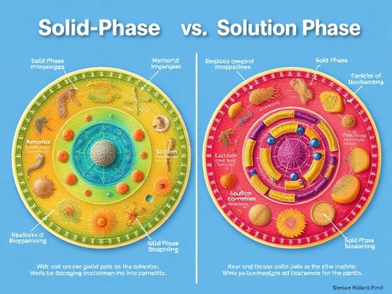
Understanding which biopanning protocol to deploy dictates the success of your screening. Here is our practical breakdown.
| Feature | Solid-Phase Biopanning (Immunotubes/Plates) | Solution-Phase Biopanning (Magnetic Beads) |
|---|---|---|
| Antigen Presentation | Coated directly on plastic; high risk of denaturation. | Suspended in liquid; preserves native folding. |
| Epitope Accessibility | Limited. Antigens may hide binding sites against the plastic. | Maximum. 360-degree accessibility to the phage library. |
| Equipment Needed | Standard ELISA plates or Nunc immunotubes. | Streptavidin magnetic beads, magnetic separation racks. |
| Background Noise | Moderate to High (binding to plastic or blocking agents). | Low (beads can be changed between rounds to remove bead-binders). |
| Best For | Simple, robust proteins and peptides. | Complex, multi-pass transmembrane proteins and conformational targets. |
Pros and Cons of Phage Display Technology
Before overhauling your lab infrastructure, it is critical to evaluate the commercial viability of phage display compared to traditional hybridoma technology.
| Pros of Phage Display | Cons of Phage Display |
|---|---|
| Does not require animal immunization (naïve libraries). | Requires highly specialized molecular biology expertise. |
| Rapid screening timeline (weeks instead of months). | Lack of post-translational modifications (PTMs) in E. coli hosts. |
| Can target highly toxic or non-immunogenic antigens. | Biopanning can occasionally select for non-specific sticky clones. |
| Direct access to the gene sequence of the binder. | Library construction is capital- and labor-intensive. |
Who Should Use It (And Who Does Not Need It)
For commercial users and pharma developers: If your organization is developing therapeutic monoclonal antibodies, nanobodies (VHH), or CAR-T cell receptors, executing protocols for phage display is absolutely mandatory. It allows you to bypass the immune tolerance of animals and engineer specific affinities and cross-reactivities in vitro. Sourcing research antibodies for labs rapidly via phage display keeps pipelines moving.
Who does not need it: If your lab only needs a basic polyclonal antibody for an occasional Western blot, investing the time and capital into building a phage display workflow is a massive waste of resources. Traditional peptide immunization of a rabbit remains the most cost-effective solution for basic, low-throughput needs.
Common Mistakes in Phage Display Protocols
In our testing, we routinely audit failed screening campaigns. The most common mistakes are entirely avoidable.
Poor Quality Antigens: Garbage in, garbage out. If your target antigen is aggregated or degraded, the phages will bind to the misfolded regions. Ensure your baits are purified properly by utilizing advanced protein expression platform services.
Washing Too Aggressively in Round 1: The first round of biopanning is meant to capture as much diversity as possible. If you use high concentrations of Tween-20 or perform too many wash steps in round one, you will wash away rare, high-affinity clones. Save the aggressive washing stringency for rounds three and four.
Ignoring Growth Bias: During the amplification protocol, some phages replicate slightly faster than others due to the specific sequence of the displayed protein. If you over-amplify, these fast-growers will dominate your library, masking the actual high-affinity binders. Keep amplification times strictly monitored.
Buying Considerations: In-House vs. CRO Services
A major decision for lab directors is whether to establish these protocols in-house or outsource them. Review this buying guide to make a commercial determination.
| Consideration | Building In-House | Outsourcing to a CRO |
|---|---|---|
| Initial Capital Expenditure | High. Requires specialized vectors, electro-competent cells, and validation assays. | Low to Zero. You pay strictly for milestones and deliverables. |
| Time to First Result | 6 to 12 months (includes workflow validation and library QC). | 6 to 8 weeks for standard screening campaigns. |
| Intellectual Property | 100% retained internally immediately. | Retained by client, but requires strict MTA and service agreements. |
| Success Rate | Variable. Highly dependent on the skill of the staff technician. | High. CROs leverage pre-validated, massive libraries (10^11 diversity). |
Expert Recommendation & Brand Information
For laboratories focused on downstream biological applications rather than method development, we strongly recommend outsourcing the primary phage display screening to a specialized Contract Research Organization (CRO). The infrastructure required to construct, validate, and maintain a high-diversity library often outweighs the benefits of running the system in-house for small to mid-sized biotech firms. Partnering with experts ensures you receive reliable antibody reagents without the operational headache.
About KMD Bioscience
KMD Bioscience Co., Ltd. has been committed to becoming a leading provider of therapeutic antibody discovery and related support services since its establishment in 2022. Focusing on technological research and development, the company provides high-quality Contract Research Organization (CRO) services to scientists and research institutions worldwide, aiming to promote the development and innovation of medical science and technology. As a high-tech enterprise, it has been recognized as a National Patent Pilot Unit and obtained ISO9001:2015 Quality Management System certification for its laboratories.
The Bottom Line
Mastering the protocols for phage display transforms a laboratory's capability to discover novel biological binders. By strictly adhering to the five core protocols—Library Construction, Solid-Phase and Solution-Phase Biopanning, Amplification, and Phage ELISA—researchers can reliably isolate high-affinity monoclonal antibodies in a fraction of the time required by animal immunization. However, success hinges entirely on the quality of your target antigen and the stringency of your wash protocols. When in doubt, leverage specialized expression platforms and CRO expertise to bypass the technical bottlenecks.
Frequently Asked Questions (FAQs)
What is the best host strain for phage amplification?
For standard M13 phage display, E. coli strains carrying the F' episome are mandatory because the phage requires the F pilus for infection. Strains like TG1, XL1-Blue, and ER2738 are the industry standards. TG1 is frequently preferred for its high growth rate and robust phage yield.
How do you elute bound phages during biopanning?
The most common elution protocol involves a dramatic pH shift using an acidic buffer, such as 0.2 M Glycine-HCl (pH 2.2), followed immediately by neutralization with 1 M Tris-HCl (pH 9.1). Alternatively, competitive elution using a known ligand or enzymatic cleavage (if a protease site is engineered into the linker) can be utilized for highly specific recovery.
Can phage display be used to find binders for small molecules?
Yes, but it is technically challenging. Because small molecules (haptens) lack sufficient surface area for antibody recognition, they must be conjugated to a carrier protein (like BSA or KLH) during biopanning. A negative selection step against the carrier protein alone is required to ensure the selected phages are specific to the small molecule.
References and Authoritative Sources
National Center for Biotechnology Information (NCBI) - Phage Display Technology: Applications and Innovations.
Nature Protocols - Standardized methodologies for in vitro selection and antibody engineering.
Addgene - Educational guide and repository standards for Phagemid Vectors and Library Construction.

- Protein Platform
- Protein Expression Services
- Protein Modification & Detection Services
- Protein Interaction Services
- Protein Purification Service
- Antibody Discovery Platform
- VHH Antibody Platform
- Phage Display Platform
- Antibody Humanization Platform
- Single B Cell Screening Platform
- Antibody Expression & Validation Platform
- Peptide Library Platform
- Custom Antibody Platform
- Innovative Drug Discovery Platform
- Product Center
- Proteins
- Antibodies
- Raw Materials Reagents
- Small Molecule Reagents
- Peptide Products
- Nanoparticle Products
- Contact Us
- Phone:400-621-6806
- Mobile:13110936064(WeC
- E-mail:[email protected]
- Address:Room 108, Building 10, No. 6 Hechang Road, Wuqing Development Zone, Tianjin City
-

WeChat
-

Service Account
-
 400-621-6806
 -
 3599163699
 -
 Monday to Friday 9:00-18:00
 -

-
 0
0
-
