


The 5 Crucial Differences Between HCS and HTS in Drug Discovery
- 2026-05-13
- 136
Authored by Tianjin KMD Bioscience Co., Ltd.
In the highly competitive and scientifically rigorous field of drug discovery and biopharmaceutical development, selecting the correct screening methodology is the cornerstone of early-stage success. Researchers are constantly evaluating massive libraries of small molecules, peptides, and antibodies to identify viable therapeutic candidates. During this critical hit-identification and hit-to-lead optimization phase, two acronyms dominate the conversation: HTS (High-Throughput Screening) and HCS (High-Content Screening). While they may sound similar, understanding the differences between HCS and HTS is absolutely vital for designing effective experimental workflows, managing research budgets, and extracting meaningful biological data.
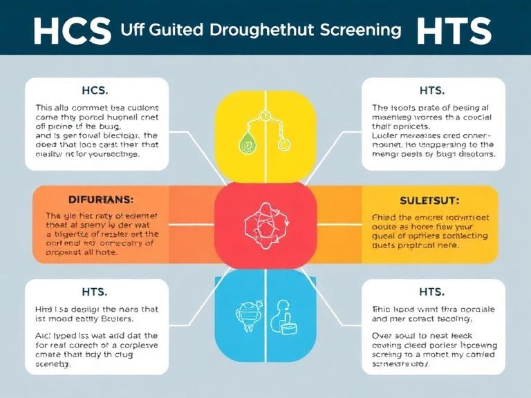
Tianjin KMD Bioscience Co., Ltd. is dedicated to becoming a leading provider of therapeutic antibody discovery and related support services. With technology research and development at our core, we deliver high-quality CRO (Contract Research Organization) services to scientists and research institutions worldwide, driving advancement and innovation in medical technology. From our experience managing complex assay developments for global clients, we frequently consult on the strategic differences between HCS and HTS. In this authoritative guide, we will explore the fundamental concepts behind these two powerful techniques and detail the 5 primary differences between HCS and HTS.
Table of Contents
1. Core Definition and Fundamental Approach
To accurately assess the differences between HCS and HTS, one must first define their foundational scientific approaches. High-Throughput Screening (HTS) is predominantly a biochemical, target-based approach. It is designed to test hundreds of thousands of compounds against a specific, isolated biological target, such as an enzyme or a receptor. The primary goal of HTS is rapid, binary decision-making: does the compound bind to or inhibit the target, yes or no? It is a blunt but highly effective instrument for finding a needle in a haystack.
Conversely, High-Content Screening (HCS), sometimes referred to as High-Content Analysis (HCA), is a phenotypic approach based on automated imaging. Instead of isolating a single protein, HCS utilizes intact, living cells to observe how a compound affects the entire cellular system. It integrates the efficiency of automated liquid handling with the depth of high-resolution fluorescence microscopy. Therefore, one of the most fundamental differences between HCS and HTS is that HTS treats biology as a simple lock-and-key mechanism, whereas HCS embraces the complex, multi-variable reality of cellular biology.
2. Data Richness and Output Complexity
When our scientific teams discuss the differences between HCS and HTS with clients, data richness is frequently the deciding factor. HTS generates single-dimensional, scalar data. The output is typically a single numerical value per well, such as total fluorescence intensity, luminescence, or absorbance. While this makes data analysis exceptionally fast and straightforward, it provides no context regarding why a compound worked or if it caused unintended off-target effects. For straightforward biochemical interactions, integrating tools from a robust molecular biology research platform can help validate these preliminary HTS results.
HCS, on the other hand, generates multi-dimensional, spatial, and temporal data. Because HCS captures high-resolution images of cells stained with multiple fluorescent probes, it can simultaneously measure dozens of parameters from a single well. This includes cell shape, nuclear size, organelle health, protein translocation, and membrane integrity. The differences between HCS and HTS regarding data output are staggering: HTS gives you a single data point, while HCS provides a comprehensive phenotypic profile of cellular health and mechanism of action.
We recommend utilizing HTS when your target is perfectly defined and you need to screen a library of over 100,000 compounds. However, if you need to understand off-target toxicity or measure subtle morphological changes, HCS is the non-negotiable choice.
3. Throughput Capacity and Processing Speed
The letters in the acronyms clearly highlight another of the major differences between HCS and HTS: throughput. HTS is engineered for pure speed and immense volume. By utilizing 384-well, 1536-well, or even 3456-well microplates, a fully automated HTS facility can screen millions of compounds in a matter of weeks. The read time per plate on a standard luminescence reader is measured in seconds. This unparalleled speed is what makes HTS the industry standard for primary hit identification.
HCS trades raw speed for deep data. Capturing multiple high-resolution images per well across several fluorescent channels is a time-intensive process. Furthermore, the massive amount of image data generated—often terabytes per screen—requires significant computational time for image analysis and feature extraction algorithms to process. While modern HCS systems are much faster than they were a decade ago, processing 10,000 to 50,000 compounds per day is generally considered high for HCS. When planning project timelines, understanding these speed differences between HCS and HTS is critical for resource allocation.
4. Instrumentation and Infrastructure Requirements
The hardware required to run these assays clearly illustrates the differences between HCS and HTS. HTS relies on automated liquid handlers, plate stackers, and multi-mode microplate readers. These instruments measure the total light output of a well without resolving any spatial details within that well. The IT infrastructure required is relatively modest, as numerical data takes up minimal storage space.
HCS requires sophisticated automated microscopes equipped with laser autofocus, environmental control chambers for live-cell imaging, and high-quantum-efficiency cameras. More importantly, HCS requires immense IT infrastructure. The data storage, data transfer speeds, and server-side processing power needed to handle thousands of high-definition images are substantial. If you are developing assays using specialized reagents, such as viral protein research products, the optical precision of HCS instrumentation allows researchers to track viral entry and replication within the host cell visually—something standard HTS plate readers simply cannot do.
5. Position in the Drug Discovery Pipeline
Because of the aforementioned differences between HCS and HTS in speed and data richness, these two methodologies occupy distinct positions within the drug discovery pipeline. HTS is positioned at the very beginning of the pipeline. It acts as the primary funnel, filtering out 99% of an enormous compound library to find a few hundred potential "hits."
HCS is typically deployed downstream as a secondary screening tool. Once HTS has identified a manageable number of hits, HCS is used for hit-to-lead optimization. It answers critical questions: Is the compound toxic to cells? Does it hit the intended target within the complex cellular environment? Does it alter cell morphology? In some advanced workflows, such as when researchers require sequence-level validation using a protein de novo sequencing service, HCS data provides the phenotypic validation that the engineered protein functions correctly in vitro. Therefore, the strategic application of these assays highlights the practical differences between HCS and HTS.
Integrating Screening with Therapeutic Antibody Discovery
At Tianjin KMD Bioscience Co., Ltd., we operate at the cutting edge of therapeutic antibody discovery. When identifying highly specific monoclonal antibodies, grasping the differences between HCS and HTS allows us to design robust screening funnels for our clients. Standard HTS methodologies are often utilized for initial binding assays, such as high-throughput ELISAs, to rapidly screen hybridoma supernatants.
However, modern antibody discovery is increasingly relying on advanced cellular technologies. For instance, our single B cell screening service allows for the rapid and high-throughput identification of antigen-specific B cells directly from immunized animals or human donors. This approach dramatically accelerates the timeline compared to traditional hybridoma technology. Through our comprehensive single B cell antibody discovery platform, we can isolate rare, high-affinity antibodies with exceptional efficiency.
From our experience, once a panel of lead antibodies has been identified, transitioning to HCS is vital. We utilize high-content imaging to verify that the customized antibody induces the desired phenotypic change in target cells, such as inducing apoptosis in cancer cell lines or blocking viral entry. Whether clients are utilizing our custom antibody development service or seeking comprehensive antibody customization solutions, understanding how to transition from high-throughput binding assays to high-content functional assays ensures the development of clinically relevant therapeutics.
Summary Comparison Table: Differences Between HCS and HTS
| Feature / Parameter | High-Throughput Screening (HTS) | High-Content Screening (HCS) |
|---|---|---|
| Assay Approach | Biochemical, Target-based | Phenotypic, Cell-based |
| Data Output | Single-dimensional (Scalar values, e.g., total fluorescence) | Multi-dimensional (Spatial, morphological, temporal images) |
| Throughput & Speed | Extremely High (Millions of compounds per campaign) | Moderate to High (Requires longer imaging and computation time) |
| Instrumentation | Multi-mode microplate readers, automated liquid handlers | Automated fluorescence microscopes, massive IT storage |
| Pipeline Position | Primary Screening (Hit generation) | Secondary Screening (Hit-to-lead, MOA, toxicity profiling) |
Frequently Asked Questions (FAQs)
Why is understanding the differences between HCS and HTS important for budget management?
HCS assays are significantly more expensive to run per well than HTS assays due to the cost of specialized imaging plates, complex fluorophores, and intensive data storage/processing requirements. Understanding the differences between HCS and HTS allows researchers to use cheap, fast HTS to filter the library first, saving the expensive HCS assays only for the most promising leads.
Can HCS completely replace HTS in modern drug discovery?
No. While technological advancements have increased HCS throughput, it still cannot match the sheer speed and low cost of HTS for screening ultra-large chemical libraries (e.g., millions of compounds). They are complementary technologies, not mutually exclusive.
Do antibody discovery workflows utilize both HCS and HTS?
Yes. As a leading CRO, KMD Bioscience routinely uses HTS methods (like automated ELISA) for initial antibody binding screens, and subsequently employs HCS to confirm that the antibody exerts the correct functional and phenotypic effect on living cells.
Authoritative References
To further explore the analytical and operational differences between HCS and HTS, we recommend consulting the following academic and governmental resources:
National Center for Biotechnology Information (NCBI) - High-Content Screening: A Decade of Evolution
Nature Reviews Drug Discovery - The impact of high-throughput screening in biomedicine
National Center for Advancing Translational Sciences (NCATS) - High-Throughput Screening
Mastering the differences between HCS and HTS empowers scientists to design more intelligent, efficient, and data-driven drug discovery pipelines. At Tianjin KMD Bioscience Co., Ltd., we stand ready to assist your research institution with advanced screening, antibody discovery, and molecular biology solutions to bring your next therapeutic breakthrough to life.

- Protein Platform
- Protein Expression Services
- Protein Modification & Detection Services
- Protein Interaction Services
- Protein Purification Service
- Antibody Discovery Platform
- VHH Antibody Platform
- Phage Display Platform
- Antibody Humanization Platform
- Single B Cell Screening Platform
- Antibody Expression & Validation Platform
- Peptide Library Platform
- Custom Antibody Platform
- Innovative Drug Discovery Platform
- Product Center
- Proteins
- Antibodies
- Raw Materials Reagents
- Small Molecule Reagents
- Peptide Products
- Nanoparticle Products
- Contact Us
- Phone:400-621-6806
- Mobile:13110936064(WeC
- E-mail:[email protected]
- Address:Room 108, Building 10, No. 6 Hechang Road, Wuqing Development Zone, Tianjin City
-

WeChat
-

Service Account
-
 400-621-6806
 -
 3599163699
 -
 Monday to Friday 9:00-18:00
 -

-
 0
0
-
