


Grouping for Immunoprecipitation Experiments
- 2026-07-09
- 238
Immunoprecipitation (IP) refers to the enrichment and purification of a specific target or antigen from a mixture of cells, tissues, or blood.
Immunocoprecipitation (CoIP) refers to the use of antibodies to enrich and purify a target antigen and its binding proteins from a mixed sample. In this context, the antigen serves as the bait protein, while its binding proteins are the interacting proteins that are co-purified through antibody-antigen interactions.
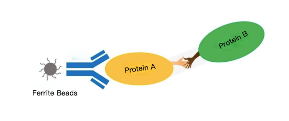
Co-IP experiments are typically used to
(1) determine whether two or more proteins interact in vivo;
(2) identify the interaction partners of a specific protein;
(3) isolate protein complexes in their native state.
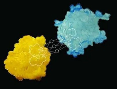
Therefore, one of the key factors in a Co-IP experiment is how to concisely and effectively set up groups to determine whether proteins interact with one another.
For a successful Co-IP experiment, it must first be demonstrated that target protein A and the predicted interacting protein B are present in the protein sample. Therefore, the extracted protein sample must be directly tested for the presence of A and B; this is generally referred to as the “input.”
Next, to verify whether the IP antibody can capture the target protein, the target protein must be detected after the IP is completed. It is important to note that a primary antibody used for Western blotting may not necessarily function as an IP antibody to capture the target protein, because during Western blotting, proteins are in a denatured state, and their three-dimensional structures are unfolded. Many antibodies are prepared using synthetic peptides as antigens; such antibodies may not necessarily recognize the target protein when it is folded in its native, undenatured state. Therefore, when selecting an IP antibody, it is essential to pay close attention to the applications listed in the antibody’s product description (e.g., ChIP, flow cytometry, ICC/IF, IHC-P, IP, WB, etc.). Furthermore, even if the antibody’s product information indicates it is suitable for IP experiments, its performance in different samples still requires actual testing to verify.
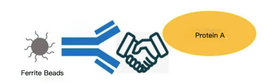
If target protein A is not detected, there may be several possible reasons:
Either the IP antibody is defective—in which case you should try a different antibody—or the beads have not bound to the antibody, which can be verified via Western blot (WB). Most commonly used antibodies are mouse- or rabbit-derived; during WB, the presence of heavy chains or light chains can confirm whether the beads are conjugated to the antibody. It is also possible that the expression level of protein A is too low; in this case, you can increase the sample volume.
If, in an endogenous Co-IP, the target protein is not detected in the input sample, this may be due to too low a concentration of the target protein in the sample itself or an improper protein extraction procedure; of course, issues with the antibody should not be overlooked. Therefore, we generally recommend first attempting a Co-IP using an overexpressed protein.
If both proteins A and B are detected after IP, that is certainly encouraging, but could this result be a false positive? A negative control must be performed. Several methods are commonly mentioned in the literature:
(1) Add only the beads without an IP antibody
(2) Use an antibody against an unrelated protein as the IP antibody
(3) Use normal IgG as the IP antibody
(4) Use a protein sample that does not express target protein A but does express protein B as a control.
Additionally, Co-IP is generally classified as endogenous or exogenous. Simply put, endogenous Co-IP detects whether proteins A and B, which are naturally present in the cells, interact; exogenous Co-IP involves overexpressing proteins A and B within the cells and then detecting whether they interact.
For endogenous Co-IP experiments, a negative result may be due to insufficient expression of the protein within the cells; in such cases, an overexpression Co-IP can be performed first as a control.
Only when proper controls are established can the results be reasonably interpreted and considered valid.
First, let’s review the relevant terms:
IB: Stands for immunoblotting, also known as conventional Western blotting, which is used to detect the target protein.
IP: Stands for immunoprecipitation. This step is primarily used to purify and enrich the target protein.
Input: Whole-cell lysate, which can be considered the positive control. This refers to the cellular sample obtained after sample processing. Before performing the IP experiment, a portion of the Input must be set aside to confirm the presence of both Protein A and Protein B in the sample while analyzing the IP results.
Anti-A: An immunoprecipitation antibody against Protein A
IgG: Isotype control antibody for Anti-A; that is, one from the same species as Anti-A. For example, if Anti-A is mouse IgG, the isotype control antibody should be mouse IgG.
Currently, we commonly use the following grouping methods:
1. Endogenous CO-IP detection
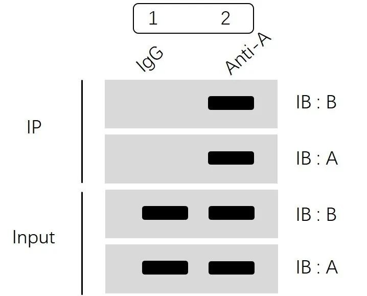
The diagram above is divided into two main sections: the IP section and the Input section.
First, let’s look at the Input section. By separately detecting the expression levels of Protein A and Protein B, we can confirm that:
Both Protein A and Protein B are expressed in Group 1 (IgG group) and Group 2 (Anti-A group), and their expression levels are consistent. This rules out false negatives or false positives caused by the absence of protein expression in some groups or significant differences in protein expression levels between groups.
Next, let’s look at the IP section:
The “IB:A” results show that Group 1 (IgG group) does not bind to Protein A, while Group 2 (Anti-A group) can specifically enrich Protein A.
The “IB:B” results show that there is no nonspecific binding in the IgG group, whereas the Anti-A group, by specifically enriching Protein A, also pulls down Protein B. This suggests that Protein A and Protein B can form a complex and interact with each other.
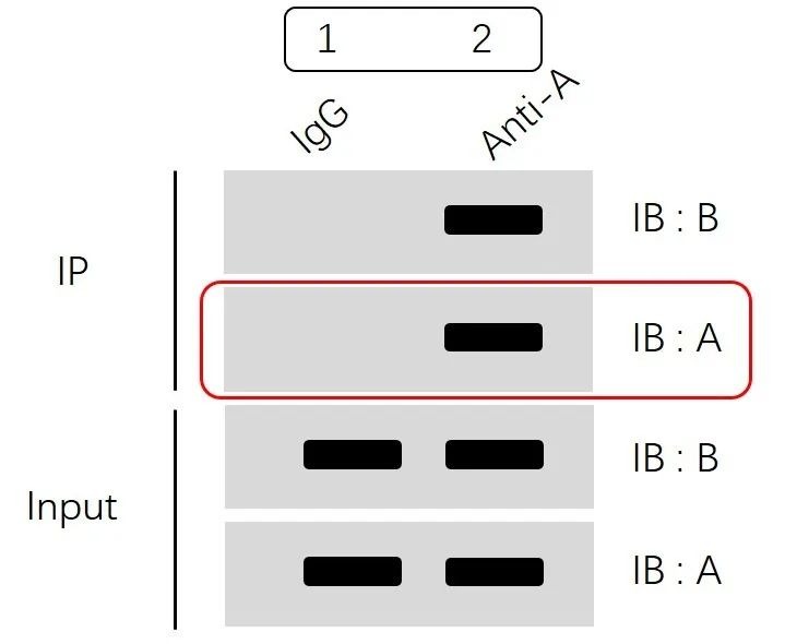
The “IB:A” step in the IP section is critical; it clearly indicates that Protein A has been successfully and specifically recognized and enriched onto the magnetic beads or gel via immunoprecipitation, meaning the immunoprecipitation procedure was successful. Whether Protein B can be detected in the next step depends primarily on whether an interaction exists between the two proteins. Of course, factors such as low protein expression levels or the need for specific stimulation conditions may also affect whether Protein B can be detected.
2. Exogenous CO-IP Assay
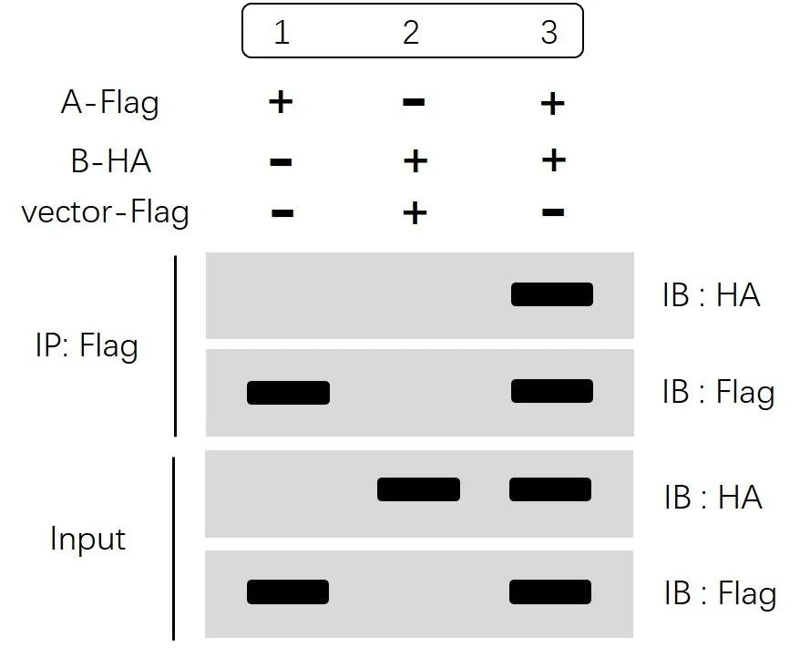
The results shown in the figure above are also divided into two main sections: the IP section and the Input section. For exogenous samples, we generally do not need to set up an IgG group (negative control); instead, we can use methods such as blank controls to establish a negative control group.
First, let’s look at the “Input” section. By measuring the expression levels of HA and Flag separately, we can confirm that:
Both A-Flag and B-HA are detectable after overexpression, and their expression levels are consistent, ruling out false negatives or false positives caused by uneven sample quantities or missing sample components.
Next, let’s examine the IP section. The “IB:Flag” results show that Groups 1 and 3 specifically recognized the Flag tag via IP:Flag and enriched Protein A. The “IB:HA” results show that Groups 1 and 2 (controls) did not capture Protein B, while Group 3 specifically enriched Protein A and subsequently captured Protein B. This indicates that Protein A and Protein B can form a complex and interact with each other.
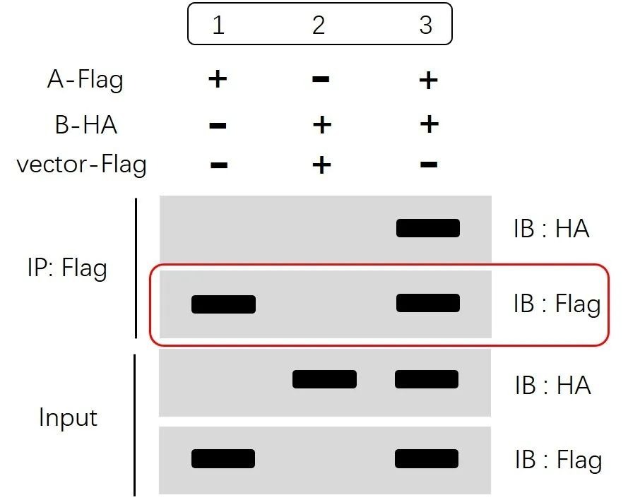
The “IP:Flag IB:Flag” step is also intended to confirm that the immunoprecipitation—specifically, the recognition and enrichment of Protein A using magnetic beads or gel—was successful.
Of course, some of our colleagues have also chosen the following grouping method:
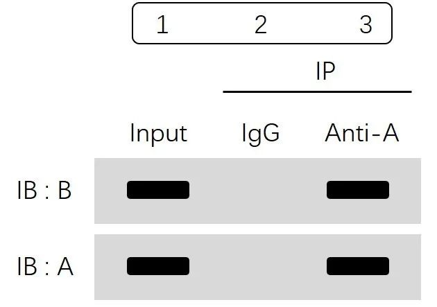
This experiment is divided into two main groups: the Input group and the IP group.
The IP group is further divided into the IgG group (negative control) and the experimental group. Western blot (WB) analysis was used to verify the presence of proteins A and B in each group.
Based on the results from the Input group, it was concluded that both Protein A and Protein B were present, confirming that the sample processing and protein extraction steps were performed correctly. This also suggests that, in this experiment, a single cell sample is typically selected: a portion is set aside in advance for the Input group, while the remainder is divided into two groups—the IgG group (negative control) and the experimental group.
Let’s continue with the IP section: The results from Group 2 (IgG group) show no bands for either Protein A or Protein B, indicating that the IgG antibody did not precipitate these proteins. This proves that Protein A and Protein B do not bind to IgG and simultaneously rules out the possibility of nonspecific binding between the proteins and the antibody; In contrast, the experimental group showed bands for both Protein A and Protein B, indicating that immunoprecipitation using the anti-Protein A antibody enriched Protein A, while Protein B was also co-precipitated. This suggests that Protein A and Protein B can form a complex and interact with each other.
The examples above are just a few of the more commonly used ones. The ultimate goal of setting up different control groups is to enable the experimental group to yield definitive results in a concise and effective manner. Therefore, you can choose the optimal approach based on your specific circumstances.
- Immunoprecipitation Assay
- Immunoprecipitation Experiment
- IP
- Co-IP

- Protein Platform
- Protein Expression Services
- Protein Modification & Detection Services
- Protein Interaction Services
- Protein Purification Service
- Antibody Discovery Platform
- VHH Antibody Platform
- Phage Display Platform
- Antibody Humanization Platform
- Single B Cell Screening Platform
- Antibody Expression & Validation Platform
- Peptide Library Platform
- Custom Antibody Platform
- Innovative Drug Discovery Platform
- Product Center
- Proteins
- Antibodies
- Raw Materials Reagents
- Small Molecule Reagents
- Peptide Products
- Nanoparticle Products
- Contact Us
- Phone:400-621-6806
- Mobile:13110936064(WeC
- E-mail:[email protected]
- Address:Room 108, Building 10, No. 6 Hechang Road, Wuqing Development Zone, Tianjin City
-

WeChat
-

Service Account
-
 400-621-6806
 -
 3599163699
 -
 Monday to Friday 9:00-18:00
 -

-
 0
0
-
