


Spot Hybridization Experiment
- 2026-07-09
- 233
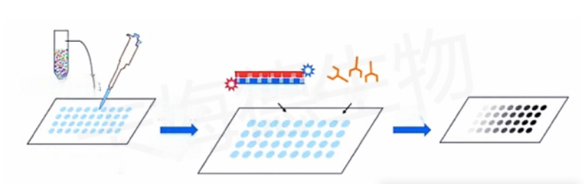
Dot blotting is a technique used to detect, analyze, and identify DNA, RNA, and proteins. In dot blotting, the biomolecules to be detected are not first separated by electrophoresis; instead, a mixture containing the target molecules is directly spotted onto a membrane and baked to fix it in place. Detection is then performed after hybridization with a nucleotide probe or antibody. The presence or absence of spots and their intensity are used to determine whether hybridization has occurred and to assess its strength. As the most abundant post-transcriptional RNA modification currently known in higher eukaryotes and plants, m6A modification regulates RNA splicing and transport, stability, nuclear export, and the efficiency of translation into proteins. Therefore, spot hybridization is frequently used in the study of m6A modification of RNA to perform qualitative or semi-quantitative analyses of m6A modification levels in total cellular RNA.
Principle
m6A dot blotting involves denaturing the RNA sample to be tested, applying it to a nitrocellulose (NC) membrane, cross-linking it with ultraviolet light, hybridizing it with an m6A antibody, washing the membrane, and performing autoradiography. The presence or absence of spots, or the intensity of their color, indicates the level of m6A modification.
Purpose
To detect changes in the level of m6A modification in total cellular RNA.
Materials and Equipment
m6A antibody (suitable for spot hybridization), 20× SSC buffer, mixed-bed resin PCR instrument, nitrocellulose membrane, medium-wave UV lamp (302 nm), PBST, secondary antibody (IgG), methylene blue powder, sodium acetate (M = 136), ECL developer, and chemiluminescence imager.
Reagent Preparation:
(1) Preparation of PBST buffer: Dissolve commercial PBS powder in 2 L of distilled water, then add 1 mL of Tween-20, mix well, and store at room temperature.
(2) Preparation of 37% deionized formaldehyde: Add 3 mL of Guoyao formaldehyde to 0.3 g of mixed-bed resin and mix thoroughly until the resin turns from blue to golden yellow. After a brief centrifugation, carefully aspirate the supernatant and aliquot it; store at room temperature.
(3) Preparation of Methylene Blue Solution: Weigh 20 mg of methylene blue powder and dissolve it in 10 mL of 0.3 mol/L sodium acetate solution. After thorough dissolution, a deep blue solution will form; store at room temperature.
(4) Preparation of 0.3 mol/L sodium acetate solution: Weigh 20.4 g of sodium acetate and dissolve it in 500 mL of deionized water; add hydrochloric acid to adjust the pH to approximately 5.2.
Procedure
1. Isolate total RNA from cells using Trizol reagent, taking care to adjust the RNA concentration to be as consistent as possible across different treatment groups.
2. Prepare the denaturation mixture (composed of 20× SSC buffer and 37% deionized formaldehyde in a 3:2 ratio); mix the denaturation mixture with the RNA in a 1:1 ratio, and incubate at 95°C for 5 minutes in a PCR machine to denature the nucleic acids.
3. Dispense 2 μL each of the diluted standards at different concentrations, samples, negative controls, and positive controls onto the nitrocellulose membrane, then irradiate under a 302 nm UV lamp for 6 minutes to cross-link the membrane.
4. After cross-linking, wash the membrane with PBST buffer for 5 min; then block with 5% non-fat dry milk at room temperature for 2 h, wash the membrane with PBST buffer for 5 min, and incubate with the m6A antibody overnight at 4 °C.
5. Subsequently, wash the membrane four times in PBST buffer for 5 minutes each time; incubate with the corresponding IgG secondary antibody at room temperature for 2 hours; then wash the membrane four times in PBST buffer for 5 minutes each time.
6. Perform ECL development using ECL development solution.
7. Methylene blue quantification of nucleic acids: Take a membrane treated in the same manner, add an equal volume of sample, and after UV cross-linking, perform methylene blue staining. Place the membrane in the solution for 10 minutes, wash with PBST for 10 minutes (replacing the PBST multiple times during this period), and then take a photograph.
Important Notes
1. Methylene blue is difficult to wash off; be sure to wear a mask and a lab coat during the experiment. When performing methylene blue staining, take care to avoid drawing up the sediment at the bottom of the solution.
2. The nucleic acid denaturation solution must be prepared fresh and used immediately.
3. Denatured RNA can be stored in a 4 °C refrigerator and used within one week.
4. When loading samples onto nitrocellulose membranes, use a water-based marker to label the membrane to avoid mixing up the order.
5. Work quickly when dispensing samples, and use RNAse-free tips; change the tip for each sample.
6. When dispensing samples, keep the volume to no more than 1 μL to prevent spreading and ensure proper spot formation.
7. There are slight differences in the selection of chemiluminescence imagers; it is best to choose one that allows you to adjust the exposure time manually.
Frequently Asked Questions
1. Is overexposure common during development?
A: Reduce the sample volume. Before conducting formal experiments, it is best to determine the optimal concentration range on your own. Generally, a range from 500 ng to 100 ng is acceptable.
2. Are the spots not clearly visible after methylene blue staining?
A: Generally, 10 minutes of methylene blue staining is sufficient. However, you should adjust the staining time based on your specific sample volume. During staining, use tweezers (preferably flat-tipped ones to avoid damaging the nitrocellulose membrane) to lift a section of the membrane for inspection; stop staining once you see clearly visible blue spots. Similarly, monitor the decolorization process closely; generally, stop when the background color and the spots are clearly distinguishable.
- Dot Blotting Experiment
- Dot Blotting
- Molecular Interactions
- dot blot

- Protein Platform
- Protein Expression Services
- Protein Modification & Detection Services
- Protein Interaction Services
- Protein Purification Service
- Antibody Discovery Platform
- VHH Antibody Platform
- Phage Display Platform
- Antibody Humanization Platform
- Single B Cell Screening Platform
- Antibody Expression & Validation Platform
- Peptide Library Platform
- Custom Antibody Platform
- Innovative Drug Discovery Platform
- Product Center
- Proteins
- Antibodies
- Raw Materials Reagents
- Small Molecule Reagents
- Peptide Products
- Nanoparticle Products
- Contact Us
- Phone:400-621-6806
- Mobile:13110936064(WeC
- E-mail:[email protected]
- Address:Room 108, Building 10, No. 6 Hechang Road, Wuqing Development Zone, Tianjin City
-

WeChat
-

Service Account
-
 400-621-6806
 -
 3599163699
 -
 Monday to Friday 9:00-18:00
 -

-
 0
0
-
