


Fab Antibody Phage Display Process
- 2026-07-09
- 322
1.What is a Fab antibody?
The Fab fragment is the region of an antibody structure capable of binding to an antigen; it is also known as the antigen-binding fragment and is responsible for characteristics such as antibody-antigen binding affinity and antibody specificity. The Fab fragment of an antibody contains one variable region (V region) and one constant region (C1). The V region contains three complementary determining regions (CDRs), which are involved in the production of unique antibodies and the hypermutation of antibodies; in particular, the CDR3 domain is the core region for antibody engineering and modification. The C1 region primarily serves a structural support function.
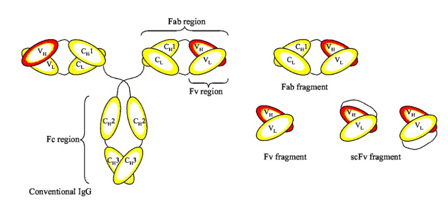
Figure 1: Schematic diagram of the fab antibody
2.Advantages of the fab Antibody Library
The advantages of recombinant Fab are quite evident: since it lacks the Fc region, it does not require post-translational modifications or glycosylation and can be expressed in both prokaryotic and mammalian systems. To produce Fab fragments, E. coli and mammalian expression systems are typically used. The E. coli expression system offers advantages such as low production costs and rapid production, but it is prone to forming inclusions, making it difficult to guarantee activity after renaturation.
3.The Difference Between SCFV Antibodies and Fab Antibodies
Table 1: Differences Between Fab Antibodies and scFv Antibodies
Difference | Fab | scFv |
Composition | VH、CH1、VL、CL | VH、VL |
Molecular weight(kDa) | 55 | 25 |
Tumor Permeability | Good | Stronger |
Stability | High stability during long-term storage | Unstable when stored for long periods |
Approachability | Very high | Higher |
4.Applications of the fab Antibody Library
Fab-type antibody fragments are characterized by their low molecular weight, high tissue-specificity, low immunogenicity, and suitability for genetic engineering, making Fab one of the key components in pharmaceutical research. Fab-based drugs have widespread applications in prevention, diagnosis, and treatment. For example, the marketed cetozumab ester (a Fab fragment, Cat: YR1612) is effective in treating rheumatoid arthritis; Mitoxantrone I, a Fab fragment, has also been used to treat lung cancer and other conditions.
5. KMD Bioscience is able to provide customers with high-quality Fab antibody phage display services.
KMD Bioscience has been dedicated to research in the field of antibodies for many years. We have accumulated extensive experience in antibody library construction and are able to provide our clients with Fab antibody library construction services based on phage display technology.
6. An Overview of the Entire Process for Fab Antibody Library Construction Services Provided by KMD Bioscience to Clients
KMD Bioscience offers clients FAB phage library construction services. The process includes: antibody cDNA preparation, gene amplification, vector construction, electroporation, antibody screening, and FAB library packaging.
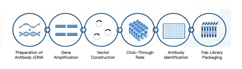
Figure 2: The Process of Constructing an Antibody Library Using Fab Phages
6.1 Preparation of Antibody cDNA
6.1.1 Cloning of Peripheral Blood Monocytes
The client provides human peripheral blood lymphocytes, lymph node cells, or spleen cells (preserved in a solution such as RNA Later to prevent RNA degradation and shipped on blue ice) for EBV monoclonalization.
6.1.2 Total RNA Extraction
After removing peripheral blood lymphocytes from the refrigerator, aliquot them, add chloroform, centrifuge, and collect the supernatant. Add isopropanol to the supernatant, centrifuge, and retain the pellet. Add 75% ethanol, centrifuge, and retain the pellet. After drying, add DEPC-treated water and incubate to ensure RNA dissolution. Combine the contents of all tubes into a single tube to obtain the total RNA. Perform electrophoresis on the total RNA and measure its concentration using a nucleic acid concentration meter.
6.1.3 cDNA Synthesis by Reverse Transcription
Prepare cDNA according to the instructions for the commercial kit, and reverse-transcribe the RNA obtained in the previous step into cDNA.
6.2 Gene Amplification
Amplify the genes encoding the antibody heavy chain (Fd segment) and light chain variable regions using cDNA as a template.
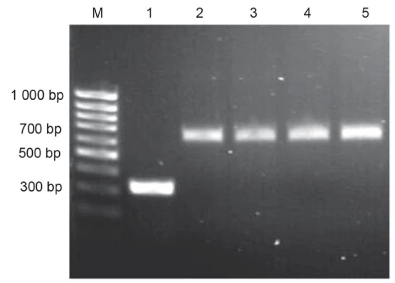
Figure 3: PCR Amplification Results for Fd and Light Chains
6.3 Vector Construction
The PCR-amplified Fd fragment (isolated by agarose gel electrophoresis) was digested with excess restriction enzymes. Typically, approximately 350 ng of the fragment was mixed with 2 μg of the Xho/Spe I-linearized pComb3 vector (isolated by agarose gel electrophoresis) in a total volume of 150 μL and incubated with ligase at 16°C for 15 hours, named P+Fd. The PCR-amplified light chain fragment and the recombinant P+Fd (isolated by agarose gel electrophoresis) were digested with excess restriction enzymes, ligated, and transformed; the phage particle containing Fd and the κ light chain was named pComb3H+κ+Fd.
6.4 Electroporation
6.4.1 Construction of an E. coli library containing the target antibody fragment via electroporation of the ligation product
Take E. coli competent cells, add the recovered ligation product, and transfer the mixture of competent cells and ligation product to a pre-chilled electroporation cup. Perform electroporation using the instrument’s preset transformation program. Immediately after electroporation, add culture medium to the electroporation cup and perform at least 20 electroporation cycles. Thaw the cells and spread them onto agarose plates for overnight growth. Rinse and scrape the cells from the plates used in the previous step with culture medium and a spreader, add glycerol, measure the OD600 nm value, and store at -80°C; this constitutes the bacterial library.
6.4.2 Screening of the fab Phage Library
Coat a 6-well plate (Nunc™) with 5 × 10⁶ peripheral blood lymphocytes, then block with BSA/PBS overnight at 4°C. Add 1 mL of the phage library to each well and incubate at 37°C for 2 h. For rounds 1, 2, 3, 4, and 5, unbound phages were washed 1, 5, 10, 10, and 10 times, respectively, with TBS/Tween 20 (TBST). Elute bound phages by adding elution buffer (0.1 M HCl, adjusted to pH 2.2 with solid glycine and containing 0.1% BSA), and neutralize the elution solution with 2 M Tris buffer, pH 8.0. Follow the VCSM13 auxiliary phage amplification protocol as described above, while simultaneously titrating the input and output phages on LB-ampicillin plates. Perform the phage binding, elution, and amplification steps for 5 rounds.
6.5 Antibody Identification
Randomly select 20–50 clones, determine the PCR positivity rate, and sequence and analyze the antibody sequences.
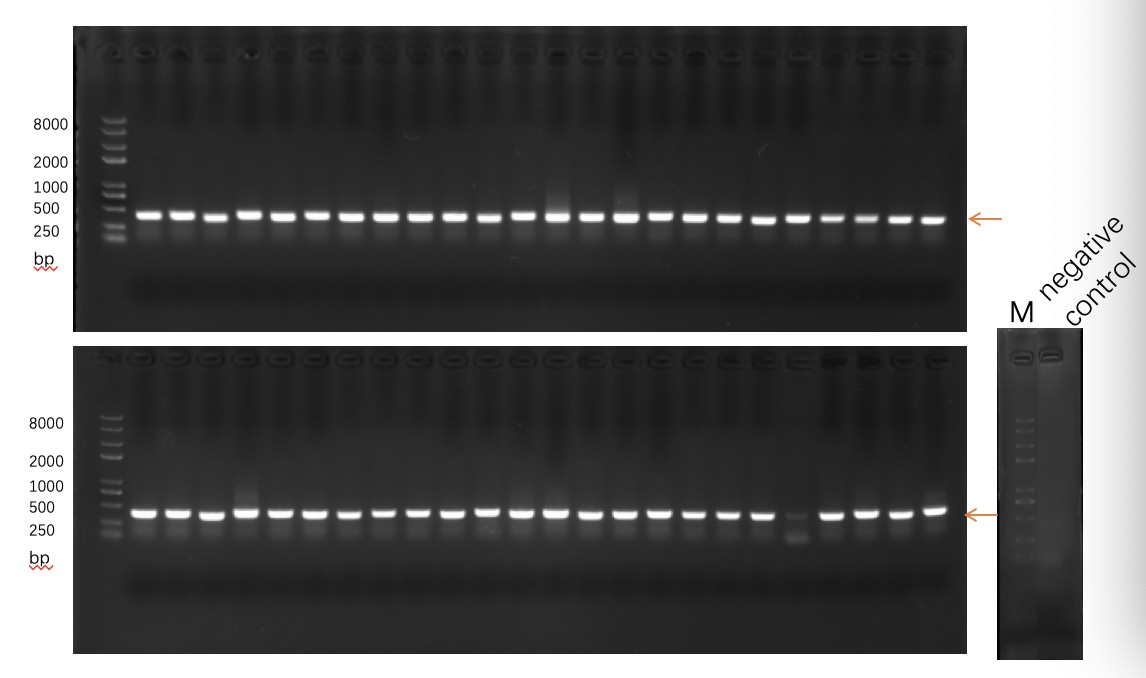
Figure 4: Positive Rate: >90%
6.6 Fab Library Packaging
Delivery: >1x10^13 phage particles/mL.
Traditional hybridoma technology for producing monoclonal antibodies is costly, and the resulting murine antibodies often trigger human anti-mouse antibody (HAMA) reactions. Therefore, recombinant Fab antibodies generated via phage display technology can serve as a suitable alternative for cancer therapy. KMD Bioscience’s humanized Fab antibody library construction service offers high affinity and high library capacity, with 10^7–10^12 independent clones. We prepare libraries according to customer requirements and tailor experimental protocols for each client.
- Fab antibody fragments
- Fab libraries
- Fab antibody libraries
- Fab antibody phage display services

- Protein Platform
- Protein Expression Services
- Protein Modification & Detection Services
- Protein Interaction Services
- Protein Purification Service
- Antibody Discovery Platform
- VHH Antibody Platform
- Phage Display Platform
- Antibody Humanization Platform
- Single B Cell Screening Platform
- Antibody Expression & Validation Platform
- Peptide Library Platform
- Custom Antibody Platform
- Innovative Drug Discovery Platform
- Product Center
- Proteins
- Antibodies
- Raw Materials Reagents
- Small Molecule Reagents
- Peptide Products
- Nanoparticle Products
- Contact Us
- Phone:400-621-6806
- Mobile:13110936064(WeC
- E-mail:[email protected]
- Address:Room 108, Building 10, No. 6 Hechang Road, Wuqing Development Zone, Tianjin City
-

WeChat
-

Service Account
-
 400-621-6806
 -
 3599163699
 -
 Monday to Friday 9:00-18:00
 -

-
 0
0
-
