


The Complete Process of Constructing a Phage Display Library
- 2026-07-09
- 394
1. What is phage display?
A phage library is a phage vector used for gene cloning. A phage display library is a new technology that fuses exogenous proteins or peptides with phage coat proteins, displays them on the phage surface while maintaining a specific spatial conformation, and utilizes specific affinity to screen for specific proteins or peptides.
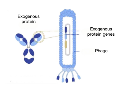
Figure 1: Phage Display Technology
2. Types of Phage Libraries
KMD Bioscience has conducted research in the field of antibodies for many years. The phage display technology services we offer cover a wide range of library types, including alpaca/ camel VHH antibody libraries, human phage libraries, human ScFV antibody libraries, human Fab antibody libraries, phage peptide libraries, and pre-engineered peptide libraries. We can tailor these high-quality antibody libraries to meet our clients’ specific needs. A phage display library is a phage vector used for gene cloning; phage display libraries are a new technology that fuses exogenous proteins or peptides with phage coat proteins, displays them on the phage surface while maintaining a specific spatial conformation, and utilizes specific affinity to screen for specific proteins or peptides. KMD Bioscience offers customized services and provides flexible and effective screening services.
3. Applications of Phage Library Screening
Phage display library technology is an emerging biotechnology. Exogenous proteins obtained through phage display retain their independent three-dimensional structure and biological activity. Currently, this technology is widely applied in the development of novel vaccines, the screening of enzyme inhibitors, medical diagnosis and treatment, the development of peptide drugs, antigen epitope analysis, monoclonal antibody screening, and the study of protein-protein interactions, and it shows great promise for future applications.
4. Technical Advantages of Phage Library Construction
(1) It offers a large screening capacity, is suitable for large-scale fermentation production, and employs a simple method. Currently, the phage primary immunological libraries we have obtained have a library capacity of 10⁸–10⁹, with a sequence abundance of >99%, ensuring that antibodies obtained through direct screening achieve affinities at the nM or even pM level.
(2) This represents a new screening technology that integrates genotype with phenotype, and molecular binding activity with phage amplifiability.
(3) Rapid preparation of soluble antigens, combined with the company’s existing recombinant protein expression system, enables the rapid production of high-purity, highly biologically active soluble antigen proteins to support phage antibody immunization and screening efforts.
5. KMD Bioscience provides customers with high-quality phage library construction services
The M13 single-stranded filamentous phage display system is currently one of the most widely used library display systems. By fusing proteins, antibodies, or peptides with the phage pIII protein for co-expression, these molecules are incorporated into phage assembly and displayed on the phage surface, forming a phage display library. Using M13 filamentous phage pIII and PVIII display technologies, KMD Bioscience can construct phage antibody display libraries from natural or immunogenic sources across various species for clients. Through antibody library technology, we produce monoclonal antibodies from multiple species, such as humans, mice, rabbits, chickens, sheep, geese, pigs, cattle, horses, donkeys, camels, llamas, sharks, and other species. Customized services are available based on client needs, and library capacity can reach 10^11.
6. Overview of the Full Process for Phage Library Construction Services Provided by KMD Bioscience
KMD Bioscience provides phage library construction services to clients. The process includes: total RNA extraction, reverse transcription to obtain cDNA, PCR amplification, restriction enzyme digestion and ligation of the vector with the PCR product, bacterial library construction, phage library construction, and phage library titer testing.
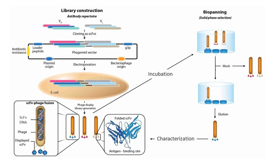
Figure 2: Workflow for Constructing a Phage Display Antibody Library
6.1 Total RNA Extraction
Remove the peripheral blood lymphocytes from the refrigerator and aliquot them. Add chloroform, centrifuge, and collect the supernatant. Add isopropanol, centrifuge to retain the pellet, add 75% ethanol, centrifuge again to retain the pellet, dry the pellet, add DEPC-treated water, and incubate to ensure RNA dissolution. Combine the contents of all tubes into a single tube to obtain total RNA. Take 1 μL of total RNA for electrophoresis, and take 2 μL to measure the concentration using a nucleic acid concentration meter.
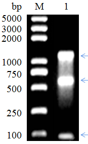
Figure 3: VHH RNA Extraction
6.2 Reverse Transcription to Obtain cDNA
Prepare cDNA according to the instructions for the commercial kit. Divide the RNA obtained in the previous step into two portions and reverse transcribe each into cDNA, using an Oligo dT primer and random 6-mers as reverse transcription primers, respectively.
6.3 PCR Amplification
6.3.1 First Round of PCR
Perform the first round of PCR using the cDNA as a template and HS Ex Taq enzyme for PCR amplification. The PCR reaction mixture is as follows:
Reagents | Usage |
| cDNA | 0.5-5μl |
| AlpVh-L/CALL 002 | 2μl/2μl |
| dNTP Mix | 4μl |
| 10×ExTaq Buffer | 5μl |
| HS Ex Taq | 0.25μl |
| ddH2O | Up to 50μl |
Perform 1% agarose gel electrophoresis on the resulting PCR amplification products, identify the target band, and amplify it using the PCR conditions described above. Perform 1% agarose gel electrophoresis on all PCR products, and use a universal DNA purification kit to recover DNA from the gel strips.
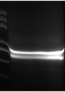
Figure 4: Results of the first round of PCR amplification
6.3.2 Second Round of PCR
Using the DNA fragment amplified and recovered in the previous PCR step as a template, specific antibody fragments were amplified again. The PCR reaction mixture was as follows:
| Reagents | Usage |
Recovery of products from the first round of PCR | 0.1-2μl |
| Rvhh FP/RvhhRP | 2μl/2μl |
| dNTP Mix | 4μl |
| 10×ExTaq Buffer | 5μl |
| HS Ex Taq | 0.25μl |
| ddH2O | Up to 50μl |
Use a DNA purification kit to recover the PCR amplification products according to the instructions.
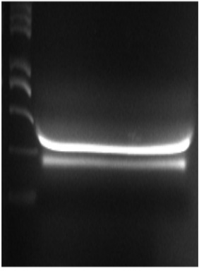
Figure 5: Results of the Second Round of PCR Amplification
6.4 Enzyme Digestion and Linkage of the Vector and PCR Products
The products from the second round of PCR were ligated into the phage plasmid pADL-10b via enzyme digestion, thereby constructing a phage plasmid library containing the amplified fragments. The ligation products were purified using a DNA purification kit according to the instructions, and 2 μL of each was taken to measure the concentration of the purified products using a nucleic acid concentration meter.
6.5 Construction of a Bacterial Library
The ligation products were subjected to electroporation to construct an E. coli library containing the target antibody fragment.
Take E. coli SS320 competent cells, add the recovered ligation products, and transfer the mixture of competent cells and ligation products to a pre-chilled electroporation cup. Perform electroporation using the instrument’s preset transformation program. Immediately after electroporation, add 950 μL of SOC medium to the electroporation cup. Perform at least 20 electroporation cycles. After resuspending the cells, spread them onto agar plates containing ampicillin resistance and allow them to grow overnight. Scrape the cells from the plates that have grown overnight using 2× YT medium and a spreader, add 20% glycerol, measure the OD₆₀₀ value, and store at −80°C; this constitutes the bacterial library.
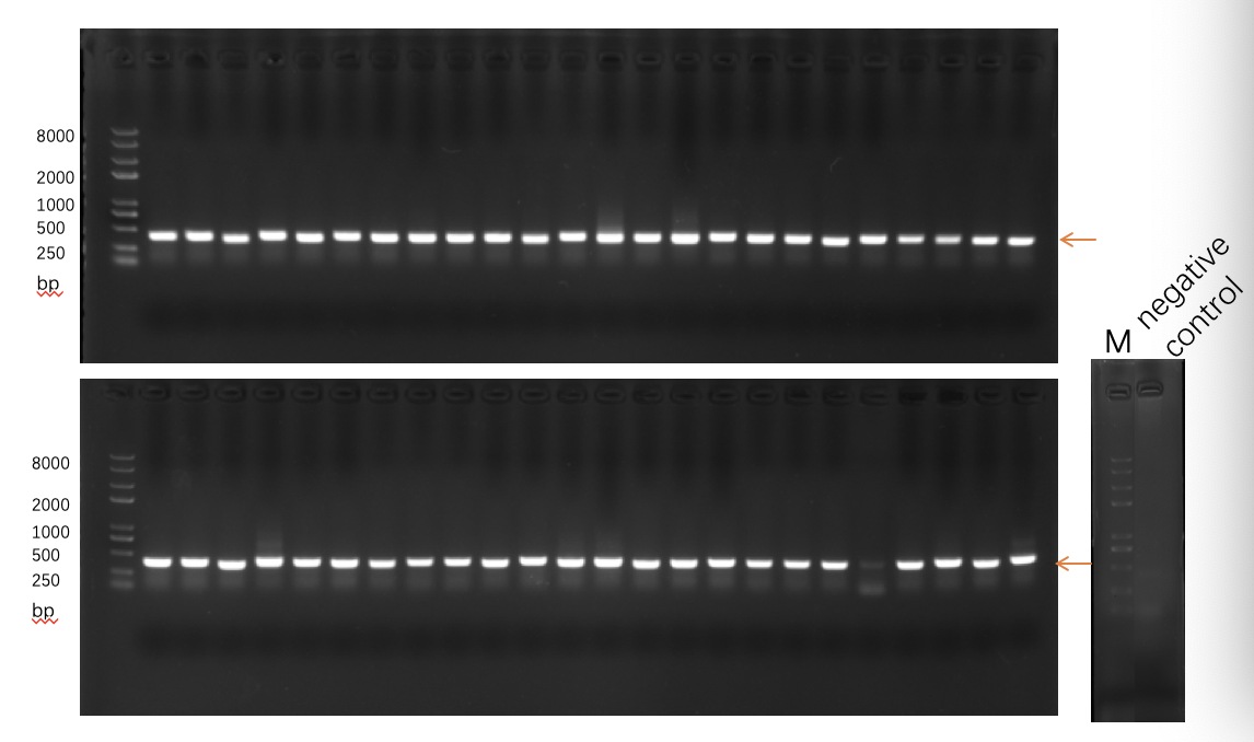
Figure 6: VHH Positivity Rate: >90%
6.6 Construction of a Phage Library
6.6.1 Amplification of the Phage Library
Mix the bacterial cells scraped from the previous step and transfer them to 100 mL of 2x YT medium pre-treated with tetracycline. Incubate at 37°C and 250 rpm until the OD₆₀₀ reaches 0.5–0.55.
Add helper phages at a ratio of 20:1 (helper phages to bacterial cells) and continue incubation at 37°C for 30 minutes. Add Kana to a final concentration of 50 µg/mL and PTG to a final concentration of 0.2 mM, then incubate overnight on a shaking incubator at 30°C.
6.6.2 Phage Library Preparation
Centrifuge the overnight bacterial culture at 4°C and 4000 rpm for 20 minutes. Transfer the supernatant to a new centrifuge tube, add 1/4 of the volume of pre-chilled 20% PEG/2.5 M NaCl, and incubate on ice for 30 minutes. Centrifuge at 4°C and 4000 rpm for 20 minutes to remove the supernatant, then add 1 mL of PBS buffer to resuspend the pellet. Add another 1/4 of the volume of pre-chilled 20% PEG/2.5 M NaCl and incubate on ice for 10 minutes. Centrifuge at 4°C and 12,000 xg for 10 minutes, remove the supernatant, and dissolve the pellet in 1 mL of PBS to obtain the phage library. Store long-term at –80°C; for short-term storage (1–2 weeks), store at 4°C.
6.7 Phage Library Titer Assay
Cultivate the SS320 strain stored at −80°C. Pick a single colony from a colony plate and inoculate it into 5 mL of 2× YT medium for overnight culture; the OD₆₀₀ of the overnight culture should be 0.5–0.55. Prepare 12 dilution gradients of the phage library. Add 90 μl of the SS320 culture to each dilution, then transfer 5 μl from each dilution tube to 2× YT solid medium (Amp) for overnight incubation. The titer can then be calculated.
- Phage display
- library construction
- phage display library construction
- VHH antibody library

- Protein Platform
- Protein Expression Services
- Protein Modification & Detection Services
- Protein Interaction Services
- Protein Purification Service
- Antibody Discovery Platform
- VHH Antibody Platform
- Phage Display Platform
- Antibody Humanization Platform
- Single B Cell Screening Platform
- Antibody Expression & Validation Platform
- Peptide Library Platform
- Custom Antibody Platform
- Innovative Drug Discovery Platform
- Product Center
- Proteins
- Antibodies
- Raw Materials Reagents
- Small Molecule Reagents
- Peptide Products
- Nanoparticle Products
- Contact Us
- Phone:400-621-6806
- Mobile:13110936064(WeC
- E-mail:[email protected]
- Address:Room 108, Building 10, No. 6 Hechang Road, Wuqing Development Zone, Tianjin City
-

WeChat
-

Service Account
-
 400-621-6806
 -
 3599163699
 -
 Monday to Friday 9:00-18:00
 -

-
 0
0
-
