


Several Systems of Conventional Phage Display Technology (T7, T4, and M13)
- 2025-02-26
- 560
I.What is phage display
Phage display technology is a new technique to display foreign proteins or peptides on the surface of phage and maintain a specific spatial conformation, and to screen specific proteins or peptides by using specific affinity.
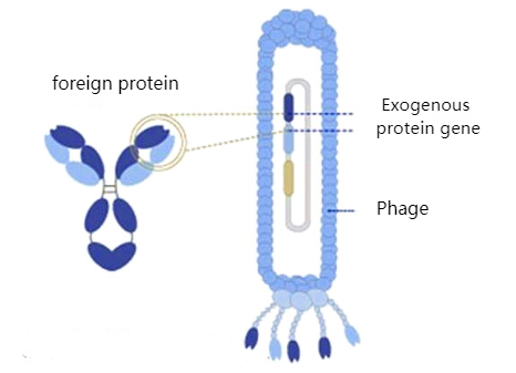
Figure 1: Phage display technology
II.Application of Phage Library Screening
Phage display library technology, an emerging biotechnology, enables the preservation of native spatial structures and biological activities in exogenous proteins obtained through phage display. It has been widely applied in various fields including novel vaccine development, enzyme inhibitor screening, medical diagnostics and therapies, peptide drug design, antigen epitope analysis, monoclonal antibody screening, and protein interaction studies, demonstrating promising application prospects.
III. Classification of Phage Display Systems
The phages commonly used in phage display are the filamentous phages M13, T4 and T7 of Escherichia coli.
1、Escherichia coli filamentous bacteriophages (f1, fd, M13)
Filamentous bacteriophages are defined by their filamentous or rod-shaped morphology. Filamentous bacteriophages typically contain single-stranded DNA genomes and infect Gram-negative bacteria. In phage display systems, the Ff, M13, fd, and f1 families are significant phage families, with M13 bacteriophages being the most commonly utilized.
The M13 bacteriophage is cylindrical in shape, measuring 880 nm in length and 6 nm in diameter. It encloses a single-stranded genome that encodes five distinct capsid proteins, including two groups: major capsid proteins (pVIII) and minor capsid proteins (pVII, pIX, pVI, and pIII).
Escherichia coli filamentous bacteriophages are commonly used for phage display. Most antibodies and polypeptides are displayed on phage proteins pIII and pVIII, thereby establishing the pIII and pVIII display systems. Additionally, hybrid phage systems can display large proteins with all five M13 coat proteins as N-terminal fusions with pIII, pVIII, pVII, and pIX, as well as C-terminal fusions with pVI, pIII, and pVIII.
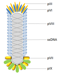
Figure 2: M13 bacteriophage
pIII is the protein responsible for determining the infectivity of viral particles. It consists of 406 amino acid residues and appears in 3 to 5 copies at the phage tip. One advantage of using pIII over pVIII is that it allows for monovalent display when phage-like particles are used to bind auxophages. Additionally, pIII permits the insertion of larger protein sequences (>100 amino acids) and is more tolerant than pVIII.
pVIII is the major coat protein of the Ff phage, expressed by gene 8 and present in 2,700 copies. Consequently, when phage-displayed antibodies bind to antigens, it is utilized to amplify the detection signal. The peptide is typically fused to the N-terminus of pVIII, usually consisting of 6-8 amino acids. This makes the use of this protein less favorable for identifying high-affinity binding partners. Furthermore, significant progress has been made in modifying pVIII to enhance display efficiency on pVIII.
pVI has been widely utilized for cDNA library display. Due to its high-throughput capability, it serves as an attractive alternative to the yeast-2-hybrid method for identifying interacting proteins and peptides. pVI is preferred over pVIII and pIII for cDNA library expression because the target protein can be added to the C-terminus of pVI without significantly compromising its role in phage assembly.
pVII and pIX are located at the phage tip opposite to pIII, which can complement the current phage display system and serve as alternative scaffolds for display and selection, thereby further improving phage display as a final combinatorial engineering platform.
2、T4 phage
Enterobacteriophage T4 (T4 phage) is a bacteriophage that infects Escherichia coli. It belongs to the T-even phage group, which includes Enterobacteriophage T2 and T6. Enterobacteriophage T4 is a relatively large phage, approximately 90 nm in width and 200 nm in length. Its double-stranded DNA genome is about 169 kbp in size and encodes 289 proteins. Enterobacteriophage T4 consists of three essential proteins: gp23, which forms the hexagonal capsid lattice; gp24, which forms a pentamer at eleven of the twelve vertices; and gp20, which forms a unique dodecameric entry vertex through which DNA enters during packaging and exits during infection.
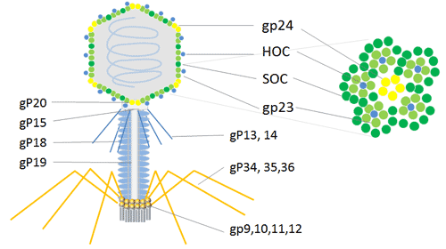
Figure 3: T4 phage
In addition to the essential capsid proteins gp23, gp24, and gp20, the T4 capsid is also decorated with two non-essential outer capsid proteins: HOC (highly antigenic outer capsid protein) and SOC (small outer capsid protein). Both HOC and SOC are dispensable and bind to the capsid after its assembly is complete.
HOC and SOC are optional T4 capsid proteins that can be utilized for phage display of polypeptides and proteins in multiple copies. The T4 phage HOC/SOC dual-display system is attractive for high-copy expression of cDNA and display peptides or proteins on the phage capsid surface. It can be applied to cDNA expression to display larger proteins in high copies, with termination codons inserted at the C-terminus of the 810-copy SOC protein or at the N-terminus of the 155-copy HOC protein. Consequently, the T4 phage dual-site display serves as a powerful method to enhance animal immune responses for the research and development of immunological products.
3、T7 phage
T7 phage is an octahedral virus belonging to the Podoviridae family, possessing a linear double-stranded (ds) DNA genome. Similar to T4, the T7 phage exhibits a head-tail structure. The octahedral head of T7, which preserves its dsDNA genome, consists of 415 copies of the capsid gp10 protein, arranged as 60 hexamers on the surface and 11 pentamers at the apex. The major capsid proteins gp10, gp10A, and gp10B exist in two isoforms in a 9:1 ratio, caused by natural translational frameshift at amino acid position 341.
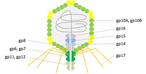
Figure 4: T7 phage
The secondary protein gp10B is induced by a frameshift at the gene terminus, resulting in an elongation of the capsid protein by 52 residues. The fusion protein exhibits the C-terminal region of gp10B with these additional 52 residues, thereby avoiding issues related to steric hindrance. T7 phage particles demonstrate high stability under various extreme conditions, including high temperatures and low pH values, which facilitates efficient high-throughput affinity washing. The ultimate application of the T7 phage display system aids in elucidating the mechanisms of molecular interactions, particularly in antigen discovery, vaccine development, protein-protein interactions, and cancer diagnosis and therapy.
IV.KMD Bioscience provides customers with a comprehensive introduction to the full-process service of phage library construction
KMD Bioscience provides clients with phage library construction services, including the following procedures: total RNA extraction, reverse transcription to obtain cDNA, PCR amplification, restriction enzyme digestion and ligation of vectors with PCR products, bacterial library construction, phage library construction, and phage library titer detection.
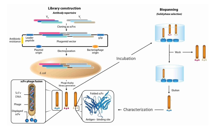
Figure 5: Construction workflow of phage display antibody library
1、Total RNA Extraction
The peripheral blood lymphocytes were collected from the refrigerator and aliquoted. They were then precipitated and resuspended using chemical reagents, followed by concentration measurement with a nucleic acid concentration analyzer.
2、Reverse transcription to obtain cDNA
cDNA was synthesized by reverse transcription of RNA obtained from the previous step according to the instructions of the commercial kit.
3、PCR Amplification
Two rounds of PCR were performed using cDNA as the template.
4、ector and PCR product ligase digestion
The PCR products were ligated to the phage plasmid pADL-10b to construct a phage plasmid library.
5、Bacterial Library Construction
The E. coli library containing the target antibody fragment was constructed by electroporation of the linker.
6、Phage Library Construction
The phage library from the previous step was amplified to prepare the phage library.
7、Phage Library Titer Detection
The phage library can be calculated for titer through dilution culture
- Phage display technology
- phage library screening
- Escherichia coli filamentous phages
- T4 phage

- Protein Platform
- Protein Expression Services
- Protein Modification & Detection Services
- Protein Interaction Services
- Protein Purification Service
- Antibody Discovery Platform
- VHH Antibody Platform
- Phage Display Platform
- Antibody Humanization Platform
- Single B Cell Screening Platform
- Antibody Expression & Validation Platform
- Peptide Library Platform
- Custom Antibody Platform
- Innovative Drug Discovery Platform
- Product Center
- Proteins
- Antibodies
- Raw Materials Reagents
- Small Molecule Reagents
- Peptide Products
- Nanoparticle Products
- Contact Us
- Phone:400-621-6806
- Mobile:13110936064(WeC
- E-mail:[email protected]
- Address:Room 108, Building 10, No. 6 Hechang Road, Wuqing Development Zone, Tianjin City
-

WeChat
-

Service Account
-
 400-621-6806
 -
 3599163699
 -
 Monday to Friday 9:00-18:00
 -

-
 0
0
-
