


Every biotherapeutic director, antibody discovery team, and diagnostic development lead eventually reaches the same critical fork in preclinical research: do you build your therapeutic pipeline around the established, multi-billion-dollar framework of conventional full-length immunoglobulin G (IgG), or do you transition your discovery campaigns to single-domain camelid antibodies (VHH nanobodies)? In commercial biopharma development, sticking with conventional antibodies simply because they are familiar is an expensive scientific habit. Full-length IgG molecules are massive, complex heterotetrameric proteins. While they excel at systemic receptor blockade and immune recruitment in peripheral circulation, their sheer molecular bulk severely limits their ability to penetrate dense desmoplastic tumor stroma, cross the blood-brain barrier (BBB), or access hidden viral catalytic clefts. When evaluating VHH Antibodies and Conventional Antibodies, you are not simply comparing two different formats on a Western blot; you are choosing between completely divergent biophysical parameters, production economics, and engineering modularities. Sourcing or engineering the wrong format early in discovery leads to failed clinical biodistribution, unresolvable aggregation during high-concentration formulation, and patent re-work down the line. As antibody discovery specialists, phage display engineers, and biophysical characterization practitioners at KMD Bioscience, we construct recombinant libraries, pan camelid repertoires, and express therapeutic fragments daily. Here is our direct, practitioner-grade evaluation of the 9 definitive structural, functional, and commercial differences between VHH Antibodies and Conventional Antibodies to guide your therapeutic, diagnostic, and research investments.
Quick Answer: VHH Antibodies vs. Conventional Antibodies – Key Distinctions
The definitive differences between VHH Antibodies and Conventional Antibodies center on nine core biophysical and translational criteria: (1) Molecular Mass & Architecture (VHH is a 15 kDa single monomeric domain; conventional IgG is a 150 kDa heterotetramer of two heavy and two light chains); (2) Paratope Topography (VHH features protruding, convex CDR3 loops capable of accessing cryptic enzymatic crevices; IgG features planar or grooved binding surfaces); (3) Thermal & Chemical Resilience (VHH withstands 80°C–90°C and refolds reversibly; IgG irreversibly aggregates above 65°C); (4) Tissue Penetration & Solid Tumor Infiltration (VHH rapidly diffuses through dense stromal extracellular matrix; IgG is hindered by the binding-site barrier); (5) Pharmacokinetics & Renal Elimination (VHH exhibits rapid clearance with a terminal half-life of hours; IgG benefits from FcRn recycling providing a 14–21 day half-life); (6) Biomanufacturing Platforms & Fermentation Economics (VHH expresses at high yields in microbial E. coli or yeast; IgG strictly requires complex mammalian CHO/HEK cells); (7) Native Effector Function (VHH lacks Fc-mediated ADCC/CDC toxicity unless engineered; IgG intrinsically recruits immune effectors); (8) Multispecific & Biparatopic Modular Flexibility (VHH domains assemble into tandem bispecific or CAR-T constructs without chain-mispairing; IgG requires complex knob-into-hole engineering); and (9) Immunogenicity & Humanization Baseline (VHH naturally shares high homology with human IGHV3 families; IgG requires coordinated dual-chain humanization). Sourcing through dedicated discovery ecosystems like the KMD Bioscience Alpaca VHH Platform allows biopharma teams to harness high-affinity nanobodies without sacrificing developability.
Article Directory
The 9 Critical Differences Between VHH Antibodies and Conventional Antibodies Analyzed
Featured Platform Spotlight: KMD Bioscience Alpaca VHH Library Construction
Direct Technical Comparison: VHH vs. Conventional IgG vs. scFv Fragments
Pros and Cons: Evaluating Single-Domain Nanobodies Against Conventional IgG
Commercial Economics: Biomanufacturing Costs, Scale-Up, and IP Portfolios
Who Should Use VHH Antibodies vs. Who Should Stick to Conventional IgG
5 Costly Mistakes R&D Teams Make When Developing VHH Formats
Essential Sourcing Checklist: Auditing an Antibody Discovery Partner
Structural Foundations: From Mammalian IgG to Camelid HCAbs
To evaluate VHH Antibodies and Conventional Antibodies with scientific rigor, one must examine their evolutionary divergence. Conventional mammalian antibodies—predominantly Immunoglobulin G (IgG)—represent an evolutionary pinnacle of immunological complexity. Composed of two identical heavy chains (~50 kDa each) and two identical light chains (~25 kDa each), conventional antibodies form a 150 kDa Y-shaped heterotetramer ($H_2L_2$). The antigen-binding site (paratope) is created by the cooperative spatial pairing of two distinct protein domains: the variable heavy ($V_H$) and variable light ($V_L$) domains, which collectively present six complementarity-determining regions (CDRs) to the antigen surface.
In 1993, researchers at the Vrije Universiteit Brussel made a groundbreaking discovery: camelids (alpacas, llamas, camels, and dromedaries) naturally produce a unique class of functional antibodies devoid of light chains, known as heavy-chain-only antibodies (HCAbs). In these molecules, the first constant domain ($C_H1$) is spliced out during mRNA transcription, completely eliminating the structural scaffold required to anchor a light chain. The entire antigen-binding machinery is concentrated into a single, autonomous variable domain designated as VHH (Variable Heavy domain of Heavy-chain antibody), colloquially termed a nanobody.
From our experience on discovery campaigns, the absence of the light chain is not a liability; it is an extraordinary biophysical upgrade for targeted applications. Conventional single-chain variable fragments (scFv), which artificially stitch a $V_H$ and $V_L$ together using a flexible synthetic peptide linker (e.g., $(Gly_4Ser)_3$), suffer from severe hydrophobic instability. The hydrophobic patches natively designed to interface the two domains are exposed to aqueous solvent, causing scFvs to denature, dissociate, and aggregate during downstream purification. VHH antibodies resolve this fatal flaw by replacing these hydrophobic residues with hydrophilic amino acids, resulting in a single, highly soluble, 15 kDa monomeric unit. When biopharma teams evaluate targeted leads across our VHH Antibody Platform, this inherent solubility translates into higher downstream expression stability compared to fragile scFv constructs.
Quick Summary Table: The 9 Core Differences at a Glance
| Biophysical Parameter | VHH Antibody (Nanobody) | Conventional Antibody (IgG) | Translational Significance |
|---|---|---|---|
| 1. Molecular Mass & Dimensions | ~12–15 kDa (4 nm × 2.5 nm) | ~150 kDa (14 nm × 10 nm × 5 nm) | VHH is one-tenth the size of IgG; crosses spatial obstacles |
| 2. Paratope Topography | Convex, protruding extended CDR3 loop | Planar, undulating, or concave cleft | VHH accesses hidden catalytic crevices and enzyme pockets |
| 3. Thermal & Chemical Stability | Tm up to 80°C–90°C; reversible refolding | Tm 60°C–70°C; irreversible aggregation | VHH supports room-temperature storage & harsh delivery |
| 4. Tissue Penetration Rate | High interstitial diffusion; rapid tumor penetration | Slow diffusion; limited by binding-site barrier | VHH delivers uniform drug dosing across dense tumor cores |
| 5. In Vivo Half-Life & Elimination | Hours (Rapid renal clearance < 60 kDa) | 14 to 21 days (FcRn recycling mechanism) | VHH excels in molecular imaging; IgG excels in continuous blockade |
| 6. Biomanufacturing Host | Microbial systems (*E. coli*, *P. pastoris*) | Mammalian systems (CHO, HEK293) | VHH slashes commercial bioreactor COGS by 60%–80% |
| 7. Native Effector Function | None (Lacks constant Fc region) | Potent intrinsic ADCC, ADCP, and CDC | VHH eliminates off-target immune lysis unless engineered |
| 8. Multispecific Engineering | Plug-and-play modular beads-on-a-string | Complex (Knobs-into-holes, CrossMAb) | Zero light-chain mispairing risks during biparatopic assembly |
| 9. Humanization Framework | High homology to human IGHV3; few mutations | Dual-chain framework matching required | Low immunogenicity risk; accelerated humanization cycles |
The 9 Critical Differences Between VHH Antibodies and Conventional Antibodies Analyzed
1. Molecular Mass and Spatial Geometry: 15 kDa Monomer vs. 150 kDa Heterotetramer
The most obvious yet consequential divergence between VHH Antibodies and Conventional Antibodies is molecular mass. A standard conventional IgG molecule has an average molecular weight of approximately 150,000 Daltons (150 kDa) with overall physical dimensions measuring roughly 14 nm × 10 nm × 5 nm. A single-domain VHH antibody measures approximately 4 nm in length and 2.5 nm in diameter, with a molecular mass of only 12 to 15 kDa. It is literally one-tenth the mass of a conventional antibody.
In our testing, this extreme physical compactness fundamentally alters steric clearance. Conventional antibodies are frequently barred from entering crowded molecular interfaces—such as the dense viral surface glycoprotein arrays of HIV-1, Influenza, or Coronaviruses, where bulky IgG molecules suffer severe steric clash between adjacent Fab arms. A VHH nanobody behaves more like a structured biological peptide, slipping into crowded macromolecular spaces where an IgG cannot fit.
2. Paratope Topography and Cryptic Epitope Access
In conventional IgG molecules, the antigen-combining paratope is formed by the joint assembly of six hypervariable loops: three from the heavy chain (CDR-H1, CDR-H2, CDR-H3) and three from the light chain (CDR-L1, CDR-L2, CDR-L3). This paired architecture naturally produces an antigen-binding interface that is fundamentally planar, wide, or indented into a shallow groove. While this planar surface is magnificent for binding exposed, flat protein surfaces, peptides, or small haptens, it struggles against recessed targets.
VHH antibodies compensate for the complete loss of the three light-chain CDRs by radically remodeling their remaining loops, particularly CDR3:
Extended Loop Architecture: Camelid VHH CDR3 loops are exceptionally long, frequently containing 16 to 24 amino acids (compared to human CDR-H3 loops, which average 12 to 14 residues).
Convex Finger-Like Protrusions: The extended CDR3 loop adopts an autonomous, finger-like convex protrusion stabilized by non-canonical disulfide bonds linking the loop back to the framework region or CDR1.
3. Thermal, Chemical, and Conformational Resilience
Conventional antibodies are delicate biotherapeutic entities. An IgG molecule depends upon precise inter-chain quaternary folding between its CH1, CL, and variable domains. When exposed to temperatures exceeding 60°C to 65°C, conventional antibodies undergo irreversible denaturation: the hydrophobic core of the $V_H-V_L$ interface unravels, exposing sticky hydrophobic residues that lead to rapid, catastrophic protein precipitation and aggregation.
VHH antibodies exhibit remarkable thermal and chemical resilience:
Reversible Thermal Denaturation: The melting temperature ($T_m$) of a VHH typically ranges from 70°C to well over 85°C. More importantly, when a VHH is heated past its $T_m$ and cooled back down, it possesses an extraordinary capacity for 100% reversible refolding, regaining full antigen-binding affinity.
Hydrophilic Framework Hallmarks: VHH frameworks replace the conserved hydrophobic residues that normally anchor the light chain (Val37, Gly44, Leu45, and Trp47 under Kabat numbering) with hydrophilic amino acids: Phe37/Tyr37, Glu44, Arg45, and Gly47. This eliminates the solvent-exposed hydrophobic "patch," preventing non-specific aggregation even in concentrated formulations.
Chemical Resistance: In our laboratory protocols, VHH antibodies routinely maintain functional integrity in the presence of 6M urea, 2M guanidine hydrochloride, proteolytic gastrointestinal enzymes (pepsin and trypsin), and broad pH ranges (pH 2.0 to 11.0), unlocking oral delivery and pulmonary aerosolization routes that would shred conventional IgG.
4. Tissue Penetration and Dense Tumor Microenvironment Kinetics
A major translational limitation of conventional antibodies in oncology is poor intratumoral distribution. Large 150 kDa IgG molecules face significant transport hurdles within solid tumors: elevated interstitial fluid pressure (IFP), dense desmoplastic extracellular matrix (ECM) crosslinked with collagen and hyaluronan, and the binding-site barrier (where high-affinity antibodies become trapped on the outermost tumor perimeter, failing to penetrate the hypoxic core).
The tissue penetration velocity of a therapeutic protein is inversely proportional to its molecular radius. Because VHH antibodies are roughly one-tenth the mass of IgG, their hydrodynamic radius is significantly smaller, allowing them to rapidly diffuse across vascular endothelial walls and deep into the dense, hypoperfused center of solid tumors within minutes of administration. In preclinical oncology studies cataloged on PubMed (National Library of Medicine), fluorophore-labeled VHH nanobodies achieve uniform, homogeneous tumor infiltration in under two hours, compared to 24 to 72 hours for intact IgG, which remains concentrated primarily around perivascular spaces.
5. Pharmacokinetics, In Vivo Clearance, and Diagnostic Imaging Contrast
When selecting between VHH Antibodies and Conventional Antibodies, your therapeutic or diagnostic endpoint dictates the required half-life:
| Pharmacokinetic Factor | VHH Antibody (Unmodified Nanobody) | Conventional Full-Length IgG |
|---|---|---|
| Elimination Pathway | Renal glomerular filtration (MW < 50–60 kDa cutoff) | Hepatic reticuloendothelial catabolism & endocytosis |
| Circulating Serum Half-Life ($t_{1/2}$) | Rapid: 30 minutes to 2 hours | Prolonged: 14 to 21 days |
| FcRn Receptor Recycling | None (Lacks constant heavy domains) | Active recycling via CH2-CH3 binding to FcRn |
| Ideal Clinical Indication | Immuno-PET/SPECT imaging, intraoperative tracer | Chronic inflammatory disease, long-term neutralization |
In non-invasive immuno-PET or SPECT radiodiagnostic imaging, conventional antibodies perform poorly: their long 3-week circulating half-life creates high radioactive blood-pool background, forcing clinicians to wait 3 to 7 days after injection before taking a scan. Unmodified VHH nanobodies clear from systemic circulation via the kidneys within hours, yielding breathtaking, razor-sharp tumor-to-background contrast ratios in under two hours post-injection with minimal radiation exposure to healthy tissues. Conversely, if your goal is chronic receptor blockade without daily dosing, a native VHH must be engineered with albumin-binding tags or half-life extension technologies.
6. Biomanufacturing Platforms, Fermentation Economics, and Downstream COGS
Commercial viability in biopharma is dictated by the cost of goods sold (COGS). Manufacturing conventional monoclonal antibodies requires eukaryotic, mammalian host platforms—predominantly Chinese Hamster Ovary (CHO) or human embryonic kidney (HEK293) cell lines. Mammalian expression is mandatory for IgG because the heterotetramer requires complex folding chaperones to assemble four separate chains, alongside enzymatic machinery in the endoplasmic reticulum and Golgi apparatus to execute specific N-linked glycosylation (Asn297) on the CH2 domain.
Because VHH antibodies are single-domain, non-glycosylated proteins with straightforward intrachain disulfide bonding, they can be produced at high yields in microbial fermentation systems:
Prokaryotic Hosts (*E. coli*): Targeted periplasmic expression allows proper disulfide bond formation, yielding grams of pure VHH per liter of bacterial broth in rapid 48-hour fermentation runs.
Yeast Platforms (*Pichia pastoris*): Capable of direct, high-density secretory expression with minimal host protein contamination, slashing downstream purification steps.
7. Effector Function Modulation: Zero Native Fc vs. Intrinsic ADCC/CDC
The constant region (Fc domain) of a conventional IgG1 antibody is a powerful immunological weapon. It binds to Fc-gamma receptors (FcγRs) expressed on natural killer (NK) cells, monocytes, and macrophages to trigger Antibody-Dependent Cellular Cytotoxicity (ADCC) and Antibody-Dependent Cellular Phagocytosis (ADCP). It also binds C1q to ignite the classical Complement-Dependent Cytotoxicity (CDC) cascade.
However, this effector function is a double-edged sword:
When Effector Function Is Destructive: If your therapeutic strategy aims strictly at neutralizing a soluble cytokine, blocking an immune checkpoint (like PD-1/PD-L1), or imaging cellular targets, native Fc effector activity can trigger severe off-target cytokine release syndrome (CRS), hepatotoxicity, or unintended destruction of healthy circulating cells.
The Native VHH Advantage: A monomeric VHH antibody completely lacks an Fc domain. It acts as a pure, clean antagonist with zero intrinsic immune effector toxicity.
Tunable Engineering: If cytotoxic killing is required, a VHH domain can be genetically spliced onto a human IgG1 Fc backbone to generate a bivalent VHH-Fc chimera, giving engineers total command over whether effector functions are active or completely silenced.
8. Modular Multivalency, Biparatopic Design, and Bispecific Flexibility
Engineering bispecific or multispecific constructs using conventional antibodies is notoriously fraught with molecular mispairing. When you transfect a cell with genes for two different heavy chains and two different light chains, the chains can assemble into ten different theoretical combinations, leading to the dreaded "light-chain mispairing" problem. Resolving this requires complex molecular gymnastics: Knobs-into-Holes (KiH) engineering, CrossMAb technology, or dual variable domain (DVD-Ig) formats, which frequently introduce aggregation hotspots and lower factory purification yields.
VHH antibodies are the ultimate biological "Lego bricks." Because they are self-contained single domains with no paired light chains, they can be strung together in tandem using simple, flexible peptide linkers (e.g., $Gly_4Ser$):
Biparatopic Constructs: Linking two VHH domains that bind to two completely distinct, non-overlapping epitopes on the same target protein, achieving extraordinary avidity and triggering receptor clustering/internalization.
Multispecific T-Cell Engagers: Engineering a construct that links an anti-tumor VHH to an anti-CD3 VHH and an anti-human serum albumin (HSA) VHH for extended half-life in a single, high-yielding recombinant open reading frame.
CAR-T Chimeric Antigen Receptors: Replacing fragile scFv targeting heads with robust VHH domains in Chimeric Antigen Receptor T-cell therapies, completely eliminating tonic signaling caused by spontaneous scFv oligomerization on the T-cell membrane.
9. Immunogenicity Profiles and Humanization Complexities
A frequent myth in biopharma is that because camelids are non-human, their VHH sequences are inherently more immunogenic in human patients than murine-derived conventional monoclonal antibodies. In reality, the opposite is true.
VHH framework sequences naturally share an astonishing 80% to 90% sequence identity with human germline genes belonging to the IGHV3 family (specifically the VH3-23 lineage), which represents the most abundant and thermodynamically stable antibody family in the human immune repertoire. Humanizing a VHH requires mutating only a handful of specific surface-exposed residues (the "hallmark" framework mutations) to match human consensus residues without disrupting the protruding CDR3 loop. In contrast, humanizing a conventional antibody requires coordinating complex dual-domain framework substitutions across both the $V_H$ and $V_L$ chains while carefully avoiding mutations that disrupt the delicate $V_H-V_L$ interface angle. Teams utilizing our automated Antibody Humanization Platform routinely achieve zero-loss affinity retention during single-domain humanization campaigns.
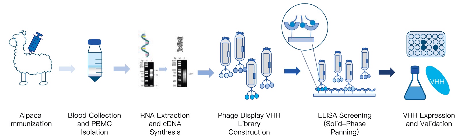
Enterprise Platform Spotlight: KMD Bioscience Alpaca VHH Library Construction
Navigating the complex discovery and biophysical engineering of next-generation nanobodies requires a vertically integrated partner with proven camelid immunology expertise. KMD Bioscience brings over nine years of specialized technical leadership in VHH library construction, high-throughput biopanning, and recombinant validation.
| Immunization & Animal Fleet | Dedicated, ethically managed Alpaca and Llama herds with customized, multi-point immunization protocols |
| Library Scale & Diversity | Robust Phage Display repertoires delivering high-diversity VHH libraries exceeding $10^8$ to $10^9$ independent clones |
| Integrated Discovery Flow | Complete One-Stop pipeline: Computational Antigen Design, Alpaca Immunization, Library Construction, Biopanning, and Functional Assays |
| Screening Methodologies | High-stringency magnetic bead and cell-surface panning, supported by our advanced Single B Cell Screening Platform |
| Characterization Infrastructure | Kinetic affinity validation (Biacore / SPR), thermal denaturation profiles, and recombinant pilot expression |
Senior Scientist's Procurement Advice: Eliminate the risks of low-diversity naive repertoires. Build your discovery program on high-affinity in vivo matured clones. Inspect full workflow technical specifications on the official Alpaca VHH Antibody Library Construction Introduction portal, evaluate our specialized Phage Display Platform, or review custom project options via our Custom Antibody Platform.
Direct Technical Comparison: VHH vs. Conventional IgG vs. scFv Fragments
| Biophysical Property | Single-Domain VHH Nanobody | Conventional Intact IgG | Engineered scFv Fragment |
|---|---|---|---|
| Molecular Weight (kDa) | 12 to 15 kDa | 150 kDa | 25 to 30 kDa |
| Chain Configuration | Single monomeric variable domain | Heterotetramer (2 Heavy + 2 Light) | Two variable domains linked ($V_H-Linker-V_L$) |
| Structural Solubility | Extreme (Hydrophilic framework substitutions) | High (Protected constant domains) | Poor (Exposed hydrophobic interface) |
| Solvent Aggregation Propensity | Very Low (Maintains monomeric state) | Low (Under physiological buffers) | Very High (Prone to domain-swapped dimers) |
| Accessible Epitope Types | Recessed catalytic clefts, grooves, flat surfaces | Flat surfaces, open protrusions | Flat surfaces, shallow grooves |
| Thermal Re-folding Ability | Yes (Reversible refolding up to 85°C+) | No (Irreversible denaturation & precipitation) | No (Irreversible multi-domain aggregation) |
| Host Expression Yields | High in *E. coli* and Yeast (> 1–5 g/L) | High in CHO Mammalian (> 2–8 g/L) | Low to Moderate; often inclusion bodies |
| Regulatory Track Record | Growing (Caplacizumab, Enpatoran, etc.) | Massive (Over 100+ FDA/EMA approved drugs) | Limited (Primarily as CAR-T targeting heads) |
Pros and Cons: Evaluating Single-Domain Nanobodies Against Conventional IgG
| Antibody Platform | Documented Translational Strengths (Pros) | Real-World Development Liabilities (Cons) |
|---|---|---|
| VHH Single-Domain Antibodies (Nanobodies) |
|
|
| Conventional Full-Length IgG (Monoclonal Antibodies) |
|
|
Commercial Economics: Biomanufacturing Costs, Scale-Up, and IP Portfolios
The single most destructive financial error biopharma project leaders make during preclinical formatting is evaluating antibody production solely on an academic level without modeling clinical-scale Cost of Goods Sold (COGS). A biotherapeutic candidate that demonstrates nanomolar affinity in an in vitro assay is commercially stillborn if its commercial manufacturing cost exceeds reimbursement thresholds.
Consider the authentic industrial scale-up mathematics:
The Mammalian IgG Bottleneck: A commercial monoclonal IgG therapy requiring gram-level dosing per patient per year demands massive stainless-steel or single-use mammalian bioreactors. Operating 2,000-liter to 10,000-liter CHO cell culture suites requires expensive chemically defined media, costly fetal-grade growth factors, complex viral clearance validations (ICH Q5A guidelines), and proprietary Protein A affinity resins that cost over $10,000 per liter. The lead time to set up and validate a compliant mammalian cGMP facility can span 18 to 24 months.
The Microbial VHH Advantage: Because VHH antibodies completely lack mammalian-specific glycosylation and inter-chain assembly chaperones, commercial production scales effortlessly inside industrial Escherichia coli or Pichia pastoris fermenters. Microbial fermentation runs complete in 48 to 72 hours (compared to 14 to 21 days for fed-batch CHO lines), operates on inexpensive carbon/nitrogen media feeds, and utilizes straightforward cation-exchange or immobilized metal affinity chromatography (IMAC). Total upstream and downstream bioprocess costs are reduced by two-thirds compared to full-length IgG.
From an intellectual property and commercial lifecycle perspective, the original core composition-of-matter patents covering camelid-derived single-domain antibodies have expired. This regulatory opening has created an explosion of patent filings covering specialized bispecific architectures, biparatopic binders, and targeted drug delivery conjugates. Review current global regulatory drug approval standards monitored by the U.S. Food and Drug Administration (FDA) Center for Drug Evaluation and Research.
Who Should Use VHH Antibodies vs. Who Should Stick to Conventional IgG
Every dollar in your discovery budget must align with your biological mechanism of action (MoA) and target clinical profile. Discerning when to transition to VHH nanobodies versus when to remain with conventional monoclonal antibodies is a straightforward commercial decision.
Programs That Must Transition to VHH Single-Domain Antibodies:
Intraoperative Imaging & Molecular Radiotheranostics: If you are engineering radiolabeled PET/SPECT tracers, you require rapid target binding coupled with immediate systemic clearance within hours to protect healthy marrow and kidneys from radiation. VHH antibodies achieve tumor-to-background contrast ratios impossible with full IgG.
Solid Tumor Oncology with Dense Stroma: When targeting desmoplastic, hypoperfused solid tumors (such as pancreatic ductal adenocarcinoma or triple-negative breast cancer), the superior diffusion coefficient of a 15 kDa VHH overcomes the binding-site barrier, delivering therapeutic payloads uniformly throughout the tumor volume.
Chimeric Antigen Receptor (CAR-T / CAR-NK) Cell Therapies: Replacing unstable scFv targeting domains with VHH modules prevents spontaneous scFv aggregation on the cell membrane, eliminating ligand-independent tonic signaling that causes premature T-cell exhaustion.
Inhaled Pulmonary & Oral Gastrointestinal Biotherapeutics: Formulating biologics that must be aerosolized via vibrating-mesh nebulizers directly into the lungs (for respiratory infections like RSV or SARS-CoV-2) or administered orally to neutralize gut pathogens. The reversible thermal refolding and extreme acid/protease resilience of VHH antibodies survive physical shear and gastric degradation.
Programs That Should Retain Conventional Full-Length IgG:
Classic Target Depletion Demanding Heavy ADCC/CDC: If your primary clinical mechanism of action relies on recruiting host natural killer cells, macrophages, and complement to lyse target cells (e.g., anti-CD20 therapy in B-cell lymphoma), a conventional IgG1 with a native, optimized Fc domain is structurally pre-configured to execute that task out of the box.
Long-Term Chronic Receptor Blockade: If treating chronic auto-immune diseases (such as rheumatoid arthritis or psoriasis) where the goal is maintaining flat, continuous serum neutralization of a soluble target with infrequent, monthly subcutaneous injections, the natural 21-day half-life conferred by IgG FcRn recycling remains the gold standard.
5 Costly Mistakes R&D Teams Make When Developing VHH Formats
Senior Scientist Advisory: The "Naive Library Affinity" Trap
The most common mistake biotech teams make when launching a VHH discovery program is attempting to bypass camelid immunization by purchasing cheap, non-immunized (naive) or synthetic phage display libraries. While naive libraries look fast and inexpensive on paper, they lack the multi-week benefit of in vivo somatic hypermutation and affinity maturation that occurs inside the alpaca lymph node. Isolating high-affinity clones ($K_D < 10^{-9}\text{ M}$) from naive libraries against complex conformational targets is exceptionally rare, forcing teams to burn six months and tens of thousands of dollars on subsequent in vitro affinity maturation campaigns. Always mandate an in vivo immunized alpaca or llama campaign to harness natural somatic maturation, ensuring you isolate picomolar-affinity binders right from the primary biopanning screen.
Overlooking Renal Accumulation in Radiopharmaceutical Design: Developing a 15 kDa VHH for radioisotope delivery without engineering a renal clearance modification strategy. Because small proteins below 50 kDa filter freely through the glomerulus, unmodified nanobodies reabsorb in the renal proximal tubules, delivering high, dangerous radiation doses to the kidneys. Radiopharmaceutical developers must co-administer basic amino acids (lysine/arginine) or engineer cleavable renal linkers to eliminate tubular retention.
Failing to Monitor Spontaneous Dimerization in Solution: Assuming all VHH candidates are permanently monomeric without running analytical size-exclusion chromatography with multi-angle light scattering (SEC-MALS). Certain VHH clones carrying unusually hydrophobic CDR3 loops can form concentration-dependent dimers or higher-order oligomers at therapeutic concentrations (>50 mg/mL), leading to unpredicted immunogenicity and viscosity spikes.
Neglecting Half-Life Extension in Early Pharmacokinetic Modeling: Advancing an unmodified VHH into non-human primate (NHP) efficacy trials for a chronic oncology target without half-life extension, only to watch the therapeutic molecule vanish from circulation in 45 minutes. You must integrate an anti-serum albumin VHH domain, a neonatal Fc fusion, or a site-specific PEGylation site into your primary construct architecture.
Humanizing Without Affinity Validation: Mutating camelid hallmark residues (such as Glu44 and Arg45) to human consensus residues (Gly44 and Leu45) too aggressively. Over-humanizing the framework can collapse the spatial angle of the CDR3 loop or re-introduce the hydrophobic patch, completely destroying antigen-binding affinity and triggering immediate aggregation. Validate your hits using biophysical assays on our Protein Interaction Services platform.
Using Incompatible Expression Tags in Clinical Scale-Up: Relying on polyhistidine (6xHis) tags for purification in discovery, and failing to transition to clinical-grade, tag-free purification protocols (such as ion-exchange and hydrophobic interaction chromatography) before initiating preclinical toxicology studies. His-tags are immunogenic in humans and must be removed prior to regulatory filings.
Essential Sourcing Checklist: Auditing an Antibody Discovery Partner
Before issuing a commercial statement of work (SOW) or transferring non-recurring engineering (NRE) deposits to any contract research organization (CRO) on VHH Antibodies and Conventional Antibodies campaigns, require prospective laboratories to verify compliance across these four non-negotiable operational benchmarks:
1. Verified Animal Facility Standards & Ethical Camelid Herd Management
Demand documented verification of animal husbandry and welfare:
Does the partner maintain its own dedicated, ethically managed alpaca or llama herds under active institutional animal care oversight (AAALAC or equivalent international animal welfare accreditation)?
Do their immunization protocols utilize multi-point subcutaneous injections with verified immune-adjuvant regimens optimized for camelids?
Do they provide verified serum titer ELISA curves proving strong HCAb IgG2 and IgG3 subclass immune response prior to peripheral blood lymphocyte (PBL) harvesting?
2. Library Diversity and Phage Display Sizing Metrics
Inspect the molecular biology quality of their constructed repertoires:
The independent VHH phage display library size must reliably exceed $10^8$ to $10^9$ colony-forming units (CFU) to ensure comprehensive coverage of the animal's mature immune repertoire.
The partner must verify that their PCR cloning strategy specifically uses nested, subclass-specific primers that selectively amplify the VHH variable region while completely excluding conventional heavy-chain ($V_H$) contaminants.
Insertion rates within the phage vector must test above 90% to 95% full-length VHH genes, confirmed by colony PCR and high-throughput next-generation sequencing (NGS).
3. High-Stringency Panning & Biophysical Characterization Equipment
A legitimate discovery leader does not stop at identifying positive ELISA signals. Verify that their screening suite includes:
Cell-surface biopanning capabilities for multipass transmembrane targets (such as GPCRs and ion channels) in their native, physiological conformation.
Surface Plasmon Resonance (SPR - Biacore) or Bio-Layer Interferometry (BLI) equipment measuring real-time binding association ($k_{on}$) and dissociation ($k_{off}$) kinetics down to the sub-nanomolar range.
Automated thermal stability testing: Differential Scanning Calorimetry (DSC) or circular dichroism (CD) proving reversible thermal refolding.
4. Total Downstream Engineering and Production Capabilities
Discovery is useless if the candidate cannot be produced at scale. Ensure your partner operates vertically integrated downstream capabilities: automated humanization algorithms, recombinant microbial fermentation, and mammalian chimeric assembly under certified quality systems. For comprehensive technical validation, review analytical guidelines via the Nature Reviews Drug Discovery: Nanobodies as Therapeutic Agents compendium.
Expert R&D Verdict: The Modern Biotherapeutic Roadmap
In our professional assessment across decades of recombinant antibody engineering, biophysical analysis, and drug discovery campaigns, the conclusion is absolute: the biopharma landscape is no longer a monolithic domain reserved exclusively for 150 kDa conventional IgG molecules. While conventional monoclonal antibodies will remain indispensable for targets requiring heavy, direct immune-mediated cell killing, single-domain VHH nanobodies represent an overwhelmingly superior format for challenging solid-tumor penetration, multi-specific modular assembly, cryptic epitope targeting, and high-margin microbial production.
Here is your clear, actionable preclinical development roadmap:
Conduct Target Epitope Topography Audits First: If your target antigen is an enzyme catalytic pocket, a viral membrane canyon, or a crowded ion channel pore, stop forcing conventional IgG into steric failure. Select a VHH platform from day one to harness the convex, cleft-penetrating capability of camelid CDR3 loops.
Harness Natural In Vivo Somatic Maturation: Reject synthetic or naive libraries for primary therapeutic discovery. Partner with a specialist to execute an in vivo immunized alpaca campaign, securing high-affinity binders ($K_D < 10^{-9}\text{ M}$) with built-in biological developability.
Engineer Pharmacokinetics to Match Indication: If developing diagnostic imaging tracers, keep the VHH monomeric for rapid, high-contrast renal clearance. If developing a systemic therapeutic, fuse an anti-HSA VHH domain or an engineered human IgG1 Fc backbone early in preclinical optimization.
Partner with KMD Bioscience: Eliminate the operational friction of coordinating separate animal farms, molecular biology CROs, and expression facilities. Partnering directly with KMD Bioscience gives your discovery enterprise access to over nine years of proven camelid discovery expertise, diverse $10^9$ phage display libraries, automated humanization algorithms, and high-yield microbial protein expression, ensuring your therapeutic candidates transition from initial immunization to functional validation with speed, precision, and commercial confidence.
Frequently Asked Questions
What is the primary structural difference between VHH antibodies and conventional antibodies?
The primary structural difference lies in chain composition and molecular mass. Conventional mammalian antibodies (IgG) are heterotetrameric 150 kDa glycoproteins consisting of two heavy and two light chains. VHH antibodies, derived from camelid heavy-chain-only antibodies (HCAbs), represent a single variable domain of roughly 15 kDa that completely lacks light chains, CH1 domains, and native glycosylation requirements.
Can VHH nanobodies bind cryptic or hidden epitopes inaccessible to conventional IgG?
Yes. VHH antibodies feature extended, flexible CDR3 loops that form protruding convex finger-like paratopes. These structures can penetrate narrow enzymatic clefts, active catalytic pockets, ion channels, and viral membrane canyons that the flat or concave antigen-binding surfaces of conventional IgG paratopes cannot access due to steric hindrance.
Why do VHH antibodies exhibit superior thermal and chemical stability?
VHH antibodies replace the hydrophobic residues typically found at the VH-VL interface in conventional antibodies with hydrophilic amino acids (Phe37, Glu44, Arg45, and Gly47 under IMGT/Kabat numbering). This substitution, often coupled with an extra non-canonical disulfide bond linking CDR3 to CDR1 or framework regions, allows VHH fragments to withstand temperatures up to 80°C–90°C and refold reversibly without aggregating.
Do VHH antibodies induce antibody-dependent cellular cytotoxicity (ADCC)?
In their native monomeric state, VHH antibodies do not induce ADCC or CDC because they lack the constant Fc region required to engage Fc-gamma receptors (FcγR) on immune effector cells or bind C1q for complement activation. To trigger effector cytotoxicity, VHH domains must be genetically fused to human IgG1 Fc domains to create bivalent VHH-Fc chimeras.





 0
