


Every principal investigator, lead discovery scientist, and biopharma program director eventually encounters the exact same high-stakes bottleneck: you have identified a validated therapeutic target or an elusive monoclonal antibody epitope, but your lead generation project stalls because your peptide screening platform yields unreplicable hits, insoluble sequences, or false-positive binders. The modern biotechnology landscape has moved far beyond the era of blindly ordering generic randomized peptides. Today, synthetic peptide libraries represent the premier structural engine for mapping discontinuous epitopes, deciphering critical protein-protein interaction (PPI) networks, engineering high-affinity peptidomimetic therapeutics, and optimizing enzyme substrate kinetics. Yet entering this field without rigorous design discipline is a recipe for wasted R&D budgets and months of unproductive screening assays. In our testing across automated peptide synthesizers, next-generation sequencing flow cells, and biopanning runs, success is never determined by sheer sequence quantity alone. It is determined by the mathematical architecture, conformational constraints, and physicochemical properties integrated into the library before a single amino acid coupling takes place. Executing the steps in peptide library design requires balancing computational molecular modeling, degenerate codon selection, and solid-phase synthesis chemistry. As structural biologists and high-throughput discovery specialists who formulate screening strategies, construct phage-display vectors, and validate macromolecular complexes daily at KMD Bioscience, we cut through academic generalities. Here is the practitioner-grade breakdown of the six essential steps in peptide library design required to generate robust, nanomolar-affinity leads and accelerate your pipeline toward clinical readiness.
Quick Answer: The 6 Critical Steps in Peptide Library Design
To successfully engineer a high-yield peptide screening platform, researchers must execute six non-negotiable steps in peptide library design: (1) Define Biological Target Geometry & Screening Objectives (differentiating linear epitope mapping, conformational pocket binding, or allosteric modulation); (2) Select the Structural Library Topology (choosing overlapping, alanine scanning, truncation, positional scanning, scrambled, or random degenerate formats); (3) Execute In Silico Physicochemical De-risking (filtering out hydrophobic aggregation motifs, unpredictable cysteines, deamidation sites, and optimizing charge distributions); (4) Determine the Presentation Modality & Synthesis Vehicle (chemical solid-phase peptide microarrays vs. biological M13/T7 phage display, ribosome display, or mRNA display); (5) Assemble the Molecular Library & Validate via NGS (deploying NNK/NNS codon strategies, high-efficiency electrotransformation into E. coli host strains like ER2738, and quantifying real diversity); and (6) Calibrate Stringency Panning & Orthogonal Validation Assays (optimizing target immobilization, surface plasmon resonance kinetics, and counter-screening against non-specific background binders). Sourcing your workflow through a vertically integrated partner like KMD Bioscience ensures your library bridges in silico design directly to wet-lab screening success.
Article Directory
Structural Mechanics: Why Arbitrary Peptide Libraries Fail in the Wet Lab
Platform Spotlight: KMD Bioscience Peptide Library Screening Service
Direct Technical Comparison: Chemical SPPS Arrays vs. Phage vs. mRNA Display
Pros and Cons Table: Custom De Novo Libraries vs. Pre-Made Off-The-Shelf Libraries
Commercial Economics: Diversity Sizing, QC Depths, and Project ROI
Who Needs Custom Peptide Libraries vs. Who Can Rely on Recombinant Fragments
Essential Sourcing Checklist: Auditing a Contract Peptide Synthesis & Screening Partner
Structural Mechanics: Why Arbitrary Peptide Libraries Fail in the Wet Lab
To master the steps in peptide library design, one must confront the staggering combinatorial mathematics of peptide space. For a simple linear 12-mer peptide constructed exclusively from the twenty canonical L-amino acids, the theoretical sequence diversity is $20^{12}$, which equals approximately $4.096 \times 10^{15}$ unique chemical permutations. No laboratory on earth can physically synthesize or screen a library of four quadrillion individual peptide species in a single vessel. The practical physical ceiling for biological systems—such as M13 filamentous phage display—sits around $10^9$ to $10^{10}$ transformants, while synthetic solid-phase peptide synthesis (SPPS) arrays typically handle $10^2$ to $10^5$ discrete immobilized sequences.
Because you can only explore an infinitesimal fraction of total sequence space, unguided, arbitrary library design is an extraordinarily inefficient strategy. In our testing across therapeutic screening campaigns, libraries assembled without structural hypothesis fail for predictable physical reasons:
Conformational Entropy Penalties: Short linear peptides in aqueous solution exist as dynamic, unstructured random coils. When a flexible linear peptide binds to a rigid protein groove, it incurs a massive entropic penalty ($\Delta S$) as it freezes into a single bound conformation. Without introducing chemical staples, disulfide cyclization, or terminal constraints during library drafting, your hits will struggle to break through the micromolar affinity threshold ($K_D > 10^{-6}\text{ M}$).
Physicochemical Collapse: Unfiltered random libraries inevitably encode runs of highly hydrophobic residues (Leu-Phe-Val-Trp). These peptides self-assemble into insoluble beta-amyloid-like aggregates in aqueous buffers, precipitating out of solution or sticking non-specifically to polystyrene screening plates, yielding endless false positives.
Translational Redundancy and Premature Termination: In biological expression libraries, using unconstrained random nucleotide triplets (NNN) guarantees that 3 out of 64 codons will code for translational stop signals (TAG, TAA, TGA). For a 15-mer peptide, this means nearly half of your transformed clones will express truncated, useless fragments.
From our experience on production and screening lines, disciplined peptide library design is the art of applying rational in silico constraints to eliminate junk sequences, ensuring that every synthesized molecule possesses the conformational flexibility and solubility profile needed to engage the target pocket.
Quick Summary Table: The 6 Steps in Peptide Library Design
| Design Phase | Primary Technical Focus | Core Computational / Molecular Tool | Critical Failure Prevented | Standard Deliverable Milestone |
|---|---|---|---|---|
| Step 1: Target Definition | Mapping binding pockets, active sites, or linear epitopes | X-ray crystallography, Cryo-EM, AlphaFold modeling | Screening blind against dynamic or buried interfaces | Target binding profile & assay specification |
| Step 2: Topology Selection | Selecting geometric architecture (Overlapping, Scanning, Random) | Mathematical sequence offsets, structural algorithms | Mismatched format (e.g., using linear peptides for structural loops) | Library architecture schematic & fragment lists |
| Step 3: Physicochemical Filtering | De-risking isoelectric point (pI), solubility, and reactive residues | In silico filtration algorithms, hydrophobic index scoring | Insoluble aggregates, spontaneous deamidation, oxidation | Curated, filtered sequence database |
| Step 4: Platform Determination | Selecting between Chemical SPPS vs. Biological Phage Display | Assay scale evaluation, non-canonical residue needs | Unachievable diversity in SPPS; inability to use D-amino acids in phage | Platform technical protocol & expression vector |
| Step 5: Assembly & Quality Control | High-diversity molecular cloning and NGS validation | NNK/NNS degeneracy, ER2738 electrotransformation, NGS | Codon bias collapse, stop codon over-representation | Validated physical library ($>10^9$ cfu/mL diversity) |
| Step 6: Screening Calibration | Establishing stringency panning and kinetic validation | Biopanning, Surface Plasmon Resonance (SPR), BLI, ELISA | Selecting plastic-binders; missing high-off-rate leads | Ranked, verified high-affinity target binders |
The 6 Key Steps in Peptide Library Design Analyzed in Depth
Step 1: Define Target Geometry and Screening Objectives
The first step in peptide library design is establishing the exact biological mechanism you intend to interrogate or modulate. You cannot design a library until you characterize the structural interface of your target molecule. Are you mapping the binding footprint of a neutralizing antibody generated via a Custom Antibody Platform? Are you searching for a competitive antagonist that interrupts a flat, undruggable protein-protein interaction? Or are you identifying the optimal cleavage sequence for an oncogenic protease?
In most professional situations, this step requires mining atomic coordinates deposited in the RCSB Protein Data Bank (PDB) or generating validated predictive models using AlphaFold2. You must establish whether the target interaction is governed by a continuous linear motif (such as a linear peptide recognized by an SH3 domain) or a complex conformational epitope formed by discontinuous amino acid loops brought together by tertiary folding. If the binding site is a deep, narrow hydrophobic cleft, a flexible linear peptide may insert cleanly; if the target is a flat, shallow surface, designing a cyclic peptide library constrained by flanking cysteine residues ($C-X_n-C$) is mandatory to stabilize beta-turn or alpha-helical secondary structures.
Step 2: Select the Structural Library Topology
Once the screening objective is locked, the architect must select the mathematical topology of the library. Different structural layouts answer distinct biological questions:
Overlapping Peptide Libraries: The gold standard for linear epitope mapping and scanning antigen surfaces. An entire protein sequence is broken down into overlapping fragments of equal length (e.g., 15-mers) shifted by a defined offset (e.g., 3 to 5 residues). A smaller offset provides single-residue resolution, identifying exact core binding motifs at the cost of higher sequence synthesis volume.
Alanine Scanning Libraries: Mandatory for structural dissection. Every native residue within a known active peptide sequence is systematically substituted, one by one, with Alanine (or Glycine if the native residue is already Alanine). Because Alanine strips away the functional side chain beyond the beta-carbon without destabilizing the secondary backbone, this library instantly reveals which specific chemical moieties are essential for binding affinity.
Truncation Libraries: Deployed during lead optimization to identify the absolute minimal functional sequence required for receptor binding. Residues are systematically trimmed from the N-terminus, C-terminus, or both, enabling medicinal chemists to eliminate non-essential amino acids and synthesize compact peptidomimetics with superior bioavailability.
Positional Scanning & Combinatorial Libraries: Systematic identification of sequence permissiveness. A single position along the peptide chain is fixed with one of the twenty amino acids while all other positions are randomized or systematically substituted. This reveals whether a specific pocket tolerates positive charges, bulky aromatics, or small aliphatics.
Random Degenerate Libraries: Deployed for de novo hit discovery when structural knowledge is absent. Fixed flanking frameworks house central random sequences of varying lengths (e.g., $X_7$, $X_{12}$, or $C-X_8-C$).
Step 3: In Silico Physicochemical De-risking and Filtration
The single most catastrophic failure mode in custom peptide screening is generating a list of promising theoretical hits that cannot be dissolved or formulated in the wet lab. In silico sequence filtering must occur prior to physical synthesis or oligonucleotide ordering to eliminate problematic chemical liabilities:
Hydrophobic Index and Aggregation Control: Peptides containing more than 40% to 50% hydrophobic residues (Trp, Phe, Tyr, Leu, Ile, Val) or unbroken runs of four or more aliphatics will self-associate into insoluble aggregates. Algorithmic scoring must limit consecutive hydrophobic blocks and ensure an optimal ratio of charged residues (Asp, Glu, Lys, Arg) to promote aqueous solubility.
Removal of Unintended Reactive Chemistries: Unpaired free cysteine residues ($Cys$) will form spontaneous, heterogeneous intermolecular disulfide bridges, cross-linking library members into scrambled multimeric artifacts. Unless cysteines are deliberately paired to form an intramolecular cyclic loop, free cysteines should be computational filtered out or substituted with Serine.
Chemical Degradation Motifs: Long-term assay incubation can trigger spontaneous chemical breakdown. Automated filters should eliminate Asparagine-Glycine ($Asn-Gly$) motifs, which undergo rapid deamidation to cyclic isoaspartate, as well as Methionine residues ($Met$), which suffer uncontrolled oxidation into sulfoxide artifacts in standard aeration buffers.
Step 4: Determine the Synthesis Vehicle & Presentation Modality
A crucial crossroad in the steps in peptide library design is deciding whether to manufacture the library via synthetic chemical methods or biological display technologies. This choice dictates the maximum achievable diversity, project timelines, and structural modifications:
For screening thousands to millions of canonical L-peptides for initial lead discovery, biological display platforms are unbeatable. Phage display (utilizing the M13 pIII or pVIII coat proteins) and mRNA/ribosome display provide vast sequence diversity ($10^9$ to $10^{12}$ distinct variants). Because each peptide is physically linked to its encoding genetic material (genotype-phenotype coupling), iterative cycles of affinity selection (biopanning) allow you to enrich picomolar binders out of an astronomical background pool.
Conversely, for fine epitope mapping, alanine scanning, or integrating non-canonical chemical modifications, chemical Solid-Phase Peptide Synthesis (SPPS) arrays are superior. Chemical synthesis allows you to incorporate D-amino acids (rendering the peptide immune to in vivo proteolysis), unnatural cyclic beta-hairpins, fluorophores, phosphorylation modifications, and synthetic hydrocarbon staples. Sourcing these complex projects requires integrating specialized Protein Modification & Detection Services to ensure chemical purity and structural integrity.
Step 5: Molecular Assembly, Degenerate Codons & NGS Quality Control
For biological libraries, translating in silico designs into high-diversity physical vectors requires precision molecular engineering. The historical practice of synthesizing completely randomized oligonucleotides using equimolar mixtures of all four bases ($N = A/T/C/G$ at every position) is an obsolete and flawed methodology.
Modern high-performance libraries deploy refined degenerate codon schemes:
The NNK / NNS Degenerate Codon Strategy: By utilizing $N$ (all four bases) in the first two positions and $K$ ($G$ or $T$) or $S$ ($C$ or $G$) in the third wobble position, you reduce the total number of codons from 64 to 32. This preserves representation of all 20 canonical amino acids while eliminating two out of the three translational stop codons (Ochre $TAA$ and Opal $TGA$). The sole remaining Amber stop codon ($TAG$) can be suppressed as Glutamine by propagating the library inside specialized supE amber-suppressor E. coli host strains, such as ER2738.
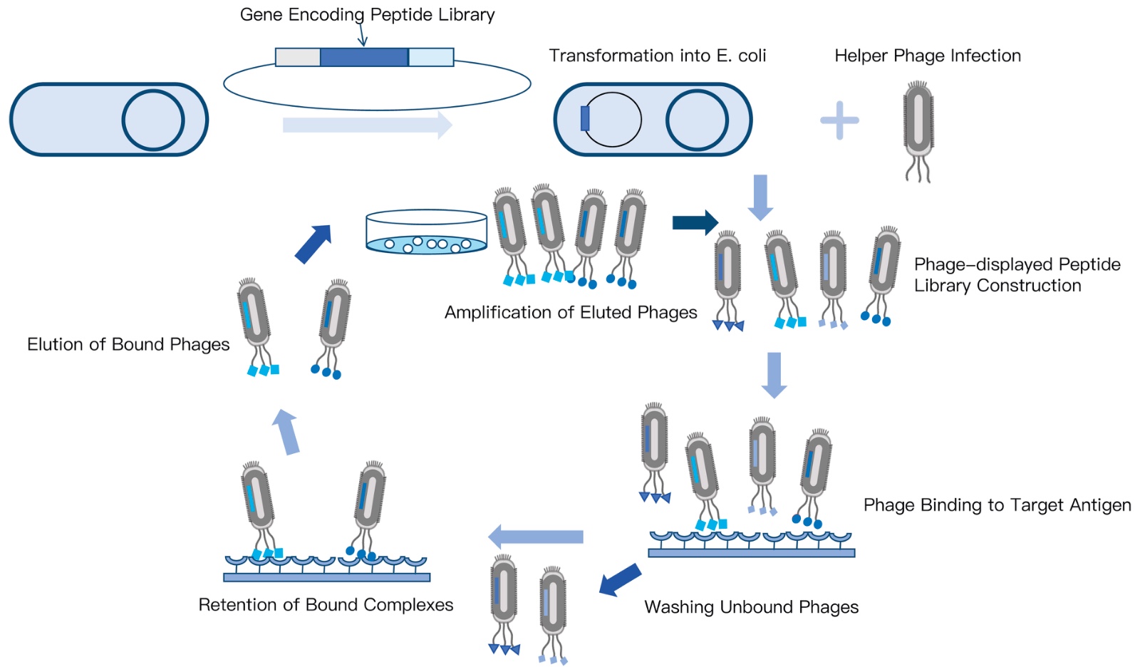
Electrotransformation and Diversity Sizing: Synthetic double-stranded DNA inserts are ligated into phagemid vectors and introduced into electrocompetent ER2738 cells via high-voltage electroporation. The functional size of your library is mathematically dictated by the number of unique colony-forming units (cfu) harvested immediately following transformation. Achieving a certified diversity of $10^9$ cfu demands ultra-high efficiency cells ($>10^9\text{ cfu/}\mu\text{g DNA}$) and precise control over electric field time constants.
Next-Generation Sequencing (NGS) Validation: Never trust a library based purely on colony counts. Modern quality control mandates running high-throughput deep sequencing across thousands of transformed clones. NGS reveals true positional diversity, identifies skewing or amino acid over-representation caused by synthesis bias, and verifies that the functional fraction of full-length, in-frame peptides exceeds 85% to 90%.
Step 6: Screening Strategy, Biopanning Pressure, and Orthogonal Validation
The final, decisive step in the peptide library lifecycle is the execution of screening assays that enrich authentic nanomolar binders while systematically stripping away non-specific artifacts. Designing a brilliant library is entirely meaningless if your screening assay selects for plastic-adhering background molecules.
A rigorous screening and validation workflow comprises:
Target Presentation and Immobilization: The target protein—frequently expressed through an E. coli protein expression system or mammalian expression—must be presented in its native, biologically active conformation. Passive adsorption onto polystyrene microplates can denature the protein, exposing non-physiological hydrophobic cores. We recommend site-specific enzymatic biotinylation coupled to streptavidin-coated magnetic beads to present the target uniformly in liquid phase.
Iterative Biopanning Stringency: Over 3 to 4 rounds of selection, panning stringency must be steadily escalated. Process engineers increase the concentration of non-ionic detergents (e.g., 0.1% to 0.5% Tween-20), shorten target incubation times, and extend washing cycles (up to 15 to 20 cycles) to force the dissociation of fast-off-rate, low-affinity binders.
Negative Depletion (Counter-Selection): Between each round, the library must be passed over naked magnetic beads, blocking proteins (BSA, ovalbumin), and irrelevant targets to deplete plastic-binders and non-specific cross-reactive peptides.
Orthogonal Biophysical Confirmation: Enriched clone populations are sequenced and synthesized as discrete soluble peptides. True hits must be validated using label-free optical biosensors: Surface Plasmon Resonance (SPR) or Bio-Layer Interferometry (BLI) via our Protein Interaction Services to determine true association rate constants ($k_a$), dissociation rate constants ($k_d$), and equilibrium dissociation constants ($K_D$).
Enterprise Platform Spotlight: KMD Bioscience Peptide Library Screening Service
Navigating the complex molecular assembly, codon calibration, and high-stringency biopanning required for modern lead discovery demands an experienced contract research partner. KMD Bioscience provides a fully integrated, turnkey peptide library platform spanning custom oligonucleotide synthesis to validated nanomolar hit delivery.
| Nucleotide Synthesis | Random or rationally constrained synthesis of client-specified length nucleotide sequences (NNK/NNS) |
| Polymerization & Digestion | Precision enzymatic polymerization and high-efficiency restriction digestion of synthetic DNA cassettes |
| Electroconversion Prep | Optimized ER2738 electrocompetent host cell preparation with high-efficiency recombinant plasmid transformation |
| Capacity & Diversity QC | Rigorous bacterial colony-forming unit assessment coupled with deep NGS-based sequence diversity verification |
| Phage Packaging & Panning | High-titre helper phage infection, liquid-phase magnetic biopanning, and negative depletion cycles |
| Cold-Chain Delivery | Direct delivery of high-titre phage libraries and sequence databases shipped on secure cold-chain ice packs |
Consultation Advisory: Accelerate your discovery timeline. Review comprehensive technical protocols on the official Peptide Library Screening Service Introduction page, evaluate our master Peptide Library Platform, or consult our specialized team on our Innovative Drug Discovery Platform.
Direct Technical Comparison: Chemical SPPS Arrays vs. Phage vs. mRNA Display
| Platform Dimension | Chemical SPPS Peptide Arrays | M13 / T7 Phage Display Libraries | Cell-Free mRNA / Ribosome Display |
|---|---|---|---|
| Maximum Library Diversity | $10^2$ to $10^5$ individual peptides | $10^9$ to $10^{10}$ unique clones (Transformation limited) | $10^{12}$ to $10^{14}$ unique peptides (Cell-free transcription) |
| Sequence Length Flexibility | 5 to 50+ residues (Length limited by SPPS coupling) | 7 to 30 residues (Limited by phage packaging & folding) | Broad (Short peptides up to large functional domains) |
| Non-Canonical Amino Acid Support | Unrestricted (D-amino acids, staples, PTMs, fluorophores) | Extremely limited (Requires specialized amber codon reassignment) | Moderate (Requires cell-free genetic code reprogramming) |
| Primary Screening Bottleneck | Synthesis cost scales linearly with sequence count | Biological expression bias and amber stop codon leakage | High sensitivity to RNase degradation; complex PCR recovery |
| Typical Discovery Phase Fit | Fine epitope mapping, alanine scans, lead optimization | De novo hit discovery, antibody paratope mapping | Ultra-high-affinity selections, ultra-large random space |
| Capital Cost Structure | Moderate-High ($$ per peptide basis) | Cost-Effective ($$$ per high-diversity pool) | High ($$$$ specialized enzymatic reagents) |
Pros and Cons Table: Custom De Novo Libraries vs. Pre-Made Off-The-Shelf Libraries
| Library Procurement Strategy | Scientific & Commercial Strengths (Pros) | Operational Liabilities & Realities (Cons) |
|---|---|---|
| Custom Rational De Novo Design (Tailored sequence space, NNK codons) |
|
|
| Pre-Made Off-The-Shelf Libraries (Generic commercial phage display kits) |
|
|
Commercial Economics: Diversity Sizing, QC Depths, and Project ROI
Every commercial director, biopharma scout, and research grant manager must evaluate peptide discovery through a rigorous financial lens. The most expensive library is not the one with the highest upfront invoice; it is the cheap library that yields twenty false-positive hits, leading your chemistry team down a six-month rabbit hole of dead-end lead optimization.
Consider the authentic economic ledger of high-throughput peptide screening:
The Fallacy of the Cheap Catalog Phage Aliquot: Purchasing a generic commercial phage library for $3,000 appears cost-effective on paper. However, because these historical libraries have been passaged through dozens of bacterial amplification cycles, fast-growing "parasitic" clones dominate the population. In our testing, up to 40% of resulting biopanning hits bind non-specifically to streptavidin, plastic polystyrene, or blocking albumins. Deconvoluting these false positives through synthetic peptide resynthesis and biophysical characterization routinely consumes $25,000 to $40,000 in wasted reagent and labor expenses.
The Economic ROI of In Silico De-risking: Investing in custom rational design—incorporating computational solubility filters, controlled cysteine architectures, and validated NNK codon assemblies—costs slightly more upfront. However, because the library has been stripped of insoluble hydrophobic motifs and premature stops, the hit-to-lead conversion rate increases by more than 300%. The resulting hits possess clean, interpretable binding kinetics on Surface Plasmon Resonance (SPR), allowing your project to advance into in vitro functional assays weeks ahead of schedule.
Who Needs Custom Peptide Libraries vs. Who Can Rely on Recombinant Fragments
Every research dollar should match your target's structural biology. Sourcing a dedicated peptide library is an indispensable strategy for specific scientific questions, while simpler recombinant protein engineering suffices for others.
Programs That Must Deploy Custom Peptide Libraries:
Monoclonal Antibody Epitope Mapping Campaigns: When filing patent claims for a newly discovered therapeutic antibody, establishing freedom-to-operate and securing composition-of-matter claims requires proving the exact linear or discontinuous amino acid residues involved in paratope binding. High-density overlapping and alanine-scanning peptide libraries provide the definitive structural proof mandated by international patent offices.
Intracellular Protein-Protein Interaction (PPI) Inhibitor Discovery: Small molecules frequently fail to inhibit protein-protein interactions because the contact surfaces are large, flat, and lack deep binding pockets. Rational peptide libraries—particularly constrained cyclic or stapled helical architectures—are the premier therapeutic modality to bridge these interfaces with nanomolar potency.
Enzyme Substrate Profiling & Chemical Biology: If you are characterizing the substrate specificity of newly identified viral proteases, kinases, or methyltransferases, positional scanning combinatorial libraries allow you to profile catalytic turnover across thousands of amino acid combinations simultaneously.
Programs That Should Use Alternative Methods:
Mapping Highly Complex Multi-Subunit Conformational Epitopes: If an antibody binds across the junction of a multi-protein quaternary complex held together by multiple trans-membrane domains, short peptides (even when cyclic) cannot recreate the tertiary folding landscape. These targets require native recombinant protein truncation domains or full-length cryo-EM structural analysis. Explore full-length targets via our comprehensive Protein Expression Services or recombinant protein expression service platforms.
Routine Antibody Quality Control: If your objective is simply verifying that a commercial polyclonal antibody recognizes its target in an everyday Western blot, basic recombinant antigen fragments are all that is required.
Costly Design Mistakes That Ruin Screening Campaigns
Factory Floor Advisory: The "Target Denaturation" Trap
The most pervasive disaster in biopanning campaigns occurs when researchers passively coat their target protein directly onto high-binding polystyrene ELISA wells at room temperature. The hydrophobic plastic surface exerts mechanical shear and interfacial tension, causing the protein to partially unfold. The library dutifully isolates high-affinity nanomolar binders—but they bind to internal, denatured hydrophobic patches that are completely hidden when the protein is correctly folded in physiological serum. Always utilize gentle, site-specific biotinylation and capture the native target in liquid phase using streptavidin-coupled magnetic beads.
Ignoring Parasitic Propagation Advantages in Phage Libraries: When an unconstrained phage library is amplified in E. coli between biopanning rounds, clones that carry wild-type-like sequences or specific mutations that allow faster bacterial infection and burst kinetics will outgrow slower-growing therapeutic clones, regardless of their binding affinity. Strict biopanning protocols must minimize amplification passages and monitor population dynamics using next-generation sequencing.
Omitting Negative Depletion (Counter-Selection) Rounds: If you biopan exclusively against target-coated magnetic beads, you will inevitably enrich peptides that bind with high affinity to the streptavidin matrix or the iron oxide bead core. A professional panning campaign must interleave negative selection rounds over naked, blocked beads to strip away matrix-binders before presenting the actual target.
Designing Rigid Helices Without Helical Propensity Modeling: Attempting to mimic an alpha-helical binding motif by simply placing two cysteines at an $i, i+7$ spacing without checking the helix-forming propensity of the intervening residues is an amateur error. Certain amino acids (Proline, Glycine) act as severe helix breakers; including them inside a constrained loop will distort the backbone, ruining binding geometry.
Failing to Account for Linker Interference: In phage display, peptides are expressed as N-terminal fusions to the viral coat protein (e.g., pIII), connected by a flexible linker (such as $Gly-Gly-Gly-Ser$). If the peptide is designed without adequate structural separation, steric hindrance from the massive phage capsid (over 900 nm long) will physically block the peptide from entering deep target clefts. Always incorporate a flexible poly-glycine-serine spacer of at least 4 to 8 residues.
Essential Sourcing Checklist: Auditing a Contract Peptide Synthesis & Screening Partner
Before issuing a purchase order or signing a master services agreement with a Contract Research Organization (CRO) for peptide library design, audit prospective partners against these four non-negotiable scientific benchmarks:
1. Verified Transformation Efficiency and Clonal Diversity Sizing
Demand documented verification of the partner's transformation capacity. A world-class CRO must demonstrate electrocompetent preparation protocols achieving bacterial transformation efficiencies exceeding $1 \times 10^9\text{ cfu/}\mu\text{g DNA}$. The physical library size must be supported by mathematical colony counts performed on serial dilution plates, establishing certified initial diversity. Review our molecular capabilities across the Molecular Platform.
2. High-Throughput Next-Generation Sequencing (NGS) Quality Control
Never accept a partner that validates library diversity by simply picking 48 colonies and running Sanger sequencing. In a pool of $10^9$ variants, 48 sequences represent a statistically meaningless snapshot. Settle for nothing less than deep NGS profiling across a minimum of $10^5$ to $10^6$ reads, generating positional amino acid frequency heatmaps, stop codon percentage metrics, and confirmation that sequence distribution matches the predicted NNK/NNS mathematical model.
3. Label-Free Orthogonal Binding Kinetic Validation (SPR / BLI)
A capable partner does not stop at delivering a list of ELISA optical density (OD) readings. Optical density is an equilibrium measurement susceptible to avidity artifacts. Settle for nothing less than full biophysical hit validation using Surface Plasmon Resonance (Biacore) or Bio-Layer Interferometry (ForteBio). The partner must provide real-time sensorgrams calculating association rate constants ($k_a$), dissociation rate constants ($k_d$), and equilibrium constants ($K_D$) on purified synthetic monomeric peptides, backed by comprehensive Detection Platform validation.
4. Total Vertical Integration: From DNA Synthesis to Protein Expression
Outsourcing your project to a broker who designs oligonucleotides with one vendor, subcontracts cloning to another, and buys recombinant target protein from a third creates communication breakdowns and timeline delays. Ensure your partner operates vertically integrated platforms—housing in-house prokaryotic and eukaryotic protein expression, automated oligonucleotide synthesis, and dedicated phage-handling laboratories under one unified quality management system.
Expert Sourcing Verdict: The Peptide Engineering Blueprint
In our professional assessment across the modern biopharmaceutical landscape, the decision to deploy a custom peptide library is the most powerful tool available for deciphering difficult biological targets—provided you treat the process as an integrated engineering workflow rather than a random lottery.
Here is your clear, actionable procurement roadmap:
Map the Structural Interface Before Ordering: Utilize crystallographic data, cryo-EM models, or AlphaFold structures to determine whether your target requires linear motifs, alanine scanning, or constrained cyclic loops ($C-X_n-C$) to match the target's physical geometry.
Enforce In Silico De-risking: Run computational filters to eliminate hydrophobic aggregation motifs, stray un-paired cysteines, and deamidation sites. Mandate the use of NNK or NNS degenerate codon schemes to eliminate wasteful stop codons and maximize functional expression.
Choose Your Modality with Precision: For structural scanning, linear epitope mapping, or integrating non-canonical D-amino acids and chemical staples, utilize Chemical SPPS Microarrays. For de novo hit discovery against complex drug targets across billions of variants, standardize on M13 Phage Display Libraries.
Partner with a Vertically Integrated Specialist: Align your research with an established biotechnology leader like KMD Bioscience. From custom oligonucleotide synthesis and ER2738 high-efficiency electroconversion to deep NGS diversity validation and high-stringency magnetic biopanning, partnering with an asset-backed contract manufacturer ensures your project transitions seamlessly from in silico concept to confirmed, nanomolar-affinity therapeutic leads.
Frequently Asked Questions
What are the primary steps in peptide library design?
The 6 core steps in peptide library design are: (1) Defining target biology and screening objectives, (2) Selecting the structural library topology, (3) In silico sequence filtering and physicochemical de-risking, (4) Selecting the synthesis or biological display platform, (5) Molecular library assembly and NGS quality control, and (6) Establishing screening assays and panning protocols.
Why is degenerate codon design (such as NNK or NNS) critical in biological peptide libraries?
Using standard NNN degenerate codons introduces all three translational stop codons (ochre, opal, amber), leading to premature termination in up to 4.7% of variants per codon. Utilizing NNK or NNS schemes eliminates two out of three stop codons (retaining only the amber stop codon UAG, which can be suppressed in specialized E. coli strains like ER2738) and dramatically reduces amino acid redundancy, balancing library diversity.
How does an overlapping peptide library differ from an alanine scanning library?
An overlapping peptide library systematically breaks down an entire primary protein antigen sequence into overlapping fragments (e.g., 15-mers with 11-residue overlaps) to identify continuous linear epitopes. An alanine scanning library sequentially substitutes each native amino acid residue of a known peptide hit with alanine to pinpoint exact functional side chains responsible for binding affinity and biological activity.
When should researchers choose chemical SPPS libraries over phage-displayed peptide libraries?
Chemical solid-phase peptide synthesis (SPPS) libraries are essential when your drug discovery campaign requires non-canonical D-amino acids, post-translational modifications (phosphorylation, methylation), macrocyclic chemical staples, or precise stoichiometric controls. Phage display is superior when screening astronomical sequence diversity (10^9 to 10^11 distinct variants) for de novo lead discovery within canonical L-amino acid space.





 0
