


Every director of biologics discovery, therapeutic program lead, and diagnostic developer eventually runs into the mechanical bottleneck of legacy antibody generation. For nearly half a century, Kohler and Milstein's hybridoma fusion served as the foundational pillar of monoclonal discovery. Yet anyone who has watched promising therapeutic campaigns stall due to unstable hybridoma chromosome loss, unviable fusion yields, low throughput, or lost cognate heavy-light chain pairing understands the operational frustration. In modern drug pipelines, spending four to six months waiting for subcloned mouse hybridomas that yield micro-molar affinities is no longer acceptable. The biopharma industry has migrated decisively toward single-cell resolution. Transitioning From Single B Cell Isolation to Antibody Validation represents the gold standard for mining the fully diversified, in vivo affinity-matured immune repertoires of immunized animals and human convalescent donors. By isolating individual antibody-secreting cells and capturing their natural genetic pairing via high-fidelity molecular biology, researchers compress discovery campaigns from months to weeks while unlocking picomolar affinities. As antibody discovery specialists who operate microfluidics sorters, construct high-throughput expression cassettes, and resolve kinetic rate constants daily, we provide an unvarnished, end-to-end technical breakdown of the nine critical steps From Single B Cell Isolation to Antibody Validation, delivering the practical benchmarks required to build a clinical-grade discovery pipeline.
Quick Answer: The 9 Steps From Single B Cell Isolation to Antibody Validation
The definitive process From Single B Cell Isolation to Antibody Validation consists of nine interconnected laboratory phases: (1) Antigen Preparation & Hyper-Immunization (generating high serum titers against native-conformation targets); (2) Lymphoid Organ Harvest & Mononuclear Cell Fractionation (isolating viable PBMCs, splenocytes, or bone marrow lymphocytes); (3) Antigen-Specific Single-Cell Sorting (deploying multi-color FACS or droplet microfluidics to isolate individual memory B cells or plasmablasts); (4) In Situ Single-Cell Lysis & Reverse Transcription (generating high-fidelity single-cell cDNA without cross-well contamination); (5) Cognate VH/VL Nested PCR Amplification (capturing naturally paired variable heavy and light chains); (6) High-Throughput Expression Vector Recombination (cloning variable domains into IgG mammalian expression cassettes via golden gate or Gibson assembly); (7) Transient Mammalian Transfection & Expression (producing recombinant monoclonal antibodies in suspension Expi293 or CHO cells); (8) Affinity Purification & Primary Binding Screening (Protein A purification followed by high-throughput ELISA/HTRF quantification); and (9) Biophysical & Functional Validation (measuring real-time SPR/BLI kinetics, thermal stability, epitope binning, and cellular bioassays). For biopharma teams requiring high developability, partnering with a proven provider like Kmd bioscience through their dedicated single B cell screening service delivers fully validated recombinant IgG leads in as little as 3 to 4 weeks.
Article Directory
Platform Mechanics: Why Single B Cell Sorting Outperforms Legacy Display and Hybridoma
Quick Summary Table: The 9 Steps From Single B Cell Isolation to Antibody Validation
The 9 Critical Steps From Single B Cell Isolation to Antibody Validation
Enterprise Technology Spotlight: Kmd Bioscience Single B Cell Discovery Engine
Direct Technical Comparison: Single B Cell vs. Hybridoma vs. Phage Display
Who Needs Single B Cell Discovery vs. Who Can Rely on Recombinant Phage Display
Critical Pitfalls in Single B Cell Campaigns That Ruin Program Yields
Essential Buying Considerations When Selecting a Discovery CRO
Platform Mechanics: Why Single B Cell Sorting Outperforms Legacy Display and Hybridoma
To grasp the strategic power of the workflow From Single B Cell Isolation to Antibody Validation, one must evaluate the biological machinery of the humoral immune response. When an animal or human host encounters a foreign antigen, naive B cells undergo iterative cycles of proliferation, somatic hypermutation (SHM), and antigen-driven clonal selection within germinal centers. This process naturally optimizes three parameters simultaneously: nanomolar or picomolar binding affinity, high thermodynamic stability, and low systemic immunogenicity.
Traditional methods discard or distort these evolutionary advantages:
Hybridoma Technology: Relies on the chemical fusion (via polyethylene glycol or electrofusion) of primary B cells with immortal myeloma lines (such as SP2/0). In our testing, fusion efficiency is notoriously poor—typically capturing fewer than 0.01% of circulating antigen-specific splenocytes. Furthermore, hybridomas are prone to genetic instability, chromosome loss, and variable-gene mutation during extended culture.
Synthetic & Naive Phage Display: While powerful for humanization and toxic antigens via a standard Phage Display Platform, phage libraries artificially scramble heavy and light chain pairings during cDNA library construction. A heavy chain derived from one B cell is randomly paired with a light chain from an entirely different clone, frequently introducing hydrophobicity, post-translational liabilities, or reduced developability.
Single B Cell Technology: Preserves the exact in vivo pairing. By interrogating individual B lymphocytes within microscopic compartments, the pipeline From Single B Cell Isolation to Antibody Validation retrieves the exact, biologically validated VH/VL pairing selected by the host immune system.
In most professional situations, this native pairing guarantees that the resulting recombinant antibodies exhibit exceptional expressibility in mammalian systems, high kinetic association rates ($k_a$), and low off-target polyspecificity. Navigating the journey From Single B Cell Isolation to Antibody Validation enables researchers to sample the rarest corners of the immune repertoire, including ultralong CDR-H3 configurations from novel species and ultra-potent neutralizers that traditional methods overlook.
Quick Summary Table: The 9 Steps From Single B Cell Isolation to Antibody Validation
| Step Number & Phase | Core Laboratory Objective | Primary Reagents & Instrumentation | Typical Phase Duration | Quality Gate / Success Criteria |
|---|---|---|---|---|
| Step 1: Antigen Design & Immunization | Generate high-affinity in vivo immune response | Target protein, adjuvant, ELISA reader | 4 to 8 Weeks | Serum titer > 1:64,000 against native antigen |
| Step 2: Cell Fractionation | Isolate viable B-cell populations from tissue | Ficoll-Paque, cell strainers, MACS magnets | 1 Day | Viability > 90%; yield > $1\times 10^7$ viable cells |
| Step 3: Antigen-Specific Sorting | Isolate individual antigen-binding B cells | Flow cytometer (FACS), fluorescent probes | 1 to 2 Days | Purity > 98% in single-cell index deposition |
| Step 4: Lysis & Reverse Transcription | Convert single-cell mRNA into stable cDNA | Oligo(dT), reverse transcriptase, RNaseOUT | 1 to 2 Days | Clean control wells; zero genomic contamination |
| Step 5: Cognate VH/VL Amplification | Amplify paired variable heavy/light genes | Nested PCR primers, high-fidelity polymerase | 2 to 3 Days | Amplification efficiency > 70%; clean gel bands |
| Step 6: Expression Vector Cloning | Insert VH/VL into human/murine IgG vectors | Gibson assembly, competent E. coli, Sanger seq | 3 to 5 Days | 100% in-frame sequencing; zero internal stops |
| Step 7: Recombinant Expression | Produce soluble full-length IgG antibodies | Expi293F or CHO-S cells, transfection lipids | 5 to 7 Days | Supernatant IgG titer > 50 mg/L in micro-scale |
| Step 8: Purification & Primary Screen | Capture pure IgG and confirm target binding | Protein A/G magnetic resin, HTRF, ELISA | 2 to 3 Days | Purity > 95% by SDS-PAGE; clear binding signal |
| Step 9: Biophysical & Functional Validation | Characterize affinity, stability, and bioactivity | Biacore/Octet (SPR/BLI), nanoDSF, cell assays | 3 to 5 Days | $K_D$ confirmed in pM to low nM range; EC50 verified |
The 9 Critical Steps From Single B Cell Isolation to Antibody Validation
Step 1: Target Antigen Preparation & Host Immunization Strategy
The success of the entire pipeline From Single B Cell Isolation to Antibody Validation is dictated by the biochemical quality of your starting target. If you immunize a host with a misfolded protein, aggregated inclusion bodies, or an incomplete peptide fragment, you will isolate antibodies that bind non-functional linear epitopes rather than the native tertiary conformation. From our experience, utilizing high-purity, mammalian-expressed Proteins with authentic post-translational modifications (glycosylation, disulfide bridging) is non-negotiable for therapeutic targets.
In most professional situations, we recommend choosing the immunization host based on evolutionary distance. While mice and rats remain common, utilizing rabbits or non-rodent hosts like camelids (for our VHH Antibody Platform) or bovines breaks immune tolerance against highly conserved human proteins. A typical hyper-immunization regimen spans four to six weeks, combining emulsion adjuvants with periodic subcutaneous or lymph-node boosts. The quality control gate for Step 1 requires serum ELISA titration: you should not proceed to harvest until binding titers exceed at least 1:64,000 against the folded antigen while exhibiting minimal cross-reactivity against negative-control carriers.
Step 2: Lymphoid Organ Harvest & Mononuclear Cell Fractionation
Once peak serum titers are established, the animal's secondary lymphoid organs are harvested under sterile conditions. Depending on the species, target tissues include peripheral blood mononuclear cells (PBMCs), the spleen, and draining lymph nodes. Splenocytes provide massive numbers of mature plasma cells, while draining lymph nodes are rich in active germinal center memory B cells.
The primary engineering goal in Step 2 is gentle mechanical dissociation and density-gradient centrifugation (using Ficoll-Paque or Percoll media) to isolate intact mononuclear cells. Enzymatic digestion must be strictly calibrated; over-digesting splenocytes cleaves delicate surface B-cell receptors (BCRs), preventing fluorescent antigen binding in downstream steps. Following density centrifugation, cells undergo magnetic-activated cell sorting (MACS) to enrich CD19+ or B220+ B-cell lineages while depleting T cells, macrophages, and dead debris. Viability must exceed 90% via trypan blue or automated cell counters before moving forward.
Step 3: Antigen-Specific Single-Cell Sorting via High-Dimensional FACS
Step 3 represents the core gating filter of the journey From Single B Cell Isolation to Antibody Validation. Here, millions of isolated B cells are routed through advanced multi-color flow cytometry (FACS) or droplet-based microfluidic cartridges to isolate the top 0.1% of high-affinity binders. The antigen itself is conjugated to clean, non-overlapping fluorophores (e.g., Alexa Fluor 488 or Phycoerythrin).
To avoid false positives, master cytometrists deploy a rigorous multi-marker gating hierarchy:
Viability & Singlet Gating: Forward scatter (FSC-A vs FSC-H) and side scatter (SSC) exclude doublets, while DAPI or 7-AAD eliminates dead, membrane-permeable cells.
Lineage Exclusion: Fluorophore-conjugated antibodies against CD3 (T cells) and CD14 (monocytes) dump non-target populations into a negative discard gate.
Antigen Specificity & Dual-Labeling: To prevent sorting sticky, polyreactive B cells, the cells are probed simultaneously with two distinct fluorophores conjugated to the same target antigen (e.g., Antigen-PE and Antigen-APC). Only dual-positive ($PE^+ / APC^+$) events are sorted.
Negative Decoy Gating: For tagged antigens (e.g., His-tagged, Fc-tagged, or biotinylated targets), an excess of uncoupled tag conjugated to a third fluorophore is included. Any B cell binding the tag is gated out.
Step 4: In Situ Single-Cell Lysis & Reverse Transcription
Once deposited, single B cells are fragile. The tiny volume of single-cell mRNA (picograms per cell) must be stabilized immediately to prevent ubiquitous environmental RNases from degrading the transcript. The sorting plates are sealed, flash-frozen on dry ice, and stored at -80°C or transitioned directly into reverse transcription.
The chemical formulation of the single-cell lysis buffer is critical: it must thoroughly disrupt the cellular membrane without inhibiting reverse transcriptase enzymes. In modern protocols, cell lysis, reverse transcription, and first-strand cDNA synthesis occur within the exact same well. High-efficiency reverse transcriptases (such as engineered SuperScript IV variants) primed with custom oligo(dT) primers or immunoglobulin constant-region-specific primers synthesize full-length cDNA copies of the variable heavy and light chains. Strict negative control wells (containing sheath fluid without sorted cells) are monitored across every plate to ensure zero cross-well nucleic acid contamination.
Step 5: Cognate VH/VL Variable Domain Nested PCR Amplification
In Step 5, the microscopic amounts of cDNA are amplified into usable, microgram-level double-stranded DNA fragments. This is executed using nested or semi-nested Polymerase Chain Reaction (PCR). Because animal and human V(D)J repertoires exhibit high sequence divergence in the framework 1 (FR1) and leader regions, standard universal primers often cause amplification dropouts.
In our testing, the highest fidelity is achieved using degenerate primer pools optimized specifically for the target species (mouse, rabbit, human, bovine, or llama). The primary PCR round amplifies from the 5' leader sequence down into the constant region ($C_H1$ and $C_L$). The secondary (nested) PCR utilizes internal primers containing engineered restriction sites or sequence overlaps for downstream cloning. High-fidelity DNA polymerases with 3'→5' proofreading activity (error rates < $1 \times 10^{-6}$) are mandatory to prevent PCR-induced point mutations that could alter antibody binding affinity. Successful amplification yields distinct 400–450 base-pair bands on agarose gel electrophoresis, preserving the natural cognate VH/VL pairing.
Step 6: High-Throughput Expression Vector Recombination
Having amplified the paired VH and VL amplicons, the genes must be cloned into standardized mammalian expression cassettes. Historically, this required labor-intensive restriction enzyme digestion and T4 DNA ligation. In the modern workflow From Single B Cell Isolation to Antibody Validation, automated biopharma pipelines utilize high-throughput seamless recombination (such as Gibson Assembly, In-Fusion, or Golden Gate cloning).
The linear variable fragments are inserted into specialized expression vectors carrying matching mammalian promoter elements (such as CMV or EF1α), signal peptides, and human or murine constant regions (e.g., Human IgG1 heavy chain and Kappa/Lambda light chain). The assembled constructs are transformed into high-efficiency competent E. coli, and individual colonies are sequenced via high-throughput Sanger or next-generation sequencing (NGS). Bioinformatic software verifies that the CDR-H3 and CDR-L3 boundaries are intact, open reading frames are maintained without internal stop codons, and sequence diversity is documented.
Step 7: Recombinant Monoclonal Antibody Transient Expression
With verified expression vectors in hand, the paired heavy and light chain plasmids are co-transfected into suspension mammalian production hosts. While bacterial systems (like E. coli) can express simple fragments, full-length IgG antibodies demand complex mammalian machinery to properly fold quaternary structures, form multiple inter-chain disulfide bridges, and add correct N-linked glycan profiles.
For high-throughput screening, suspension human embryonic kidney (Expi293F) or Chinese hamster ovary (ExpiCHO) cells are cultured in serum-free, chemically defined media inside 24-well or 96-well deep-well microplates. Using advanced cationic lipid or polyethyleneimine (PEI) transfection formulations, cells are cultured in high-humidity orbital shakers at 37°C with 8% $CO_2$. Within 5 to 7 days, recombinant antibody yields reach between 50 mg/L and 300 mg/L in micro-volume supernatants—more than sufficient for comprehensive multi-tier characterization. If transfection yields lag, review our internal protein expression troubleshooting tips to optimize cell viability and feed strategies.
Step 8: High-Throughput Purification & Primary Binding Screening
The crude cell culture broth contains host cell proteins, spent media components, and metabolic waste that interfere with kinetic biosensor assays. In Step 8, the expressed IgG antibodies are cleared via high-speed centrifugation or sterile micro-filtration and subjected to automated purification.
Magnetic Protein A or Protein G beads are deployed directly into 96-well deep-well plates. The magnetic resin binds the constant Fc domain of the IgG molecules with high specificity. After automated wash cycles on a magnetic separator manifold to remove non-specific proteins, pure antibodies are eluted using low-pH glycine buffer (pH 2.8–3.0) and neutralized into physiological PBS. Primary binding screening is executed immediately via high-throughput ELISA or Homogeneous Time-Resolved Fluorescence (HTRF). Only clones exhibiting high optical density ($OD_{450} > 1.5$) at low concentrations (<10 ng/mL) against the native target protein are selected for advanced biophysical validation. Explore our verified catalog of purified Antibodies for benchmark binding references.
Step 9: Biophysical Characterization & Functional In Vitro/In Vivo Validation
The final phase From Single B Cell Isolation to Antibody Validation separates true clinical-grade therapeutic leads from simple binding reagents. A high ELISA signal only proves that an antibody binds under static conditions; it does not guarantee that the molecule will perform inside a living biological system. Comprehensive validation requires multi-dimensional characterization:
Real-Time Surface Plasmon Resonance (SPR) or Bio-Layer Interferometry (BLI): Utilizing Biacore or Octet biosensors to calculate real-time association rate constants ($k_a$), dissociation rate constants ($k_d$), and equilibrium dissociation constants ($K_D = k_d / k_a$). Clinical-grade candidates should exhibit picomolar to low-nanomolar affinities with very slow off-rates ($k_d < 10^{-4}\text{ s}^{-1}$).
High-Resolution Epitope Binning: Cross-blocking competitive assays determine whether lead candidates bind distinct, non-overlapping spatial epitopes on the antigen, enabling the selection of synergistic antibody cocktails.
Thermal Stability via Differential Scanning Fluorimetry (nanoDSF): Measuring melting temperatures ($T_m$) and aggregation onset temperatures ($T_{agg}$) to confirm that the antibody remains thermodynamically stable without unfolding during storage.
Cell-Based Functional Assays: Flow cytometry (FACS) confirms binding to native antigen expressed on living target cell surfaces. Neutralization assays, antibody-dependent cellular cytotoxicity (ADCC), and receptor-ligand inhibition assays prove biological efficacy. Consult our extensive antibody validation guide to audit cross-reactivity and off-target safety panels.
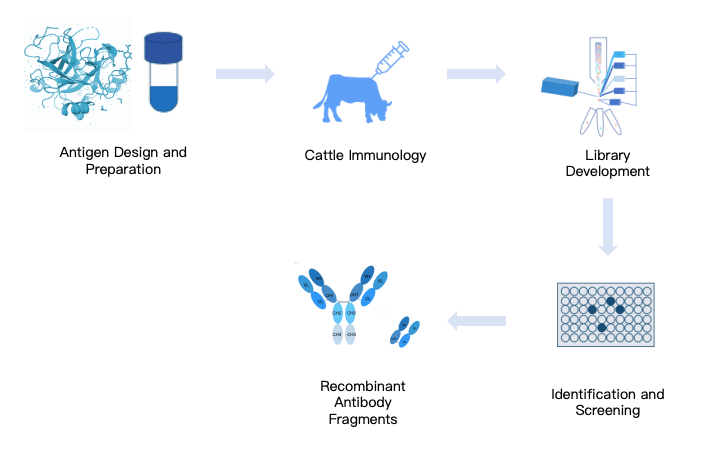
Enterprise Platform Spotlight: Kmd Bioscience Single B Cell Discovery
Bridging evolutionary biology with high-throughput microfluidic screening, Kmd bioscience delivers an end-to-end contract research platform From Single B Cell Isolation to Antibody Validation, specializing in challenging therapeutic targets, conserved antigens, and diagnostic master clones.
| Species Capabilities | Bovine, Rabbit, Camelid (VHH), Mouse, Rat, and Human Donor Repertoires |
| Sorting Technology | High-dimensional multi-color FACS and microfluidic droplet screening |
| VH/VL Pairing Fidelity | 100% native cognate pairing preserved; zero combinatorial chain scrambling |
| Turnaround Timeline | Rapid 3 to 5 weeks from tissue harvest to purified, sequence-verified recombinant IgG |
| Specialized Modalities | Bovine ultralong CDR-H3 engineering (targeting GPCRs and cryptic ion channel clefts) |
| Downstream Validation | Comprehensive Biacore SPR kinetics, nanoDSF thermal profiling, and cell-based potency assays |
Biologics Procurement Insight: Accelerate your pipeline by exploring our full single B cell antibody discovery architecture, evaluate custom screening protocols via our Single B Cell Screening Platform, or review project documentation through the COA Download portal.
Direct Technical Comparison: Single B Cell vs. Hybridoma vs. Phage Display
| Discovery Dimension | Single B Cell Discovery (e.g., Kmd Bioscience) | Traditional Hybridoma Fusion | Recombinant Phage Display Library |
|---|---|---|---|
| Heavy/Light Chain Pairing | 100% Native in vivo cognate pairing | Native pairing (if clone remains stable) | Artificial combinatorial scrambling |
| Campaign Discovery Timeline | 3 to 5 Weeks (Rapid) | 3 to 6 Months (Slow, multi-subcloning) | 6 to 10 Weeks (Iterative panning) |
| Affinity Profile ($K_D$) | Naturally picomolar ($10^{-10}$ to $10^{-12}\text{ M}$) | Nanomolar ($10^{-8}$ to $10^{-9}\text{ M}$) | Micromolar to nanomolar (Needs maturation) |
| Repertoire Sampling Depth | Massive (>100,000 isolated single cells) | Severely restricted (<0.01% fusion rate) | High ($>10^{10}$ synthetic transformants) |
| Cellular Genetic Stability | 100% Sequence locked (Recombinant IgG) | Unstable (Prone to chromosome loss) | 100% Sequence locked (Recombinant) |
| Applicability to Exotic Hosts | Broad (Bovine, Rabbit, Camelid, Human) | Severely limited (Requires partner myelomas) | Moderate (Requires complex library build) |
| Initial Capital Investment | Moderate to High ($$$$) | Moderate ($$$) | Moderate ($$$) |
Pros and Cons of the Single B Cell Discovery Pipeline
| Evaluation Vector | Technical & Commercial Advantages (Pros) | Inherent Liabilities & Challenges (Cons) |
|---|---|---|
| Biophysical & Clinical Value |
|
|
| Program Timeline & Cost |
|
|
Who Needs Single B Cell Discovery vs. Who Can Rely on Recombinant Phage Display
Every commercial program must align its technological approach with budget, timeline, and biological targets. Navigating From Single B Cell Isolation to Antibody Validation represents a high-performance engine, but choosing the right platform requires strategic discretion.
Programs That Must Deploy Single B Cell Discovery:
Therapeutic Programs Targeting Complex Membrane Proteins (GPCRs, Ion Channels): GPCRs possess tiny extracellular loops embedded within hydrophobic membranes. Synthetic libraries struggle to produce binders against these conformations. Utilizing non-rodent hosts (such as bovine platforms with ultralong CDR-H3 loops) through the single B cell pipeline allows the host's immune system to generate knob-and-stalk antibodies that reach deep into the receptor cleft.
Fast-Track Infectious Disease & Pandemic Neutralization: When discovering neutralizing antibodies from human convalescent patient blood, single B cell sorting directly isolates potent IgG-secreting memory cells within days of sample collection, preserving authentic human heavy-light pairing without requiring humanization.
High-Affinity Diagnostic Master Clones: For IVD diagnostic platforms (chemiluminescence, lateral flow, turbidimetry) where picomolar detection limits are required to identify trace cancer or cardiac biomarkers, rabbit single B cell discovery produces clones with affinities ten to one hundred times tighter than traditional mouse monoclonal lines.
Programs That Can Comfortably Use Synthetic Phage Display:
Toxic Antigens or Lethal Toxins: If the target antigen is an active toxin (e.g., botulinum, snake venoms, or highly cytotoxic proteins) that kills the host animal upon immunization, in vivo single B cell sourcing is impossible. Naive or synthetic phage display libraries are required because the panning occurs entirely in vitro.
Academic Research Reagents on Highly Constrained Budgets: If your laboratory only needs a simple Western blot or immunohistochemistry primary antibody where high-nanomolar affinity is acceptable and timeline is secondary, standard display panning or traditional hybridoma generation offers a low-cost, serviceable alternative.
Critical Pitfalls in Single B Cell Campaigns That Ruin Program Yields
Bench Advisory: The Cross-Well PCR Contamination Hazard
The most disastrous failure in the workflow From Single B Cell Isolation to Antibody Validation occurs during single-cell RT-PCR. Because you are amplifying single-digit copies of mRNA transcripts across thousands of individual wells, a single aerosolized micro-droplet containing high-copy PCR product can contaminate an entire 96-well plate. This results in identical heavy-chain sequences pairing artificially with different light chains, destroying true clonal diversity. Always enforce dedicated pre-PCR and post-PCR spatial separation with UV decontamination and unidirectional workflow protocols.
Sorting with Aggregated or Impure Antigens: If your fluorescently labeled antigen contains 5% soluble aggregate, the high avidity of the aggregate will bind non-specific, low-affinity B-cell receptors during FACS. You will sort hundreds of "positive" cells that yield completely inactive antibodies once expressed as monomeric IgGs. Always purify sorting antigens via Size-Exclusion Chromatography (SEC-HPLC) on the day of sorting.
Overlooking Somatic Hypermutation Primer Mismatches: In hyper-immunized animals, extensive somatic hypermutation alters the framework 1 (FR1) region of the antibody gene. If your nested PCR primer library relies on rigid, non-degenerate sequences, the primers will fail to anneal to the mutated template, causing up to 50% sequence dropout. Use optimized degenerate primer cocktails calibrated to your specific host species.
Relying Solely on ELISA Without Kinetic Off-Rate Screening: Selecting lead candidates based purely on high $OD_{450}$ readings in an end-point ELISA is an industry trap. A high ELISA signal can simply reflect high expression titer or non-specific stickiness. Deploy real-time biosensors (Biacore or Octet) early in Step 8 to measure real-time dissociation rate constants ($k_d$); clones that drop off the target within seconds must be purged immediately.
Neglecting Developability & Liability Sequence Audits: Failing to run computational sequence scans on your amplified VH/VL genes before expression leads to downstream manufacturing disasters. Clones containing unpredicted N-linked glycosylation motifs (N-X-S/T) in the CDRs, free cysteines, or deamidation hotspots (NG, NN) will exhibit batch-to-batch heterogeneity during therapeutic scale-up. Learn how to validate these parameters using our foundational biotech lab techniques guide.
Essential Buying Considerations When Selecting a Discovery CRO
Outsourcing your program From Single B Cell Isolation to Antibody Validation requires auditing potential contract research organizations (CROs) against four rigorous technical benchmarks:
1. True Single-Cell Isolation Platform Transparency
Ensure the provider operates certified, high-resolution single-cell isolation equipment (e.g., BD FACSAria III, Sony SH800, or microfluidic beacon platforms) and provides raw index-sorting data files. Reject vendors who utilize bulk panning or multi-cell pooling, as this destroys native cognate pairing and introduces synthetic re-assortment.
2. Multi-Species Repertoire Capability
A capable discovery partner should not restrict your project to standard mice. Inquire whether the facility has validated primer architectures and expression systems for rabbits, camelids (VHH), and bovine hosts. Sourcing specialized reagents—including Peptide Products, Nanoparticle Products, or Small Molecule Reagents—ensures that non-protein or hapten targets can be properly conjugated and presented to diverse animal immune systems.
3. Recombinant Deliverable Guarantees
Demand that the CRO deliver sequence-verified recombinant plasmids (heavy and light chain constructs) alongside purified IgG protein. Never accept hybridoma cell vials or unsequenced lysates as final deliverables. Owning the exact digital nucleotide sequence ensures your intellectual property is locked, reproducible, and permanently protected from genetic drift.
4. Comprehensive Biophysical Characterization Instrumentation
Verify that the provider's validation facility houses gold-standard surface plasmon resonance (GE Healthcare / Cytiva Biacore) or bio-layer interferometry (Sartorius Octet) equipment, alongside high-grade Raw Materials Reagents for assay design. A full kinetic report detailing $k_a$, $k_d$, and $K_D$ across multi-cycle kinetics must be provided for every lead candidate.
Expert Biologics Practitioner's Final Sourcing Verdict
In our professional assessment across decades of therapeutic and diagnostic antibody engineering, continuing to rely on legacy hybridoma fusion or unrefined synthetic phage display for high-value targets is an unnecessary developmental risk. The time lost to subcloning failures, paired with the downstream expenses of in vitro affinity maturation, dwarfs the upfront investment of single-cell discovery.
Navigating From Single B Cell Isolation to Antibody Validation is the most efficient, biologically elegant, and robust methodology available today. It respects the billions of years of evolutionary engineering refined inside the host's germinal centers, capturing pristine, picomolar-affinity antibodies with native cognate pairing in a matter of weeks.
For research teams and biopharma enterprises looking to accelerate their pipelines, we recommend partnering with a vertically integrated provider like Kmd bioscience. Their industrial platforms—spanning murine, rabbit, and bovine single B cell discovery—deliver complete sequence transparency, rapid recombinant mammalian expression, and rigorous SPR biophysical validation, empowering your organization to transition from immunized tissue to validated clinical leads with total confidence.
Frequently Asked Questions
How does the workflow from single B cell isolation to antibody validation preserve native cognate pairing?
Unlike standard hybridoma fusion or synthetic phage display libraries, the sequence from single B cell isolation to antibody validation isolates individual B lymphocytes into physical micro-compartments before cell lysis. Single-cell reverse transcription PCR (RT-PCR) amplifies the variable heavy (VH) and variable light (VL) chains from that exact single cell, permanently preserving the natural, in vivo affinity-matured pairing without combinatorial mismatching.
What is the typical timeline from single B cell isolation to antibody validation compared to hybridoma technology?
A standard hybridoma campaign requires 3 to 6 months due to fusion inefficiencies, repeated subcloning, and slow cellular expansion. In our testing, executing the protocol from single B cell isolation to antibody validation compresses this timeline down to 3 to 5 weeks. High-throughput FACS or microfluidics sorting isolates thousands of functional candidates in a single day, followed by rapid molecular cloning and recombinant expression.
Why is bovine or rabbit single B cell technology advantageous for difficult therapeutic targets?
Bovine single B cell platforms utilize unusually long CDR-H3 loops (up to 60 amino acids forming protruding 'knob-and-stalk' architectures) that access hidden, cryptic epitopes or catalytic pockets on GPCRs and ion channels that conventional rodent antibodies cannot reach. Similarly, rabbit platforms provide distinct somatic gene conversion mechanisms that yield picomolar affinities against small molecules and low-immunogenicity antigens.
What critical analytical assays are required during antibody validation?
Comprehensive antibody validation requires multi-tiered characterization: surface plasmon resonance (SPR) or bio-layer interferometry (BLI) for real-time kinetic on/off rates (Kon, Koff, KD), differential scanning fluorimetry (nanoDSF) for thermal stability (Tm), size-exclusion chromatography (SEC-HPLC) for monomeric purity, and functional target-cell assays (flow cytometry, neutralization, or immunohistochemistry).





 0
