


Too many discovery programs stumble early because research directors treat primary screening like a simple numbers race. High-throughput campaigns routinely process hundred-thousand-compound chemical libraries or massive single-chain variable fragment (scFv) outputs through basic binding screens, celebrating hundreds of primary hits. Yet, when these hits transition to secondary functional validation or lead optimization, over 80% disappear as assay artifacts, colloidal aggregators, or non-specific pan-assay interference compounds (PAINS). Screening millions of molecules means nothing if your screening funnel is built on unstable assay biology.
In modern biopharma, discovering viable small-molecule leads and therapeutic biologics requires an orchestrated cascade of orthogonal testing. Navigating assays commonly used in early drug discovery demands rigorous biophysical and biochemical scrutiny. A discovery pipeline must balance phenotypic cellular relevance, cell-free target engagement, kinetic on/off rates, and early in vitro ADMET flags. Relying on a single readout or picking an assay platform simply because the kit is cheap leads directly to dead-end lead series. We dissect the ten indispensable assays commonly used in early drug discovery, evaluating their operational mechanisms, data fidelity, and decision gates to help biotech teams advance genuine leads toward clinical candidacy.
Quick Answer: What Are the Key Assays Commonly Used in Early Drug Discovery?
The ten foundational assays commonly used in early drug discovery form a structured screening hierarchy: 1) Surface Plasmon Resonance (SPR) for real-time label-free kinetic binding (Ka, Kd, KD); 2) Differential Scanning Fluorimetry (DSF / nanoDSF) for thermal shift target engagement; 3) Homogeneous Time-Resolved Fluorescence (HTRF / TR-FRET) for high-throughput biochemical enzymatic and protein-protein interaction (PPI) inhibition; 4) Cell Viability & Cytotoxicity Assays (CellTiter-Glo ATP bioluminescence) for broad phenotypic compound safety; 5) Enzyme-Linked Immunosorbent Assay (ELISA) for high-sensitivity antibody-antigen endpoint titrations; 6) Cellular Reporter Gene Assays for functional pathway modulation and transcription factor engagement; 7) High-Content Imaging (HCS) for phenotypic multiplexed spatial cellular readouts; 8) Flow Cytometry (FACS) for cell-surface target binding and immune cell phenotyping; 9) In Vitro ADME Assays (PAMPA, Caco-2, Microsomal Clearance) for early metabolic stability and permeability screening; and 10) Automated Patch Clamp (hERG liability) for early off-target cardiotoxicity de-risking. In most professional situations, pairing a primary homogenous screen (like HTRF) with label-free biophysical validation (SPR) provides the fastest, most dependable route to lead series identification.
Table of Contents
Screening Cascade Architecture: The Filter from Millions to Leads
In-Depth Evaluation: 10 Assays Commonly Used in Early Drug Discovery
Assay Comparison Matrix: Biochemical vs. Biophysical vs. Phenotypic Cellular
Pros and Cons of Cell-Free Biochemical Screens vs. Phenotypic Cellular Screens
Who Needs Biophysical SPR Kinetics vs. Who Can Rely on Endpoint ELISA
Featured Discovery Partner: KMD Bioscience Discovery Platforms
Costly Mistakes Discovery Teams Make in Early-Stage Screening
Screening Cascade Architecture: The Filter from Millions to Leads
Early drug discovery is an attrition funnel. Whether engineering recombinant monoclonal antibodies, developing bispecific formats, or optimizing small-molecule fragment libraries, early discovery operates across three distinct biological stages: target identification and validation, hit identification (HTS), and hit-to-lead progression. Running high-content phenotypic assays on a million compounds is commercially cost-prohibitive. Conversely, advancing a compound based purely on an isolated enzymatic tube assay without cellular permeability data guarantees failure in animal efficacy models.
A rigorous screening cascade moves compounds sequentially through defined biological checkpoints:
Primary High-Throughput Screening (HTS): Highly miniaturized 384-well or 1536-well homogenous assays (like TR-FRET or AlphaLISA) screen broad compound collections to flag initial binders or functional inhibitors at single-point concentrations (typically 10 micromolar).
Secondary Orthogonal & Counter-Screening: Confirmed hits are re-tested using a completely different assay physics readout—such as swapping a fluorescence-based enzymatic assay for a label-free mass-sensitive Surface Plasmon Resonance (SPR) assay—to eliminate optical interferers, autofluorescent molecules, and non-specific aggregators.
Cellular Functional & Mechanistic Validation: Verified physical binders enter intact mammalian cell models to confirm cellular target engagement, pathway modulation, and downstream transcriptional changes.
Early In Vitro Developability & ADMET Profiling: Hits are screened for metabolic clearance in liver microsomes, artificial membrane permeability (PAMPA), and off-target toxicities (hERG channel inhibition, general cytotoxicity) before medicinal chemists synthesize analogue series.
In our testing across diverse therapeutic projects, programs that integrate biophysical hit validation early in the screening cascade save millions in wasted medicinal chemistry cycles. Validating binding constants and kinetic profiles early ensures discovery resources focus strictly on series that exhibit clean target engagement.
Quick Summary: The 10 Essential Assays at a Glance
The table below provides a quick reference to the ten assays commonly used in early drug discovery, outlining their detection mechanisms, throughput capacity, and optimal pipeline positioning:
| Assay Platform | Detection Mechanism | Throughput Tier | Target Class Focus | Optimal Stage in Cascade |
|---|---|---|---|---|
| 1. Surface Plasmon Resonance (SPR) | Label-free optical refractive index change | Medium to High | Proteins, antibodies, small-molecule hits | Hit confirmation, kinetic validation (Ka, Kd, KD) |
| 2. Differential Scanning Fluorimetry (DSF) | Thermal shift (ΔTm) protein unfolding dye | High (384-well) | Recombinant proteins, kinases, enzymes | Fragment screening, direct target engagement |
| 3. TR-FRET / HTRF | Time-resolved fluorescence resonance transfer | Very High (1536-well) | Kinases, GPCRs, protein-protein interactions | Primary high-throughput screening (HTS) |
| 4. Cell Viability (CellTiter-Glo) | Luciferase bioluminescent ATP quantification | Very High (1536-well) | Oncology lines, primary cells, toxicity panels | Compound cytotoxicity, anti-proliferation screens |
| 5. Direct & Sandwich ELISA | Colorimetric / Chemiluminescent antibody tag | Medium to High (96/384) | Monoclonal antibodies, cytokines, soluble targets | Hybridoma screening, titer checks, affinity ranking |
| 6. Reporter Gene Assays | Luciferase/GFP driven promoter transcription | High (384-well) | Nuclear receptors, GPCRs, signaling cascades | Functional cellular agonism and antagonism |
| 7. High-Content Screening (HCS) | Automated confocal multi-channel microscopy | Medium (384-well) | Subcellular translocation, organelle pathology | Phenotypic discovery, complex disease modeling |
| 8. Flow Cytometry (FACS) | Multi-laser single-cell fluorophore scattering | Medium | Cell-surface receptors, CAR-T, immune panels | Conformational cell-surface binding validation |
| 9. In Vitro ADME Panels | LC-MS/MS parent compound disappearance | Medium to High | Small molecules, peptide mimetics | Metabolic stability, PAMPA/Caco-2 permeability |
| 10. Automated Patch Clamp (hERG) | Microfluidic planar patch electrophysiology | Medium | Ion channels, secondary cardiac safety targets | Early safety profiling, cardiotoxicity de-risking |
In-Depth Evaluation: 10 Assays Commonly Used in Early Drug Discovery
1. Surface Plasmon Resonance (SPR) and Biolayer Interferometry (BLI)
Surface Plasmon Resonance stands as the gold standard for label-free real-time molecular interaction analysis. In an SPR biosensor, a target protein or antibody is immobilized onto a sensor chip surface coated with carboxymethylated dextran or specialized capture chemistries (e.g., Ni-NTA for His-tags, Protein A/G for antibodies). As analyte molecules in solution flow across the chip surface, binding increases mass concentration at the sensor surface, altering the refractive index and shifting the plasmon resonance angle in real time.
In our testing across biotherapeutic programs, SPR provides what static endpoint assays cannot: detailed kinetic rate constants. It delivers the association rate (Ka / Kon), dissociation rate (Kd / Koff), and calculates the equilibrium dissociation constant (KD = Koff / Kon). Knowing the off-rate is essential; two lead candidates might share identical 10 nM affinity values, but one may feature a slow off-rate that maintains sustained target occupancy in vivo, while the other dissociates too quickly to exert therapeutic efficacy. Sourcing recombinant targets via proven Protein Expression Services and integrating dedicated Protein Interaction Services ensures biosensor chips receive monomeric, correctly folded antigens with minimal background noise.
2. Differential Scanning Fluorimetry (DSF and nanoDSF)
Differential Scanning Fluorimetry—often termed a thermal shift assay (TSA)—operates on a fundamental biophysical principle: when a small molecule or peptide ligand binds to a protein's active site or allosteric pocket, it stabilizes the protein's tertiary structure, shifting its thermal denaturation melting temperature (Tm). In conventional DSF, an environmentally sensitive dye (like SYPRO Orange) quenches in aqueous solution but fluoresces when binding to hydrophobic protein cores exposed during thermal unfolding.
Modern discovery labs increasingly favor label-free nanoDSF, which monitors intrinsic tryptophan and tyrosine fluorescence emission shifts (350nm vs. 330nm) without fluorescent dyes. DSF is widely used in fragment-based drug discovery (FBDD). Because fragment compounds typically exhibit weak millimolar-to-micromolar affinities that are difficult to measure in functional cellular systems, thermal shifts (ΔTm > 1–2 degrees Celsius) provide fast, high-confidence evidence of direct target engagement. For structural biology workflows, validating folded targets across our Protein Expression Platform confirms consistent protein stability prior to screening.
3. Homogeneous Time-Resolved Fluorescence (HTRF / TR-FRET)
When screening libraries containing 50,000 to 500,000 compounds, wash steps destroy screening velocity. Homogeneous Time-Resolved Fluorescence (HTRF) combines standard Förster Resonance Energy Transfer (FRET) with time-resolved fluorescence spectroscopy, completely eliminating plate washing steps. The system pairs a long-lifetime europium or terbium cryptate donor fluorophore with an organic acceptor fluorophore (such as d2 or XL665).
The operational strength of HTRF lies in its temporal delay. Standard organic compounds frequently exhibit short-lived autofluorescence (under 10 nanoseconds) when excited by laser light, blinding conventional fluorescent plate readers. HTRF introduces a 50-to-100-microsecond delay before measuring acceptor emission, allowing background autofluorescence to decay completely. This yields exceptional signal-to-background ratios and high Z-prime factors (Z' > 0.7), making it a top choice for screening kinase autophosphorylation, GPCR secondary messengers (cAMP, IP-One), and protein-protein interactions. Discover targeted peptides for assay displacement via our comprehensive Peptide Library Platform.
4. Cell Viability and Cytotoxicity Assays (Bioluminescent ATP Quantitation)
Early drug discovery requires establishing compound cytotoxicity profiles before investing in deeper functional screens. The industry gold standard for cellular viability screening is the homogenous ATP bioluminescence assay (such as CellTiter-Glo). Because metabolically active cells tightly regulate intracellular ATP pools, lysing cells in the presence of recombinant luciferase and luciferin produces a glow-type luminescent signal directly proportional to the number of viable cells in culture.
From our experience on automated screening platforms, ATP bioluminescence is vastly superior to legacy tetrazolium reduction assays (like MTT, MTS, or WST-1). MTT assays require multi-hour metabolic incubations and crystal solubilization steps where test compounds can reduce tetrazolium salts chemically without living cells, producing false-positive viability reads. CellTiter-Glo lyses cells within ten minutes, delivers linear readouts across four orders of magnitude, and quickly flags non-specific cytotoxicity across early lead molecules.
5. Enzyme-Linked Immunosorbent Assay (ELISA)
Despite the rise of complex biophysical biosensors, ELISA remains an essential workhorse in biologics discovery, antibody engineering, and biomarker quantification. Whether run in direct, indirect, or sandwich formats, an immobilized target captures antibody or protein analytes, which are detected via enzyme-conjugated secondary antibodies (such as Horseradish Peroxidase, HRP) driving chromogenic (TMB) or chemiluminescent substrates.
In antibody hit discovery, high-throughput 384-well ELISA panels provide the primary filter for screening thousands of hybridoma supernatants or phage display outputs. It delivers reliable qualitative binding confirmation and concentration-dependent EC50 values. While ELISA lacks real-time kinetic on/off rate data, its high dynamic range and tolerance for crude cell lysates make it indispensable for initial screening campaigns. Discover end-to-end antibody generation platforms through our Custom Antibody Platform and validate clone specificity on our Antibody Expression & Validation Platform.
6. Cellular Reporter Gene Assays
Purified proteins in a test tube do not replicate the complex signaling dynamics of an intact mammalian cell. Cellular reporter gene assays measure whether a compound crossing the cell membrane successfully modulates target signal transduction pathways. Host cell lines are stably transfected with a promoter-reporter construct containing response elements (such as NF-kappa-B, CREB, or STAT3) linked upstream of a reporter gene like firefly luciferase, Renilla luciferase, or Green Fluorescent Protein (GFP).
When an agonist or antagonist engages a cell-surface receptor (e.g., GPCRs, RTKs) or nuclear hormone receptor, the resulting intracellular signaling cascade drives or represses reporter enzyme transcription. Dual-luciferase systems allow multiplexed normalization: one luciferase reports target pathway modulation while an orthogonal Renilla luciferase driven by a constitutive promoter controls for compound cytotoxicity. To build robust reporter cell lines, research teams leverage our dedicated Gene Editing Platform and broad Molecular Platform.
7. High-Content Screening (HCS) and Automated Phenotypic Imaging
High-Content Screening merges automated multi-channel confocal microscopy with quantitative machine-learning image analysis. While microplate readers output a single averaged signal for an entire well, HCS collects spatial single-cell data across hundreds of individual cells simultaneously, quantifying parameters like nuclear translocation, neurite outgrowth, mitochondrial membrane potential, and receptor internalization.
In our testing of phenotypic disease models, HCS identifies subtle biological phenotypes that homogenous plate readers miss entirely. For example, when tracking an antibody-drug conjugate (ADC), HCS tracks the exact kinetics of receptor-mediated internalization and lysosomal trafficking. The limitation is data storage and analysis bandwidth; processing terabytes of image data requires robust computational infrastructure, making HCS a specialized secondary validation tool rather than an initial primary screening engine.
8. Flow Cytometry and High-Throughput Cell Binding (FACS)
For therapeutic biologics targeting membrane-spanning proteins—such as multi-pass G-protein coupled receptors, ion channels, or immune checkpoint markers (PD-1, CTLA-4)—recombinant soluble proteins often fail to mimic native three-dimensional membrane conformations. Flow cytometry evaluates antibody binding directly on intact living cells in suspension.
High-throughput flow cytometry systems utilize autosamplers that process 96-well and 384-well plates, interrogating cells passing through focused laser beams to measure forward scatter (size), side scatter (granularity), and multi-spectral fluorescent emission. This ensures candidate antibodies bind to genuine cell-surface antigens in their native membrane environments rather than non-functional linear epitopes. Accelerate native B-cell lead identification using our specialized Single B Cell Screening Platform, and verify downstream functional impacts through our comprehensive Detection Platform.
9. In Vitro ADME Screening Panels (Metabolic Stability & Permeability)
Potency in the absence of favorable drug-like properties leads to failure in animal pharmacokinetic studies. Modern early discovery programs run early absorption, distribution, metabolism, and excretion (ADME) profiling in parallel with target potency screening. Primary ADME assays include:
Liver Microsomal Stability: Incubating compounds with human, rat, or mouse liver microsomes supplemented with NADPH to calculate in vitro intrinsic clearance (CLint) and half-life via LC-MS/MS.
Parallel Artificial Membrane Permeability Assay (PAMPA): Assessing passive transcellular permeability across a lipid-infused synthetic membrane filter.
Caco-2 Monolayer Permeability: Measuring transport across human colorectal adenocarcinoma cell monolayers to evaluate both passive absorption and active efflux transporter liabilities (like P-glycoprotein).
10. Automated Patch Clamp (hERG Cardiotoxicity Liability)
Sudden cardiac death caused by drug-induced Long QT syndrome is a major historical reason for pharmaceutical clinical trial holds and market withdrawals. Cardiotoxicity commonly stems from off-target inhibition of the human Ether-à-go-go-Related Gene (hERG) voltage-gated potassium channel (Kv11.1), which coordinates cardiac action potential repolarization.
Historically, manual patch-clamp electrophysiology was a slow, low-throughput technique reserved for late preclinical stages. Automated planar patch clamp platforms now screen hundreds of compounds daily against hERG-expressing HEK293 cells. Flagging micromolar hERG inhibition early in lead optimization allows chemists to modify basic lipophilic amines and mitigate cardiac safety liabilities long before filing an Investigational New Drug (IND) application.
Assay Comparison Matrix: Biochemical vs. Biophysical vs. Phenotypic Cellular
To help you select the right assay combination for your pipeline, compare the core strengths, limits, and applications across the three primary assay classes:
| Evaluation Metric | Biochemical Assays (e.g., HTRF, ELISA) | Biophysical Assays (e.g., SPR, nanoDSF) | Phenotypic & Cellular (e.g., HCS, Reporter) |
|---|---|---|---|
| Primary Biological Output | IC50 / EC50 percentage functional inhibition | Direct KD, Kon, Koff, and ΔTm binding values | Cellular pathway modulation, IC50, toxicity |
| Artifact & Interference Risk | High (Autofluorescence, quenching, PAINS) | Very Low (Direct physical mass/heat readout) | Moderate (Off-target cytotoxicity, uptake bias) |
| Throughput Capacity | Very High (Up to 100,000+ compounds/day) | Medium to High (Hundreds of sensor cycles/day) | Medium (Plates require multi-hour cell incubation) |
| Physiological Relevance | Low (Purified proteins in artificial buffers) | Low to Moderate (Cell-free direct interactions) | High (Intact cellular membranes, native biology) |
| Recombinant Reagent Demands | Requires labeled antibodies or tag substrates | Requires high-purity, monomeric target protein | Requires stable, characterized cell line banks |
Pros and Cons of Cell-Free Biochemical Screens vs. Phenotypic Cellular Screens
Balancing isolated cell-free testing against living cellular models requires evaluating key research trade-offs:
| Assay Class | Operational Advantages (Pros) | Practical Limitations (Cons) |
|---|---|---|
| Cell-Free Biochemical & Biophysical Screens (HTRF, SPR, DSF, Enzymatic) |
|
|
| Phenotypic & Functional Cellular Assays (CellTiter-Glo, Reporter, HCS, FACS) |
|
|
Who Needs Biophysical SPR Kinetics vs. Who Can Rely on Endpoint ELISA
In our experience consulting for biopharma startups, choosing the wrong analytical platform often leads to poor data quality or unnecessary screening expenses.
Who Needs Real-Time Biophysical SPR / BLI Analysis:
Antibody Engineering & Affinity Maturation Teams: When optimizing monoclonal antibodies to picomolar affinities, static endpoint ELISA signals saturate completely. SPR is mandatory to measure slow dissociation rates (Koff values slower than 10^-4 s^-1).
Fragment-Based Drug Discovery (FBDD) Programs: Small-molecule fragments (MW < 300 Da) bind with weak affinities that fail to generate robust enzymatic inhibition curves. High-sensitivity SPR detects micro-refractive index shifts from low-molecular-weight binding.
Competitive Epitope Binning & Multi-Specific Formats: Engineering bispecific antibodies or multi-protein complexes requires biosensor platforms to verify whether two antibodies bind distinct non-competing epitopes simultaneously.
Who Can Rely on Standard High-Throughput ELISA:
Early Hybridoma & Phage Supernatant Screening: When screening thousands of crude clones to distinguish strong binders from non-binders, automated 384-well ELISA provides high throughput without fouling expensive biosensor microfluidic chips.
Standard Pharmacokinetic (PK) Serum Quantitation: Measuring drug concentrations in preclinical animal serum samples is handled reliably and cost-effectively by sandwich ELISA.
Antibody Affinity Maturation Services: KMD Bioscience
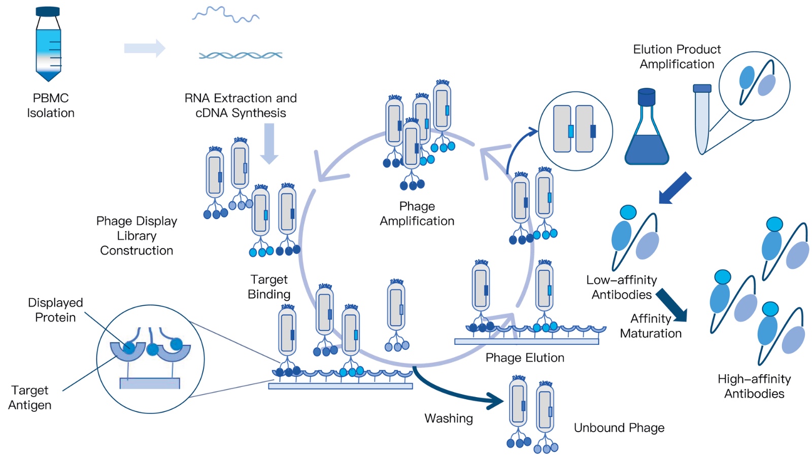
KMD Bioscience has been dedicated to antibody research for many years. Affinity is a critical parameter for antibody therapeutics, typically influencing both antibody function and efficacy. Affinity often decreases after antibody humanization. Therefore, restoring the affinity of humanized antibodies or elevating it to higher levels is a necessary step. In vitro affinity maturation technology is a commonly used technique to improve the affinity of antibodies for their antigens.
KMD Bioscience provides clients with multi-species Antibody Affinity Maturation Services based on our proprietary phage display antibody library technology. Staffed by a highly skilled antibody team with extensive experience in antibody affinity maturation, we can enhance the affinity of client-provided monoclonal antibodies by 2 to 3 orders of magnitude, achieving levels of up to 10-9 M.
Core Technology: High-Capacity Phage Display & Mutagenesis Libraries
Affinity Improvement: Increases binding affinity by 2 to 3 orders of magnitude (up to 10^-9 M)
Species Versatility: Human, mouse, rabbit, llama (nanobody), and camelid formats
Biophysical Validation: Complete SPR, BLI, and ELISA kinetic characterization
Turnkey Pipelines: Integrated humanization, expression, and stability testing
Downstream Support: Pilot scale bioprocessing via our Process Development Platform
Explore comprehensive discovery options on the Antibody Affinity Maturation Service Introduction portal, evaluate our broad Innovative Drug Discovery Platform, explore specialty systems like our Plant Genetic Transformation Platform, review our Protein Modification & Detection Services, and access technical literature via Technical Resources.
Costly Mistakes Discovery Teams Make in Early-Stage Screening
In our audits of early-stage discovery programs, research teams frequently run into four common screening errors that waste budget and derail timelines:
Relying on Single-Point Concentration Screens to Rank Hits: Ranking primary hits based on percentage inhibition at a single compound concentration (e.g., 10 μM) is inherently flawed. Compounds with steep hill slopes, poor solubility, or optical interference frequently generate false-positive rankings. Always perform rapid 8-to-10-point dose-response curves in duplicate to confirm true pharmacological behavior.
Ignoring Pan-Assay Interference Compounds (PAINS): Failing to filter out chemical substructures that produce assay artifacts leads programs to chase false leads. Common PAINS molecules—such as rhodanines, quinones, and catechols—interfere with fluorescent readouts, generate hydrogen peroxide in solution, or form colloidal aggregations that non-specifically inhibit proteins. Early counterscreens with detergent (0.01% Triton X-100) or non-related target proteins help flag these colloidal aggregators immediately.
Skipping Recombinant Protein Monomer Quality Checks: Immobilizing aggregated or partially unfolded recombinant protein onto an SPR chip or ELISA plate produces erratic binding data. Proteins must be validated via Analytical Size-Exclusion Chromatography (SEC-HPLC) and Dynamic Light Scattering (DLS) to confirm >95% monomeric purity before initiating screening.
Overlooking DMSO Tolerance Boundaries in Cellular Assays: Screening libraries are typically dissolved in 100% dimethyl sulfoxide (DMSO). Transferring compounds into 384-well cell plates without testing cellular DMSO tolerance can compromise cell membrane integrity. Keep final assay DMSO concentrations strictly at or below 0.5% (v/v) to avoid solvent-induced artifacts.
Commercial Sourcing & CRO Partner Selection Criteria
Before outsourcing screening campaigns to a contract research organization (CRO), research directors should verify these four technical capabilities:
Statistical Assay Robustness (Z-Prime Factor Standards): Ensure the CRO contractually verifies an assay Z' factor ≥ 0.6 across pilot plates before launching library screening. A Z' below 0.5 indicates excessive well-to-well variability, leading to unacceptably high false-positive and false-negative rates.
End-to-End Orthogonal Validation Platforms: Sourcing biochemical screening from one vendor and biophysical kinetics from another creates logistical bottlenecks. Partner with CROs that maintain integrated biochemical (HTRF), biophysical (Biacore SPR/BLI), and cellular screening platforms under one roof to maintain momentum.
Independent Raw Data & Sensorgram Delivery: Require the CRO to deliver complete raw instrument data files—including raw sensorgrams and reference-subtracted binding curves—rather than isolated summary spreadsheets, ensuring your internal biostatisticians can independently audit baseline drifts and fitting models.
Recombinant Reagent Sourcing Traceability: Verify whether the CRO expresses its own recombinant target proteins and antibodies in-house. CROs with dedicated expression platforms can rapidly re-generate fresh protein batches, avoiding supply delays if assay development requires modified tags or mutations.
Expert Recommendation and Lead Optimization Verdict
In most professional situations, we recommend avoiding single-assay screening funnels. Successful early drug discovery relies on building a structured, orthogonal screening cascade early in your program.
For high-throughput primary screening, deploy Homogeneous Time-Resolved Fluorescence (HTRF) to rapidly screen broad chemical or biologic collections with minimal background interference. Advance confirmed primary hits directly into Surface Plasmon Resonance (SPR) to eliminate optical artifacts, verify target engagement, and extract genuine binding kinetic rate constants (Kon, Koff, KD). Finally, validate lead series using Cellular Reporter Gene Assays and early in vitro ADME/hERG panels to ensure compounds demonstrate functional efficacy and safe pharmacology in living systems. Partnering with an experienced, integrated CRO like KMD Bioscience gives your team access to high-capacity phage display libraries, precision antibody affinity maturation, and comprehensive biophysical screening, ensuring you advance high-quality candidates toward clinical success.
Frequently Asked Questions (FAQ)
What are the most common assays used in early drug discovery?
The most common assays used in early drug discovery include Surface Plasmon Resonance (SPR), Differential Scanning Fluorimetry (DSF), Homogeneous Time-Resolved Fluorescence (HTRF/TR-FRET), ATP bioluminescent cell viability assays (CellTiter-Glo), Enzyme-Linked Immunosorbent Assays (ELISA), cellular reporter gene assays, High-Content Screening (HCS), flow cytometry (FACS), in vitro ADME clearance panels, and automated patch clamp (hERG) electrophysiology.
What is the difference between an orthogonal assay and a primary screen?
A primary screen is a high-throughput assay (such as TR-FRET or AlphaLISA) designed to rapidly evaluate thousands of compounds at a single concentration to flag potential hits. An orthogonal assay uses a completely different physical or biological readout (such as switching from a fluorescence-based assay to label-free SPR biophysics) to test the same hits, confirming direct target engagement while eliminating assay-specific artifacts like autofluorescence or chemical aggregation.
Why is Surface Plasmon Resonance (SPR) preferred over ELISA for lead characterization?
While ELISA provides endpoint binding titers, it cannot measure real-time kinetics. Surface Plasmon Resonance (SPR) is a label-free technology that tracks binding interactions in real time, delivering association rate constants (Kon), dissociation rate constants (Koff), and precise equilibrium dissociation constants (KD). Knowing the off-rate is critical for predicting target residence time and in vivo efficacy.
What is a Z-prime factor in drug discovery screening?
The Z-prime (Z') factor is a statistical parameter that measures the quality and dynamic range of a high-throughput screening assay. It incorporates the signal separation band between positive and negative controls alongside their standard deviations. A Z' factor between 0.5 and 1.0 indicates a robust, reliable assay suitable for high-throughput screening campaigns.
Why do discovery programs run early hERG safety assays?
Off-target inhibition of the hERG voltage-gated potassium channel is a primary cause of drug-induced cardiac arrhythmias (Long QT syndrome) and clinical trial attrition. Screening compounds early via automated patch clamp electrophysiology allows discovery teams to identify and modify cardiotoxic chemical structures during lead optimization rather than discovering safety liabilities during expensive regulatory animal toxicology studies.





 0
0
