


10 Essential Methods for Antibody Characterization and Validation
- 2026-09-23
- 138
The reproducibility crisis in biological research and diagnostic development can frequently be traced back to a single, catastrophic point of failure: poorly validated antibodies. If you are advancing a therapeutic candidate or engineering a critical in vitro diagnostic (IVD) kit, deploying an uncharacterized antibody is the fastest way to hemorrhage R&D capital. From our experience at KMD Bioscience, we routinely see biotechnology firms lose months of clinical progress simply because their primary detection reagent exhibited off-target cross-reactivity that was not caught during the initial screening phases.
In most professional situations, a solitary Enzyme-Linked Immunosorbent Assay (ELISA) is completely insufficient to prove an antibody's commercial or clinical viability. High-affinity binding to a recombinant protein in a plastic well does not guarantee performance in complex biological matrices. You must execute a rigorous, multi-tiered validation matrix to confirm specificity, affinity, stability, and functional neutralization. In this comprehensive industry guide, we take a definitive position on the optimal methods for antibody characterization, dissecting the exact biochemical assays you must utilize, and providing practical commercial judgment so your downstream applications succeed without costly false positives.
Quick Answer: The Core Validation Matrix
There are ten indispensable methods for antibody characterization that form the foundation of a robust validation pipeline. The most critical include:
ELISA: Rapid confirmation of target binding and preliminary titer assessment.
Western Blotting (WB): Verification of molecular weight and target specificity in denatured states.
Flow Cytometry (FC): Confirmation of binding to native conformations on the cell surface.
Surface Plasmon Resonance (SPR): The gold standard for measuring precise binding kinetics and affinity (KD).
Functional Assays: Evaluating the antibody's ability to neutralize, activate, or inhibit biological pathways.
We recommend a minimum three-pillar validation approach (WB, FC, and SPR) before advancing any candidate generated through a Single B Cell Screening Platform or hybridoma process.
Table of Contents
What It Is: The Definition of Characterization
Antibody characterization is the exhaustive analytical process of determining an antibody's biochemical properties, binding kinetics, target specificity, and functional efficacy. It answers three fundamental questions: Does the antibody bind precisely what it is supposed to bind? Does it ignore everything else? And does it perform the required biological action once bound? When a laboratory engages in Protein Expression Services to generate an antigen, the subsequent antibody raised against it must be profiled against both the recombinant target and the endogenous cellular target.
How It Works: The Validation Funnel
How it works in a commercial setting is structured as an exclusionary funnel. You begin with high-throughput, low-cost screening methods for antibody characterization (like ELISA) to eliminate the weakest binders. The surviving candidates are then pushed through increasingly stringent, lower-throughput assays (like Flow Cytometry and SPR) to map their exact kinetic profiles. This funnel prevents you from wasting expensive functional testing on antibodies that suffer from severe cross-reactivity.
The 10 Methods for Antibody Characterization
In our testing and routine deployment for global biopharma clients, these are the ten non-negotiable methods for characterizing your immunoglobulins.
1. Enzyme-Linked Immunosorbent Assay (ELISA)
ELISA remains the high-throughput workhorse of early-stage discovery. It provides rapid, quantitative data regarding antigen-antibody binding affinity and concentration. However, it is an artificial environment. The target antigen is coated onto a plastic plate, often altering its native conformation. Therefore, while ELISA is mandatory for initial screening from a Phage Display Platform, it cannot stand alone as a final validation.
2. Western Blotting (WB)
Western Blotting is critical for demonstrating specificity. By separating proteins by molecular weight via gel electrophoresis and transferring them to a membrane, WB allows you to visualize exactly which protein your antibody recognizes. If you see multiple non-specific bands across the blot, the antibody is promiscuous and should be rejected for precision applications.
3. Flow Cytometry (FC)
Unlike WB, which tests denatured proteins, Flow Cytometry is paramount for verifying that the antibody recognizes the target antigen in its native, three-dimensional conformation on the surface of living cells. If you are developing therapeutics, FC validation is non-negotiable.
4. Surface Plasmon Resonance (SPR) & Biolayer Interferometry (BLI)
To truly understand how tightly an antibody holds its target, you must measure its kinetic rates. SPR provides real-time, label-free analysis of the association rate (kon) and dissociation rate (koff), calculating the overall equilibrium dissociation constant (KD). For heavy-duty applications like therapeutic dosing, you need sub-nanomolar KD values validated through SPR.
5. Immunohistochemistry (IHC)
IHC is the cornerstone of clinical diagnostics and pathology. It utilizes antibodies to detect specific antigens in tissue sections, preserving spatial and cellular context. If you are designing a diagnostic companion assay for oncology, passing rigorous IHC testing on human tissue microarrays is an absolute requirement.
6. Immunofluorescence (IF)
Similar to IHC but utilized primarily in cell culture, IF maps the subcellular localization of the target. Whether the antigen resides in the nucleus, cytoplasm, or cell membrane, IF provides high-resolution spatial characterization. This is vital when the antibody is intended for mechanistic academic research.
7. Epitope Binning and Mapping
You must know precisely where the antibody binds. Epitope binning groups antibodies that compete for the same binding footprint. This is essential for developing sandwich ELISAs (which require non-competing antibody pairs) and for securing intellectual property via a Custom Antibody Platform.
8. Mass Spectrometry (MS) & Isotype Sequencing
Hybridoma cell lines can suffer from genetic drift, leading to the loss of antibody secretion or sequence mutations over time. Mass Spectrometry and Next-Generation Sequencing (NGS) are deployed to confirm the exact amino acid sequence of the variable regions (VH and VL), ensuring batch-to-batch reproducibility and allowing for future recombinant expression.
9. Chromatin Immunoprecipitation (ChIP)
A highly specialized assay used primarily for antibodies targeting transcription factors and histone modifications. ChIP determines whether the antibody can successfully pull down the target protein while it is cross-linked to DNA in vivo.
10. Functional Cell-Based Assays
Binding affinity is useless if the antibody does not trigger the desired biological response. Functional assays measure Neutralization (blocking viral entry or receptor-ligand interaction), Antibody-Dependent Cellular Cytotoxicity (ADCC), and Complement-Dependent Cytotoxicity (CDC). If your goal is an Innovative Drug Discovery Platform, the candidate must pass functional neutralization.
Benefits of a Rigorous Pipeline
The primary commercial benefit of deploying comprehensive methods for antibody characterization is risk mitigation. Identifying off-target binding or poor stability early in the discovery phase saves hundreds of thousands of dollars in wasted animal testing and clinical trial failures. Furthermore, fully validated antibodies command a premium in the reagent catalog market, as researchers increasingly demand data-backed guarantees of performance.
Limitations and Analytical Bottlenecks
We must use commercial judgment: exhaustive validation is expensive and time-consuming. The primary limitation is the availability of suitable positive and negative control cell lines (e.g., CRISPR knockout lines). If you do not have a reliable negative control, your Flow Cytometry and Western Blot data are structurally compromised. Additionally, assays like SPR require highly specialized equipment and trained biophysicists, which many smaller labs cannot support internally.
Who Should Use Intensive Validation
For Commercial Users & Biopharma: Any entity developing therapeutic monoclonal antibodies, nanobodies via a VHH Antibody Platform, or FDA-cleared diagnostic kits must execute the full 10-step characterization matrix. Regulatory bodies will not accept submission dossiers that lack comprehensive kinetic and functional validation data.
Who Does Not Need Full Characterization
For Beginners & Basic Academic Screening: If an academic lab is conducting preliminary screenings of hundreds of clones to simply isolate a few binders for a basic Western Blot, running SPR and IHC on every clone is a catastrophic waste of grant funding. Stick to ELISA and WB until the primary candidates are narrowed down.
Common Procurement and Testing Mistakes
From our experience, the most devastating mistake researchers make is assuming an antibody validated for one application will work for another. An antibody that excels in Western Blot (recognizing linear, denatured epitopes) will frequently fail entirely in Flow Cytometry or Immunoprecipitation (which require recognition of 3D conformational epitopes). You must explicitly characterize the antibody for the exact downstream assay you intend to use.
Expert Buying Considerations
When selecting a contract research organization (CRO) to handle your antibody generation and characterization, you must evaluate their in-house capabilities. Do they offer Protein Modification & Detection Services? Can they transition your hybridoma sequence into an Antibody Humanization Platform? Always demand transparency. A reputable partner will allow a comprehensive COA Download (Certificate of Analysis) proving the validation metrics of the final lot.
Summary and Comparison Tables
Quick Summary Table: Primary Validation Assays
| Validation Method | Primary Function | Antigen State | Throughput |
|---|---|---|---|
| ELISA | Titer & Basic Binding | Coated / Partially Denatured | Very High |
| Western Blot (WB) | Molecular Weight & Specificity | Fully Denatured / Linear | Medium |
| Flow Cytometry (FC) | Cell Surface Binding | Native / 3D Conformation | High |
| Surface Plasmon Resonance (SPR) | Exact Kinetic Affinity (KD) | Native / Label-Free | Low to Medium |
| Immunohistochemistry (IHC) | Tissue Spatial Localization | Fixed / Cross-linked | Low |
Pros and Cons of Kinetic Characterization (SPR vs. ELISA)
| Method | Pros | Cons |
|---|---|---|
| SPR / BLI | Provides real-time association (kon) and dissociation (koff) rates. Label-free. | High equipment and consumable costs. Requires expert data analysis. |
| ELISA (EC50) | Highly scalable, automated, very low cost per sample. | Only provides endpoint apparent affinity. Cannot distinguish between fast-on/fast-off and slow-on/slow-off binders. |
Expert Recommendation & Integrated Solutions
Is it actually worth investing heavily in comprehensive methods for antibody characterization? Yes. Uncharacterized antibodies will inevitably generate irreproducible data, halting drug development programs and ruining diagnostic product launches. We strongly recommend centralizing your antibody discovery and validation with a partner that controls the entire pipeline—from antigen synthesis via a Peptide Library Platform or Protein Expression Platform, all the way through functional validation.
Antibody Expression & Validation Platform
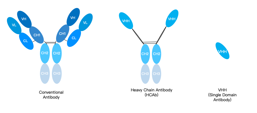
KMD Bioscience boasts an advanced antibody expression and validation platform, delivering efficient and stable antibody production and functional validation services to our clients. With a team of experts possessing profound technical expertise and extensive industry experience, we provide customized research solutions tailored to diverse project requirements. Our mature recombinant antibody preparation technology utilizes phage display, yeast display, and other systems to produce high-quality scFv, Fab, VHH, and other recombinant antibodies, ensuring exceptional specificity and binding affinity.
For antibody expression, we employ mammalian cell systems such as HEK293 and CHO for transient antibody gene expression and stable cell line development, guaranteeing high-yield production and superior purity in large-scale manufacturing. Our antibody functional validation services encompass a wide range of assays, including: Western Blot (WB); Enzyme-Linked Immunosorbent Assay (ELISA); Flow Cytometry (FC); Chromatin Immunoprecipitation (ChIP); Immunofluorescence (IF); Immunohistochemistry (IHC). These techniques ensure the efficacy, stability, and bioactivity of antibodies in their intended applications.
Explore Our Validation Capabilities
For specialized therapeutic programs investigating targeted delivery, ensuring your antibodies are thoroughly validated before conjugation via our Protein Interaction Services or integration with Nanoparticle Products is essential to preserve the integrity of the conjugate.
Frequently Asked Questions
Why is ELISA not sufficient for complete antibody characterization?
While ELISA is excellent for determining basic binding and titer, it occurs in an artificial environment where the antigen is adhered to a plastic plate, often altering its native structure. It does not provide real-time kinetic data (koff/kon rates) and cannot confirm if the antibody will bind the target on a live cell surface or in complex tissue structures.
What is the best method to determine precise antibody affinity?
In most professional situations, Surface Plasmon Resonance (SPR) or Biolayer Interferometry (BLI) are the gold standards for determining precise binding affinity. These methods provide label-free, real-time kinetic data, allowing researchers to calculate the exact Equilibrium Dissociation Constant (KD).
Why do some antibodies work in Western Blot but fail in Flow Cytometry?
Western Blots require the antibody to recognize denatured, linear peptide sequences. Flow Cytometry requires the antibody to recognize complex, 3D conformational epitopes on native cell surfaces. If an antibody recognizes a hidden (cryptic) linear sequence, it will work in WB but fail in FC because that sequence is buried inside the folded protein on a live cell.
What is the role of knockout cell lines in antibody validation?
CRISPR/Cas9 knockout cell lines act as the ultimate negative control. If you test your antibody on a wild-type cell line and observe a signal, and then test it on a knockout line (where the target protein is entirely absent) and the signal disappears, you have definitively proven the antibody's on-target specificity.
Industry References & Authoritative Guidelines
To ensure our validation protocols meet the highest standards of scientific integrity, KMD Bioscience aligns with the criteria established by the following authoritative organizations:
National Institutes of Health (NIH): Guidelines on enhancing reproducibility through rigorous authentication of key biological and chemical resources, specifically addressing antibody validation. Review NIH Reproducibility Guidelines
The International Working Group for Antibody Validation (IWGAV): Proposed scoring systems and conceptual pillars for validating antibodies for use in specific biomedical applications. Review IWGAV Standards in Nature Methods
The Human Protein Atlas (HPA): Extensive open-access data demonstrating the application of enhanced validation strategies across thousands of antibodies utilizing orthogonal and genetic validation methods. Review HPA Validation Protocols

- Protein Platform
- Protein Expression Services
- Protein Modification & Detection Services
- Protein Interaction Services
- Protein Purification Service
- Antibody Discovery Platform
- VHH Antibody Platform
- Phage Display Platform
- Antibody Humanization Platform
- Single B Cell Screening Platform
- Antibody Expression & Validation Platform
- Peptide Library Platform
- Custom Antibody Platform
- Innovative Drug Discovery Platform
- Product Center
- Proteins
- Antibodies
- Raw Materials Reagents
- Small Molecule Reagents
- Peptide Products
- Nanoparticle Products
- Contact Us
- Phone:400-621-6806
- Mobile:13110936064(WeC
- E-mail:[email protected]
- Address:Room 108, Building 10, No. 6 Hechang Road, Wuqing Development Zone, Tianjin City
-

WeChat
-

Service Account
-
 400-621-6806
 -
 3599163699
 -
 Monday to Friday 9:00-18:00
 -

-
 0
0
-
