


Example of a Bacteriophage Display Diagram 2026: Professional Analysis and Guide
- 2026-07-22
- 94
In the highly competitive landscape of recombinant antibody development, relying on outdated discovery methodologies is a fast track to commercial failure. When clients approach us looking to develop novel therapeutics or highly specific diagnostic reagents, they often ask about the underlying technology driving our rapid turnaround times. The answer inevitably points to one foundational concept: the physical linkage of genotype to phenotype. To truly grasp this, you must understand the architecture illustrated in a standard bacteriophage display diagram.
From our experience, many researchers understand the broad strokes of in vitro selection, but they fail to appreciate the critical nuances that make or break a biopanning campaign. Looking at a bacteriophage display diagram is one thing; executing the methodology flawlessly in a high-throughput laboratory environment is entirely different. At KMD Bioscience, our extensive experience in recombinant antibody development is supported by a robust technical system that provides a distinct edge within the industry. We deliver premium scientific services, and we have achieved outstanding results in preparing scFv, Fab, and VHH antibodies, earning the trust of clients globally.
This guide will dissect an example of a bacteriophage display diagram, explaining not only what the technology is and how it works, but whether your specific project actually warrants utilizing it over traditional hybridoma or B cell cloning techniques. We prioritize helping you make informed, practical decisions for your pipeline.
Quick Answer: Decoding the Diagram
A bacteriophage display diagram visually maps the core mechanism of phage display technology: the direct, physical link between a functional protein displayed on the outside of a virus (the phenotype) and the genetic blueprint encoding that protein housed inside the virus (the genotype).
By inserting foreign DNA into the phage genome, the resulting viral particle expresses the corresponding peptide or antibody fragment on its coat protein. This allows researchers to screen libraries containing billions of variants against target antigens, isolate the highest affinity binders, and immediately possess the DNA required to mass-produce that antibody.

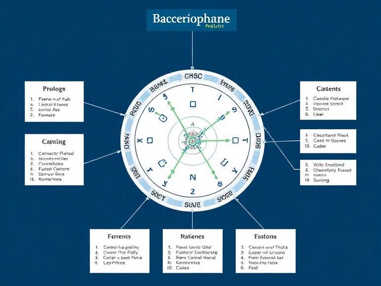
Figure 1: Example of a bacteriophage display diagram illustrating the expression of recombinant antibodies on the M13 phage surface.
Table of Contents
What is a Bacteriophage Display Diagram?
When you examine a well-constructed bacteriophage display diagram, you are looking at the ultimate biological screening tool. The diagram typically features a filamentous phage (most commonly M13, fd, or f1). Inside the tubular structure lies the single-stranded DNA. On the surface, you will see various coat proteins (pIII, pVIII, pVI, pVII, pIX).
The magic happens at the genetic level. By splicing exogenous DNA sequences into the gene encoding one of these coat proteins—usually the pIII minor coat protein at the tip of the phage—the virus is forced to display the foreign protein as a fusion product. In most professional situations dealing with antibody discovery, the displayed protein is a single-chain variable fragment (scFv), a fragment antigen-binding (Fab), or a single-domain antibody (VHH). The bacteriophage display diagram perfectly illustrates how billion-member libraries can be physically screened, because isolating the winning phenotype automatically isolates the winning genotype.
How it Works: The Biopanning Process
A static bacteriophage display diagram only tells half the story. The true value of the technology lies in the iterative screening process known as biopanning. Utilizing phage display antibody library construction followed by multiple rounds of ELISA screening, we efficiently identify high-affinity antibodies for clients.
The biopanning process consists of four critical steps:
Binding: The diverse phage library is incubated with the immobilized target antigen.
Washing: Non-binding and weak-binding phages are washed away. This requires immense precision; overly aggressive washing loses rare clones, while weak washing yields false positives.
Elution: The strong binders are stripped from the antigen using pH changes or competitive elution strategies.
Amplification: The eluted phages are used to infect a host, typically an E. coli protein expression system, to amplify the winning clones for the next round.
| Component | Function in the System | Professional Consideration |
|---|---|---|
| Phage Vector (e.g., M13) | Acts as the vehicle displaying the phenotype and housing the genotype. | Requires optimized helper phages to ensure stable valency and display efficiency. |
| Target Antigen | The molecule you want the antibody to bind to. | Must be correctly folded and properly immobilized without masking epitopes. |
| Host Bacteria | Used to amplify the recovered phages between panning rounds. | Using a highly competent E. coli strain is non-negotiable for library maintenance. |
| Antibody Library | The diverse pool of 10^9 to 10^11 unique variants. | Size matters, but diversity and quality of the initial repertoire are paramount. |
Benefits of Phage Display Systems
Why do commercial pharmaceutical entities and top-tier biotech labs rely so heavily on this technology? The benefits are overwhelming when compared to traditional in vivo immunization methods.
Firstly, it is entirely in vitro. Because you bypass the animal immune system, you can develop antibodies against highly toxic antigens, non-immunogenic molecules, and highly conserved human proteins that would otherwise trigger self-tolerance mechanisms in a host animal. Secondly, the speed is unparalleled. While hybridoma generation takes months, a skilled team can execute a biopanning campaign and isolate high-affinity binders in a matter of weeks.
Furthermore, because you possess the DNA sequence immediately upon isolation, transitioning into downstream protein expression platform services is seamless. You can easily engineer the sequence to improve affinity or reformat an scFv into a full-length IgG.
Limitations and Common Mistakes
Despite its power, a bacteriophage display diagram does not show the immense technical challenges of the methodology. In our testing, the most severe bottleneck is library quality. If your initial library lacks diversity, no amount of sophisticated biopanning will yield a world-class antibody.
| Pros (Why it Excels) | Cons (Where it Struggles) |
|---|---|
| Bypasses animal immunization tolerance mechanisms. | Requires highly specialized molecular biology expertise. |
| Generates fully human antibodies out of the gate. | Loss of natural heavy/light chain pairing (in synthetic libraries). |
| Incredibly fast timelines (weeks vs. months). | Some proteins fail to express well on the E. coli/phage membrane. |
| Direct access to the antibody's genetic sequence. | High risk of background binding (false positives) if washing is poor. |
Common Mistakes: From our experience, amateur laboratories routinely over-wash their plates in round one, destroying the rare, high-affinity clones before they can be amplified. Additionally, failing to subtract the library against the immobilization matrix (e.g., streptavidin beads or BSA) results in isolating phages that bind to the plastic or the beads rather than the actual antigen. We strongly advise conducting rigorous negative selection steps.
Who Should Use It (And Who Does Not Need It)
For commercial users and therapeutics developers: If you are developing a targeted biologic, an antibody-drug conjugate (ADC), or a CAR-T cell therapy, you absolutely should use a Phage Display Platform. The ability to screen massive human synthetic or naive libraries allows you to bypass the need for an Antibody Humanization Platform later in development, saving vast amounts of time and capital.
Who does not need it: For beginners in basic academic research who simply need a standard reagent to detect a common protein on a Western blot, engineering a custom recombinant antibody via phage display is severe overkill. In these scenarios, simply purchasing an off-the-shelf product from a reputable primary antibody supplier makes much more financial and practical sense.
Comparing Antibody Discovery Platforms
To make a concrete buying decision, you must compare phage display against competing technologies. While the bacteriophage display diagram highlights the elegance of viral screening, how does it stack up against physical B cell isolation?
| Feature | Phage Display | Hybridoma Technology | Single B Cell Screening |
|---|---|---|---|
| Methodology | In vitro panning of genetic libraries | In vivo immunization & cell fusion | Direct isolation of memory B cells |
| Timeline | 3 to 6 weeks | 4 to 6 months | 2 to 4 weeks |
| Natural Pairing | No (unless immune library used) | Yes (native VH/VL pairing) | Yes (perfect native pairing) |
| Best Use Case | Toxic antigens, human therapeutics | Standard diagnostic monoclonals | Rapid isolation of infectious disease neutralizing antibodies |
For heavy-duty applications requiring native antibody pairing and the highest possible affinity straight from a host, utilizing a Single B Cell Screening Platform is often the premium choice. However, phage display remains the undisputed king for sheer library size and the ability to engineer synthetic, highly specific binding characteristics.
Buying Considerations for Antibody Services
If you are looking to outsource your antibody discovery, do not simply accept a generic bacteriophage display diagram in a sales pitch. You must scrutinize the Contract Research Organization's (CRO) underlying technical metrics.
| Evaluation Criteria | What to Demand from a CRO | Why it is Critical |
|---|---|---|
| Library Size and Diversity | Minimum 10^9 independent clones. | Small libraries severely limit the probability of finding a high-affinity binder (KD in the nanomolar/picomolar range). |
| Validation Capabilities | In-house SPR, BLI, and flow cytometry. | A binder on an ELISA plate is meaningless if it lacks the actual kinetic affinity required for your specific end-use application. |
| Format Flexibility | Ability to output scFv, Fab, or VHH. | Different assays require different formats. VHH (nanobodies) are ideal for penetrating dense tissues. |
| Intellectual Property | Full transfer of sequences and rights. | For commercial users, you must own the sequence of the antibody you paid to discover, free of downstream royalties. |
Expert Recommendation and Final Verdict
In most professional situations involving 2026 commercial drug discovery, we recommend adopting phage display as a primary engine for pipeline development. The ability to visualize the genotype-phenotype link via a bacteriophage display diagram translates into real-world, high-throughput discovery power. It is absolutely worth the investment for companies pursuing novel therapeutics, CAR-T binders, and highly specific diagnostics.
Our Verdict: Do not compromise on your discovery platform. KMD Bioscience conducts diverse antibody screening assays tailored to specific client requirements to generate highly specific and stable antibodies. Whether you need a massive naive human library screened or require an immunized alpaca VHH library constructed, our robust technical system ensures you receive the exact sequences and validated proteins you need to advance your research to the clinic.
Frequently Asked Questions
What exactly does a bacteriophage display diagram show?
A bacteriophage display diagram illustrates the genotype-phenotype linkage of a phage. It shows the exogenous protein (phenotype) displayed on the outer coat of the virus, directly linked to the engineered DNA (genotype) encapsulated inside the viral particle.
Is investing in phage display technology worth it in 2026?
Yes. For commercial users and therapeutic developers, it is highly worth it. Unlike hybridoma technology, phage display allows for the generation of fully human antibodies, targets toxic and non-immunogenic antigens, and compresses development timelines from months to mere weeks.
What are the most common fragments shown in a bacteriophage display diagram?
The most common antibody fragments illustrated in a bacteriophage display diagram are scFv (single-chain variable fragments), Fab (fragment antigen-binding), and VHH (single-domain antibodies or nanobodies).
Authoritative References
To ensure our clients have access to the highest standards of scientific methodology and molecular biology principles, we reference the following industry and academic authorities:
National Center for Biotechnology Information (NCBI) / PubMed: For peer-reviewed literature on advanced biopanning protocols and recombinant antibody generation methodologies.
Nature Methods: The premier academic journal detailing breakthrough techniques in molecular display technologies, library construction, and high-throughput screening.
U.S. Food and Drug Administration (FDA): Providing regulatory standards and clinical approvals for therapeutic biologics derived from in vitro display technologies.

- Protein Platform
- Protein Expression Services
- Protein Modification & Detection Services
- Protein Interaction Services
- Protein Purification Service
- Antibody Discovery Platform
- VHH Antibody Platform
- Phage Display Platform
- Antibody Humanization Platform
- Single B Cell Screening Platform
- Antibody Expression & Validation Platform
- Peptide Library Platform
- Custom Antibody Platform
- Innovative Drug Discovery Platform
- Product Center
- Proteins
- Antibodies
- Raw Materials Reagents
- Small Molecule Reagents
- Peptide Products
- Nanoparticle Products
- Contact Us
- Phone:400-621-6806
- Mobile:13110936064(WeC
- E-mail:[email protected]
- Address:Room 108, Building 10, No. 6 Hechang Road, Wuqing Development Zone, Tianjin City
-

WeChat
-

Service Account
-
 400-621-6806
 -
 3599163699
 -
 Monday to Friday 9:00-18:00
 -

-
 0
0
-
