


How Does Phage Display Help Discover New Therapeutic Agents?
- 2026-05-20
- 141
The landscape of modern medicine has transitioned from accidental small-molecule breakthroughs to deliberate macromolecular engineering. As global healthcare systems face complex oncological mutations, autoimmune multi-pathways, and emerging viral threats, traditional drug screening methods often prove insufficient. To achieve localized, high-affinity targeting without triggering systemic toxicity, biopharmaceutical research depends on robust molecular display platforms. Among these, phage display technology stands as an essential foundation for modern drug discovery, linking a protein phenotype directly to its underlying genotype.
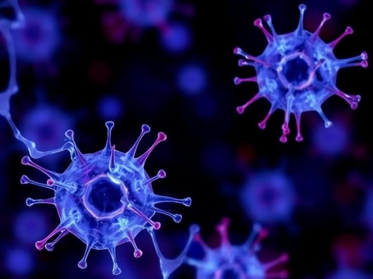
At KMD Bioscience, established in 2022, we operate as a high-quality Contract Research Organization (CRO) delivering advanced therapeutic antibody discovery and related support services to scientists and research institutions worldwide. From our experience, many research pipelines suffer from unnecessary delays because they rely heavily on traditional animal hybridoma workflows, which frequently struggle with highly conserved, non-immunogenic, or cytotoxic target antigens. Implementing a standardized, high-diversity display platform bypasses biological limitations, providing rapid access to fully human leads and specialized antibody formats. This comprehensive analysis details exactly how phage display help discover new therapeutic agents, breaking down biopanning kinetics, library architectures, and multi-format downstream integrations.
Table of Contents
2. Genotype-Phenotype Linkage: The Core Mechanism of Phage Display
4. Therapeutic Agent Modalities Discovered via Phage Display
5. Downstream Execution: Affinity Maturation and Reagent Quality Control
1. Summary Matrix: Biologics Discovery Platforms Compared
Before deep-diving into structural molecular engineering, it is helpful to compare phage display against alternative discovery approaches. The table below outlines how specific experimental parameters shape lead generation outcomes.
| Discovery Platform | Library Diversity Capacity | Animal Immunization Required? | Screening Environment Control | Primary Limitations / Challenges |
|---|---|---|---|---|
| Phage Display Platform | High (10^9 to 10^13 unique variants) | No (完全 in vitro process for naïve/synthetic) | Excellent (Custom pH, salt, and temperature controls) | Requires robust expression hosts; size limits for large proteins |
| Mouse Hybridoma | Limited by host immune response | Yes (Requires host animal stimulation) | Poor (Governed by host biological tolerance) | Humanization required; fails on toxic or highly conserved targets |
| Ribosome / mRNA Display | Ultra-High (10^12 to 10^14 unique variants) | No (Cell-free transcription/translation) | Good (Sensitive to RNA degradation forces) | Complex laboratory handling; unstable cell-free matrix parameters |
| Transgenic Animal Hubs | In vivo repertoire generation | Yes (Demands long immunization timelines) | Moderate (Relies on natural immune tolerance loops) | High cost barrier; restricted commercial licensing access |
2. Genotype-Phenotype Linkage: The Core Mechanism of Phage Display
To understand how phage display help discover new therapeutic agents, one must examine the molecular biology of the filamentous bacteriophage, typically the M13 system. The true breakthrough of this technology, pioneered in 1985 by George P. Smith, lies in the physical connection between phenotype and genotype. The structural gene encoding a specific protein or antibody fragment is cloned into a phagemid or phage vector, fused directly to the sequence of a native coat protein—most commonly the minor coat protein pIII or the major coat protein pVIII.
When the modified phage replicates inside an Escherichia coli host cell, it expresses the foreign peptide or antibody fragment on its outer surface while encapsulating the matching single-stranded DNA sequence safely inside its viral core. From our experience, this dual profile provides an invaluable advantage for high-throughput screening. Because the binding molecule is physically tied to its genetic blueprint, isolating a single target-binding phage allows researchers to instantly sequence the underlying DNA, revealing the exact primary amino acid structure responsible for the therapeutic interaction.
From Our Experience: Scaffolding Integrity Controls Failures Upfront
We recommend paying close attention to framework design during initial library construction. Many low-grade synthetic libraries exhibit excellent theoretical diversity scores but fail in practice because they contain structural liabilities, such as unpaired cysteines or hidden N-glycosylation motifs. Utilizing drug-ready, pre-validated human frameworks with naturally observed CDR patterns ensures that discovered leads possess high thermal stability and lowered immunogenicity right out of the gate.
3. The Biopanning Cycle: Streamlining Lead Enrichment
The core process used to isolate therapeutic leads from a diverse molecular library is an iterative selection protocol known as biopanning. This process effectively replicates natural selection in a highly controlled, test-tube environment, compressing months of natural immune maturation into a few days.
A typical high-throughput biopanning workflow consists of four well-defined mechanical steps:
Antigen Immobilization & Incubation: The target disease antigen—such as a purified tumor marker, viral spike protein, or extracellular receptor dome—is coated onto a solid surface, such as a microtiter plate well or magnetic beads. The high-diversity phage library is then introduced and allowed to incubate, enabling specific clones to bind.
Stringent Washing: Non-binding or weakly attached phages are systematically washed away using customized buffer solutions containing varying concentrations of detergents like Tween-20. This removes low-affinity variants and non-specific background binders from the pool.
Elution of Positive Clones: The remaining high-affinity bound phages are released from the target antigen using controlled biochemical shifts, such as an acidic pH drop (using glycine-HCl) or competitive ligand displacement.
Bacterial Amplification: The eluted phages are introduced into fresh E. coli cultures. The bacteria amplify the isolated clones exponentially, preparing a refined, enriched pool for the next selection cycle.
By repeating this cycle 3 to 5 times under increasingly stringent conditions, the background noise is eliminated, allowing researchers to isolate rare, high-affinity binders that might only represent one in ten billion initial variants. KMD Bioscience leverages this mechanism through our proprietary Phage Display Platform, which streamlines selection timelines and delivers high-affinity candidates to challenging targets in as few as 6 to 8 weeks.
4. Therapeutic Agent Modalities Discovered via Phage Display
The versatility of direct surface presentation allows scientists to screen for multiple distinct classes of therapeutic agents simultaneously, making the technology a valuable multi-tool for biopharmaceutical drug pipelines.
Fully Human Monoclonal Antibodies (mAbs)
Historically, animal-derived antibodies required extensive chemical humanization to prevent severe anti-drug antibody (ADA) responses in clinical patients. Phage display completely altered this landscape by enabling the construction of massive naïve human libraries derived directly from the B-cell repertoires of healthy donors. Discovered fragments—commonly formatted as single-chain variable fragments (scFv) or Fragment antigen-binding (Fab) modules—are 100% human from inception, accelerating regulatory transition pathways. Landmark therapeutics like Adalimumab (Humira) demonstrate the immense clinical value of display-derived fully human therapies.
Single-Domain Antibodies and Nanobodies (VHH)
Beyond traditional multi-chain immunoglobulins, the modern therapeutic pipeline focuses heavily on single-domain architectures derived from Camelidae species. These miniaturized binders, known as VHH antibodies or nanobodies, offer exceptional physical stability, strong tissue penetration, and the unique ability to target hidden enzyme clefts or complex ion channels. Operating our dedicated VHH Antibody Platform allows us to construct high-diversity immune or synthetic camelid libraries, delivering stable scaffolds optimized for multi-specific targeting or advanced CAR-T applications.
Specialized Peptide and Small-Molecule Discoveries
Phage display is not restricted to full antibody formats. By inserting randomized oligonucleotides into the vector, researchers can construct diverse peptide libraries. These short amino acid strings can act as effective targeting keys, guiding diagnostic payloads or drug-loaded nanoparticles to localized disease sites. To browse matching chemical inputs for targeted conjugate assemblies, you can review our catalog of Small Molecule Reagents and related organic components.
5. Downstream Execution: Affinity Maturation and Reagent Quality Control
Isolating an initial binding hit is only the first step in creating a viable drug candidate. To ensure successful therapeutic performance, discovered binders often undergo directed in vitro evolution to maximize their target affinity and operational stability.
Through error-prone PCR or targeted sub-library randomization, researchers can introduce minor amino acid mutations into the complementarity-determining regions (CDRs) of an isolated clone. Displaying this mutated family on the phage surface and performing a high-stringency biopanning screen allows for the selection of optimized variants featuring increased target affinity, a process known as affinity maturation.
| Therapeutic Component Category | Downstream Optimization Pathway | Primary Quality Metric Target | KMD Bioscience Resource Link |
|---|---|---|---|
| Recombinant Peptides | Synthetic sequence alignment and solid-phase synthesis | High purity, precise molecular mass verification | Peptide Products |
| Targeted Delivery Matrix | Surface conjugation and lipid encapsulation mapping | Uniform diameter, stable dispersion coefficients | Nanoparticle Products |
| Bulk Discovery Reagents | High-yield mammalian transient expression profiling | Low endotoxin, structural binding validation | Raw Materials Reagents |
Strict Validation and Analytical Certification
Every therapeutic candidate or support reagent introduced into a clinical discovery program must meet strict purity and identity criteria. Ensuring absolute batch uniformity prevents experimental anomalies from disrupting downstream workflows. We recommend that principal investigators routinely verify their material lots by utilizing our direct query portal to access certified COA Download records, guaranteeing that all raw materials match established biological and chromatographic benchmarks before starting cell assays.
6. Frequently Asked Questions (FAQs)
Why is the genotype-phenotype link so important in phage display drug discovery?
The genotype-phenotype link means that each bacteriophage presents a unique therapeutic protein on its outer surface while carrying the matching DNA sequence inside its core. When a rare binder is isolated during biopanning, researchers can instantly sequence this internal DNA, revealing the exact primary amino acid sequence needed to reproduce and manufacture the therapeutic agent.
Can phage display identify therapeutic agents against toxic or highly conserved antigens?
Yes. Because phage display is an entirely in vitro (test-tube) process, it bypasses the biological constraints of host animal immune systems. This allows researchers to successfully isolate high-affinity binders against antigens that are highly conserved across species or too toxic to be safely used in animal immunization workflows.
What is the difference between naïve and immune libraries in phage display?
Naïve libraries are constructed from the B-cell repertoires of non-immunized human donors, providing an unbiased collection of billions of unique variants suitable for targeting any generic disease. Immune libraries are sourced from individuals or animals that have been exposed to a specific antigen, yielding a pre-enriched pool of highly specialized binders focused on that target.
What does the term biopanning mean in molecular biology?
Biopanning is an iterative affinity-selection cycle used to isolate target-binding phages from a diverse library. It consists of four primary steps: incubating the library with an immobilized target antigen, washing away non-binding variants, eluting the high-affinity bound phages, and amplifying those positive clones inside bacterial hosts for subsequent screening rounds.
How are single-domain VHH nanobodies advantageous compared to standard monoclonal antibodies?
Single-domain VHH nanobodies, derived from camelid heavy-chain antibodies, represent a miniaturized antibody format consisting of a single variable domain. Due to their small physical footprint, they offer exceptional thermal stability, deep tissue penetration, and the unique capability to bind hidden or conformationally restricted disease epitopes that large, standard antibodies cannot access.
7. Academic and Regulatory References
For more detailed technical data, protocols, and clinical safety studies regarding bacteriophage display systems, consult these authoritative international resources:
National Center for Biotechnology Information (NCBI). The Role of Phage Display in Therapeutic Antibody Discovery and Biologics Engineering. National Institutes of Health.
The Nobel Prize in Chemistry. Pioneering Work on the Phage Display of Peptides and Antibodies. Royal Swedish Academy of Sciences (2018).
International Organization for Standardization (ISO). ISO/TC 276 Biotechnology – Analytical Standards and Bioprocessing Quality Infrastructure Frameworks.
- Phage Display Help Discover New Therapeutic Agents

- Protein Platform
- Protein Expression Services
- Protein Modification & Detection Services
- Protein Interaction Services
- Protein Purification Service
- Antibody Discovery Platform
- VHH Antibody Platform
- Phage Display Platform
- Antibody Humanization Platform
- Single B Cell Screening Platform
- Antibody Expression & Validation Platform
- Peptide Library Platform
- Custom Antibody Platform
- Innovative Drug Discovery Platform
- Product Center
- Proteins
- Antibodies
- Raw Materials Reagents
- Small Molecule Reagents
- Peptide Products
- Nanoparticle Products
- Contact Us
- Phone:400-621-6806
- Mobile:13110936064(WeC
- E-mail:[email protected]
- Address:Room 108, Building 10, No. 6 Hechang Road, Wuqing Development Zone, Tianjin City
-

WeChat
-

Service Account
-
 400-621-6806
 -
 3599163699
 -
 Monday to Friday 9:00-18:00
 -

-
 0
0
-
