


Authoritative Guide to the Phage Display Screening Procedure in Antibody Discovery
- 2026-05-19
- 192
Tianjin KMD Bioscience Co., Ltd. has been committed to becoming a leading provider of therapeutic antibody discovery and related support services since its establishment in 2022. Focusing on technological research and development, the company provides high-quality contract research organization CRO services to scientists and research institutions worldwide, aiming to promote the development and innovation of medical science and technology. As a premier biotech research service provider, we recognize that mastering core methodologies is essential for accelerating drug discovery. Among these methodologies, the Phage display screening procedure stands out as one of the most robust and versatile tools available to modern molecular biologists and immunologists.
Table of Contents
1. Introduction to the Phage Display Screening Procedure
The Phage display screening procedure is a high-throughput laboratory technique utilizing bacteriophages to connect proteins with the genetic information that encodes them. By inserting a gene coding for a protein of interest into a phage coat protein gene, the target protein is displayed on the outside of the virion, while the genetic material resides inside. This powerful linkage allows researchers to screen vast libraries of proteins, peptides, or antibodies to identify those with high affinity and specificity for a given target molecule.
In the highly competitive landscape of therapeutic antibody discovery, the Phage display screening procedure is indispensable. It bypasses the limitations of traditional hybridoma technology, allowing for the rapid isolation of fully human antibodies, synthetic antibodies, and specialized fragments such as single-chain variable fragments (scFv) and variable domains of heavy chain antibodies (VHH or nanobodies). As a comprehensive provider of life science research solutions, our laboratory has executed thousands of biopanning cycles, refining our approach to maximize recovery and specificity.
2. The Prerequisite: High-Quality Library Construction
Before initiating the actual Phage display screening procedure, an immune, naive, or synthetic library must be constructed. The success of the entire Phage display screening procedure is inherently limited by the diversity and quality of this starting library. Library sizes typically range from 10^8 to over 10^11 independent transformants. A larger, more diverse library increases the statistical probability of identifying high-affinity binders.
From our experience, meticulous attention must be paid during the RNA extraction, cDNA synthesis, and PCR amplification stages to prevent the introduction of bias. When dealing with specialized molecules like nanobodies, customized primer designs are required to accurately capture the entire repertoire of VHH genes. The amplified fragments are then ligated into phagemid vectors and transformed into specialized E. coli strains, such as TG1 or SS320. Following transformation, helper phages (such as M13KO7 or VCSM13) are introduced to facilitate the packaging of the phagemid DNA and the extrusion of fully assembled phage particles displaying the target protein.
3. Core Steps of the Phage Display Screening Procedure
The Phage display screening procedure operates through an iterative process commonly referred to as biopanning. This cycle is typically repeated three to five times to enrich the phage pool for binders with the highest affinity. Below is a detailed breakdown of the Phage display screening procedure.
3.1 Antigen Immobilization
The initial step in the Phage display screening procedure involves presenting the target antigen. Antigens can be immobilized on various solid supports, including immunotubes, microtiter plates, or magnetic beads. From our experience, utilizing streptavidin-coated magnetic beads combined with biotinylated antigens often yields superior results, as it maintains the antigen's native conformation and allows for in-solution biopanning, thereby reducing steric hindrance.
3.2 Binding
Once the antigen is prepared, the prepared phage library is incubated with the target. During this phase of the Phage display screening procedure, phages displaying proteins with an affinity for the antigen will bind, while non-binding phages remain in suspension. We recommend incorporating blocking agents, such as BSA or skim milk, to minimize non-specific interactions between the phages and the solid support.
3.3 Washing
Washing is arguably the most critical step in the Phage display screening procedure. The goal is to remove weak binders and non-specific phages while retaining high-affinity candidates. As the biopanning rounds progress, the washing stringency must be systematically increased. This is achieved by raising the concentration of detergents (like Tween-20 in PBS), increasing the number of wash cycles, or extending the wash duration.
3.4 Elution
Bound phages must be decoupled from the antigen without destroying their infectivity. Standard elution techniques in the Phage display screening procedure utilize acidic buffers (e.g., Glycine-HCl at pH 2.2) followed by immediate neutralization with Tris-HCl. Alternatively, competitive elution utilizing an excess of soluble target antigen or specific proteases can be employed to selectively recover phages that bind to desired epitopes.
3.5 Amplification
The eluted phages represent an enriched, yet extremely small, sub-population of the original library. To proceed to the next round of the Phage display screening procedure, these phages must be amplified. They are used to infect exponentially growing E. coli (typically TG1). The bacteria are superinfected with helper phages, leading to the production of a new, enriched phage library ready for the subsequent round of panning.
4. Optimization Strategies and Expert Recommendations
From our experience, one of the most common pitfalls in the Phage display screening procedure is over-panning, which can lead to the dominant selection of fast-growing phages (parasitic phages) rather than genuine high-affinity binders.
To optimize the Phage display screening procedure, scientists must carefully monitor the input and output phage titers at every round. A successful Phage display screening procedure will demonstrate a significant enrichment factor—the ratio of output phages to input phages should exponentially increase in later rounds.
We recommend employing depletion strategies, also known as negative selection, prior to the binding phase. By incubating the phage library with an irrelevant protein or the empty solid support, non-specific binders are effectively removed from the pool. Additionally, if cross-reactivity is desired (e.g., finding an antibody that binds both human and murine homologues), alternating the target antigen between rounds within the Phage display screening procedure is a highly effective strategy.
5. Applications in Nanobody Discovery
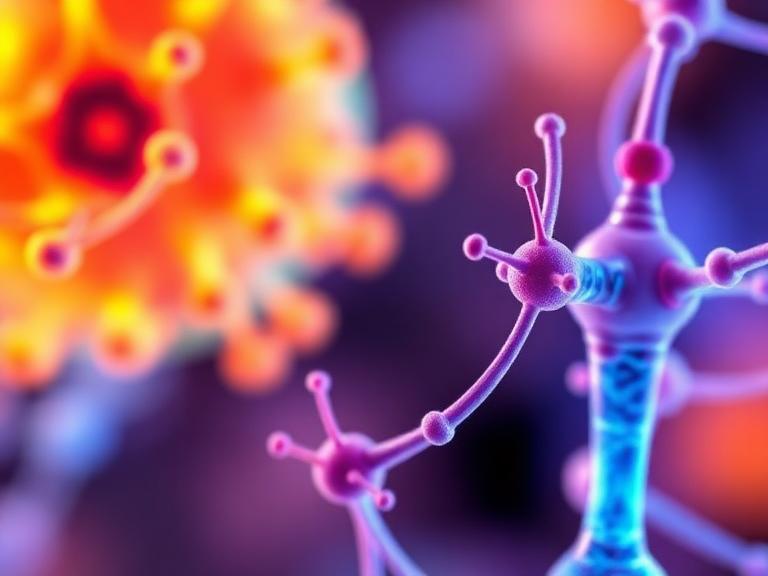
The Phage display screening procedure has revolutionized the discovery of VHH antibodies, commonly known as nanobodies. Derived from the heavy-chain-only antibodies found in camelids, nanobodies offer exceptional stability, deep tissue penetration, and the ability to access cryptic epitopes inaccessible to traditional monoclonal antibodies.
At KMD Bioscience, our specialized nanobody library construction service integrates seamlessly with our advanced Phage display screening procedure. Following the immunization of alpacas or llamas, we isolate peripheral blood mononuclear cells (PBMCs) and amplify the VHH repertoire. Through rigorous biopanning, we isolate nanobodies with picomolar affinities. For researchers seeking off-the-shelf solutions, our catalog of nanobody antibody products provides thoroughly validated reagents discovered utilizing this exact Phage display screening procedure.
6. Summary Table: The Phage Display Screening Procedure
| Procedure Phase | Objective | Key Reagents & Equipment | Expert Recommendation |
|---|---|---|---|
| Antigen Immobilization | Present the target in a native-like conformation. | Immunotubes, Magnetic Beads, Biotinylated Antigen | Utilize in-solution biopanning with magnetic beads to reduce steric hindrance and preserve epitopes. |
| Library Binding | Allow specific phages to recognize and attach to the target. | Phage Library, Blocking Buffer (BSA/Milk), PBS | Perform negative selection (depletion) prior to positive binding to eliminate background noise. |
| Stringent Washing | Remove non-specific and low-affinity background phages. | PBST (PBS + Tween-20) | Gradually increase Tween-20 concentration (0.05% to 0.5%) across successive panning rounds. |
| Target Elution | Recover high-affinity phages from the antigen. | Glycine-HCl (pH 2.2), Trypsin, or Soluble Antigen | Use competitive elution if you require antibodies specific to a known binding pocket. |
| Bacterial Amplification | Multiply the enriched phage pool for the next round. | TG1 E. coli, Helper Phage (M13KO7), 2xYT Media | Strictly monitor the input/output ratio (titration) to quantify the enrichment factor. |
7. Frequently Asked Questions (FAQs)
What is the optimal number of rounds for the Phage display screening procedure?
Typically, 3 to 4 rounds of biopanning are sufficient. Proceeding beyond 5 rounds often leads to the amplification of non-specific background phages or clones with growth advantages rather than superior affinity.
How do you prevent the loss of sequence diversity during the Phage display screening procedure?
To maintain diversity, we recommend maintaining high phage titers throughout the process and avoiding excessive washing stringency in the very first round. The first round should aim to capture as many diverse binders as possible, while subsequent rounds apply selective pressure.
Can the Phage display screening procedure be used for membrane proteins?
Yes, but membrane proteins are notoriously difficult to maintain in their native conformation outside a lipid bilayer. From our experience, utilizing whole-cell biopanning or incorporating the membrane proteins into virus-like particles (VLPs) or nanodiscs yields the best results during the Phage display screening procedure.
Why is E. coli TG1 the preferred strain for amplification?
TG1 is an amber suppressor strain (supE). This allows for the read-through of the amber stop codon located between the inserted antibody gene and the gene III coat protein in many phagemid vectors, ensuring that the target protein is actively displayed on the phage surface.
8. References
Smith, G. P. (1985). Filamentous fusion phage: novel expression vectors that display cloned antigens on the virion surface. Science, 228(4705), 1315-1317.
McCafferty, J., Griffiths, A. D., Winter, G., & Chiswell, D. J. (1990). Phage antibodies: filamentous phage displaying antibody variable domains. Nature, 348(6301), 552-554.
Bradbury, A. R., Sidhu, S., Dübel, S., & Franks, J. (2011). Beyond natural antibodies: the power of in vitro display technologies. Nature Biotechnology, 29(3), 245-254.
Hoogenboom, H. R. (2005). Selecting and screening recombinant antibody libraries. Nature Biotechnology, 23(9), 1105-1116.

- Protein Platform
- Protein Expression Services
- Protein Modification & Detection Services
- Protein Interaction Services
- Protein Purification Service
- Antibody Discovery Platform
- VHH Antibody Platform
- Phage Display Platform
- Antibody Humanization Platform
- Single B Cell Screening Platform
- Antibody Expression & Validation Platform
- Peptide Library Platform
- Custom Antibody Platform
- Innovative Drug Discovery Platform
- Product Center
- Proteins
- Antibodies
- Raw Materials Reagents
- Small Molecule Reagents
- Peptide Products
- Nanoparticle Products
- Contact Us
- Phone:400-621-6806
- Mobile:13110936064(WeC
- E-mail:[email protected]
- Address:Room 108, Building 10, No. 6 Hechang Road, Wuqing Development Zone, Tianjin City
-

WeChat
-

Service Account
-
 400-621-6806
 -
 3599163699
 -
 Monday to Friday 9:00-18:00
 -

-
 0
0
-
