


How Does Phage Display Work? A Comprehensive Scientific Guide by KMD Bioscience
- 2026-05-06
- 136
In the rapidly advancing field of biopharmaceuticals and antibody engineering, few technologies have been as transformative as phage display. First described in 1985 by George Smith, this elegant technique has revolutionized how we discover and engineer antibodies, peptides, and proteins. But for many researchers entering the field of therapeutic discovery, a fundamental question remains: exactly how does phage display work?
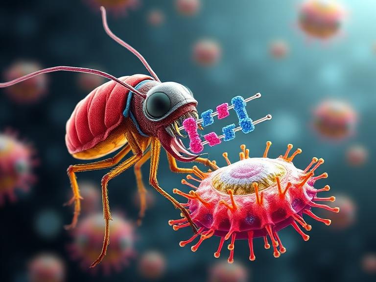
As a leading biotech research service provider, KMD Bioscience possesses an advanced phage display technology platform, offering customized high-quality phage display library construction services. We can create diverse natural libraries (mouse, rabbit, human, etc.), immune libraries (such as VHH libraries, scFv libraries, Fab libraries), synthetic libraries (e.g., peptide libraries), and semi-synthetic libraries for our clients, along with professional library panning and screening services. Through this platform, clients can rapidly identify antibodies or ligands with high affinity and specificity for their target of interest.
In this comprehensive guide, we will delve deep into the molecular mechanics of how phage display work operates, the systematic steps involved in biopanning, and the myriad of ways this high-throughput technology accelerates the drug discovery pipeline.
Table of Contents
1. The Biological Foundation: What is Phage Display?
To fully grasp how phage display work succeeds in identifying highly specific binding molecules, one must first understand the biology of the bacteriophage. A bacteriophage (or phage) is a virus that infects and replicates within bacteria. In the context of phage display work, the most commonly utilized phages are filamentous phages, such as M13, fd, and f1, which infect Escherichia coli (E. coli).
Filamentous phages have a relatively simple structure: a circular single-stranded DNA genome encased in a long, flexible cylinder of coat proteins. The major coat protein is pVIII (present in thousands of copies), while the minor coat proteins, such as pIII, are located at the tips of the phage particle. The brilliance of phage display work lies in the genetic manipulation of these coat proteins. By inserting a foreign DNA sequence into the gene encoding a coat protein (most frequently the pIII gene), the phage will express the foreign peptide or protein fragment as a fusion on its external surface.
2. The Core Mechanism: How Phage Display Work Functions
The defining characteristic of successful phage display work is the physical linkage between genotype and phenotype. The phenotype—the physical protein or antibody fragment being displayed on the outside of the phage—is directly linked to the genotype—the DNA sequence encoding that protein encapsulated within the phage particle.
This linkage is what makes high-throughput screening possible. When an entire library of phages is generated, representing billions of different protein variants, researchers can expose this library to an immobilized target molecule (such as a disease-associated receptor). Only the phages displaying a protein that binds tightly to the target will adhere. Because the DNA sequence instructing the synthesis of that specific binding protein is safely stored inside the adhered phage, researchers can recover it, sequence the DNA, and immediately know the exact genetic code of the successful binder.
From our experience at KMD Bioscience, ensuring a seamless genotype-phenotype linkage requires meticulous vector design and optimal E. coli host strain selection. We recommend using phagemid vectors combined with helper phages to maximize the efficiency and stability of your phage display work, particularly when dealing with large antibody libraries.
3. The Biopanning Process: A Step-by-Step Breakdown
The actual execution of phage display work in the laboratory is driven by a cyclic process known as biopanning. This is an affinity selection technique designed to enrich a vast library of random clones down to a select few high-affinity binders. The standard phage display work protocol typically involves the following four steps, repeated over three to five rounds:
Step A: Target Immobilization and Binding
The target antigen (a protein, peptide, or even whole cells) is immobilized on a solid surface, such as a microtiter plate, magnetic beads, or a chromatography column. The diverse phage library is then incubated with the target. During this incubation, phages displaying complementary sequences bind to the target, while non-binders remain in the solution.
Step B: Washing Away Non-Binders
This is arguably the most critical step in phage display work. The surface is subjected to stringent washing to remove unbound and weakly bound phages. From our experience, we recommend progressively increasing the washing stringency in subsequent rounds of biopanning (e.g., by adding detergents like Tween-20 or increasing the number of wash cycles) to effectively eliminate background noise and isolate only the highest-affinity binders.
Step C: Elution of Bound Phages
The phages that successfully remained bound to the target must now be recovered. This is achieved through elution, which disrupts the interaction between the displayed protein and the target antigen. Elution is typically performed using acidic solutions (which are subsequently neutralized), competitive ligands, or enzymatic cleavage if a specific cleavage site was engineered into the fusion construct.
Step D: Amplification in E. coli
The eluted phages, which now represent an enriched pool of target-specific binders, are used to infect fresh E. coli cultures. The bacteria act as microscopic factories, amplifying the phages to produce a massive yield of the enriched pool. This amplified output becomes the input for the next round of biopanning. With each successive round of this phage display work, the pool becomes increasingly dominated by the strongest binders.
4. Types of Phage Display Libraries
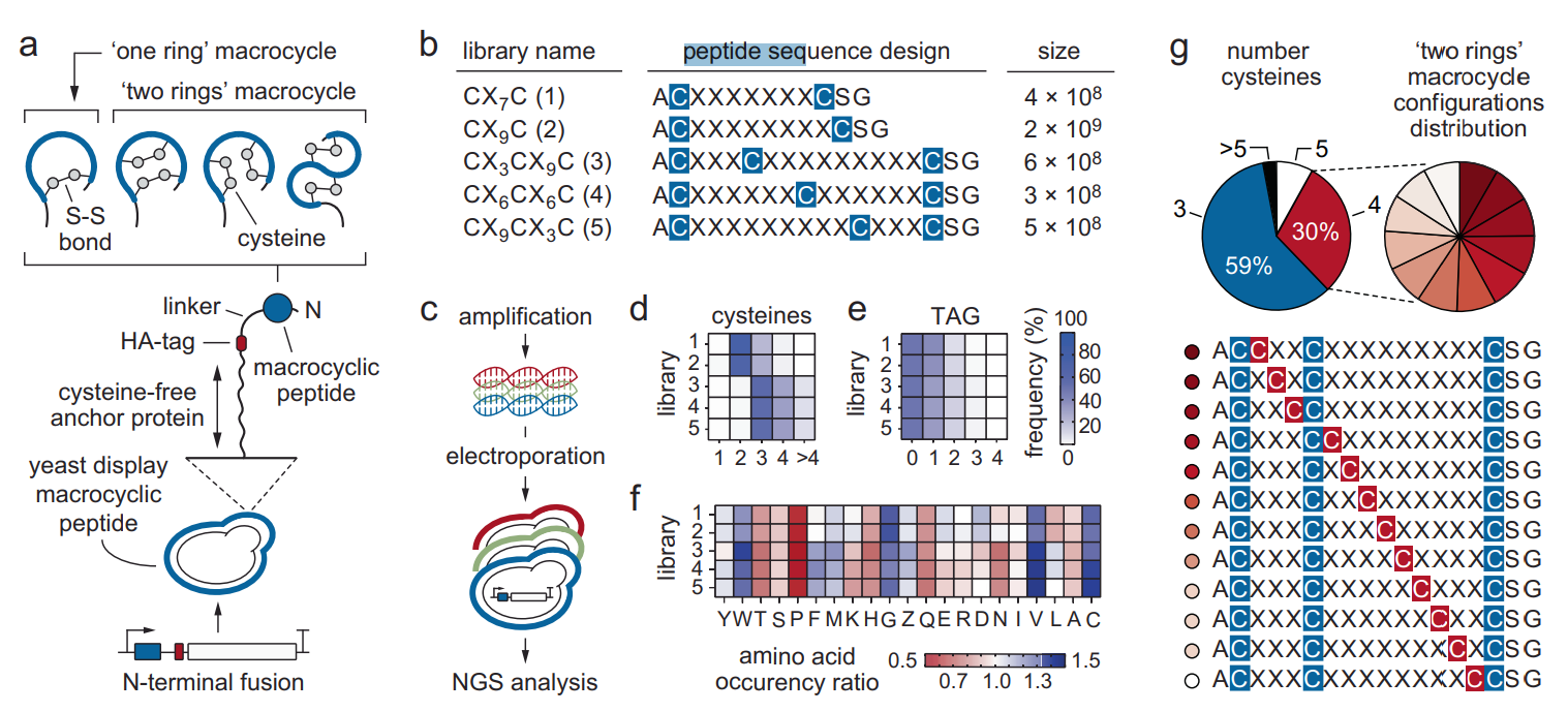
The success of any phage display work relies heavily on the quality and diversity of the initial library. As a premier contract research organization CRO, KMD Bioscience excels in creating customized libraries tailored to the unique goals of our clients.
Natural / Naive Libraries: Constructed from the B-cells of unimmunized donors. These libraries are vast and unbiased, making them highly versatile for discovering antibodies against virtually any antigen, including toxic or non-immunogenic targets.
Immune Libraries: Generated from donors (such as humans, mice, rabbits, or alpacas) that have been actively immunized or exposed to a specific pathogen. These libraries are inherently biased toward the target antigen and typically yield antibodies with naturally matured, high affinities. Our nanobody library construction service relies heavily on immunized alpacas to generate premium single-domain antibodies.
Synthetic and Semi-Synthetic Libraries: These libraries feature artificially designed frameworks into which randomized complementarity-determining regions (CDRs) are introduced. This approach allows researchers to optimize the structural stability and humanization profile of the resulting antibodies from the very beginning of the phage display work.
The advantages of phage display technology lie in its high throughput and flexibility, enabling rapid responses to various demands. Regardless of the library type selected, comprehensive life science research solutions require libraries with sizes often exceeding 109 to 1011 independent transformants to ensure a high probability of finding rare, potent binders.
5. Tackling Challenging Targets in Phage Display Work
While discovering antibodies against soluble proteins is relatively straightforward, the frontiers of modern biopharmaceuticals demand more. Additionally, we have extensive experience in discovering antibodies against challenging targets, particularly in areas such as GPCRs (G-protein-coupled receptors), ion channel-related proteins, small molecules, and peptides, delivering tailored solutions for our clients.
Targets like GPCRs and ion channels are notoriously difficult because their native conformation is heavily dependent on the lipid bilayer of the cell membrane. Traditional soluble antigen panning often fails here. In our specialized phage display work, we recommend utilizing whole-cell panning, virus-like particles (VLPs), or nanodisc technologies to present these multi-pass transmembrane proteins in their natural, functional state. When paired with high-quality nanobody antibody products, which are small enough to penetrate cryptic epitopes on these complex receptors, the success rates of therapeutic discovery increase dramatically.
6. Post-Discovery Optimization and Engineering
Identifying a binder is only the first phase of successful phage display work. To transition a discovered antibody into a viable clinical candidate, rigorous optimization is required. In the antibody discovery process, we not only provide a range of optimization services such as antibody sequence expression, affinity maturation, and antibody labeling but also offer antibody humanization to further enhance antibody stability and clinical application value.
Affinity maturation mimics the natural immune system's somatic hypermutation in vitro. By introducing targeted mutations into the CDRs of the initially discovered antibody and performing highly stringent rounds of phage display work, we can enhance the binding affinity by several orders of magnitude. Through KMD Bioscience’s phage display platform, clients can not only obtain high-quality, highly specific antibodies but also receive comprehensive support in antibody optimization and engineering, facilitating smooth progress in research. We are committed to providing innovative and precise technical services to drive continuous advancements in the biopharmaceutical field.
7. Summary Table: The Lifecycle of Phage Display Work
| Phase of Phage Display Work | Process Description | Key Deliverable |
|---|---|---|
| Library Construction | Isolating mRNA, synthesizing cDNA, and cloning diverse antibody/peptide genes into phagemid vectors. | A diverse library of 109-1011 unique phage clones. |
| Biopanning (Rounds 1-4) | Iterative cycles of binding, stringent washing, elution, and bacterial amplification. | An enriched pool of target-specific binding phages. |
| Screening and Sequencing | ELISA screening of individual clones to confirm specific binding; DNA sequencing of positive clones. | Verified unique genetic sequences of high-affinity binders. |
| Recombinant Expression | Subcloning the identified sequences into expression vectors for mammalian or bacterial cell production. | Purified, functional antibodies or peptides. |
| Affinity Maturation & Humanization | Engineered mutations to increase affinity and reduce immunogenicity for therapeutic use. | Clinical-grade, optimized antibody candidates. |
8. Frequently Asked Questions (FAQs)
Why is M13 the most common bacteriophage used in phage display work?
M13 is a non-lytic filamentous phage, meaning it does not kill the E. coli host cell upon exit. Instead, it is continuously secreted, allowing for a sustained and massive yield of phage particles without the cellular debris and protein contamination associated with lytic phages like T7. This makes M13 incredibly efficient and clean for standard phage display work.
What is the difference between an scFv and a Fab library in phage display work?
Both are common formats for antibody display. An scFv (single-chain variable fragment) consists of the heavy and light chain variable regions connected by a flexible peptide linker. It is smaller and easier to display. A Fab (Fragment antigen-binding) includes both the variable and the first constant domains of the heavy and light chains. Fab fragments are more stable and closer in structure to full-length IgG, though they require more complex cloning strategies in phage display work.
How does phage display compare to hybridoma technology?
From our experience, while traditional mouse hybridoma technology is proven, it is limited by the animal's immune tolerance, time-consuming immunization schedules, and difficulties with humanization. Phage display work bypasses immune tolerance (especially with naive or synthetic libraries), allows for rapid in vitro screening in weeks rather than months, and provides the DNA sequence immediately, significantly accelerating downstream engineering.
9. Academic References
For researchers seeking to deepen their understanding of how phage display work operates at a molecular level, we recommend consulting the following foundational and contemporary academic sources:
Smith, G. P. (1985). Filamentous fusion phage: novel expression vectors that display cloned antigens on the virion surface. Science, 228(4705), 1315-1317. View on PubMed
McCafferty, J., Griffiths, A. D., Winter, G., & Chiswell, D. J. (1990). Phage antibodies: filamentous phage displaying antibody variable domains. Nature, 348(6301), 552-554. View on PubMed
Winter, G., Griffiths, A. D., Hawkins, R. E., & Hoogenboom, H. R. (1994). Making antibodies by phage display technology. Annual Review of Immunology, 12, 433-455. View on PubMed

- Protein Platform
- Protein Expression Services
- Protein Modification & Detection Services
- Protein Interaction Services
- Protein Purification Service
- Antibody Discovery Platform
- VHH Antibody Platform
- Phage Display Platform
- Antibody Humanization Platform
- Single B Cell Screening Platform
- Antibody Expression & Validation Platform
- Peptide Library Platform
- Custom Antibody Platform
- Innovative Drug Discovery Platform
- Product Center
- Proteins
- Antibodies
- Raw Materials Reagents
- Small Molecule Reagents
- Peptide Products
- Nanoparticle Products
- Contact Us
- Phone:400-621-6806
- Mobile:13110936064(WeC
- E-mail:[email protected]
- Address:Room 108, Building 10, No. 6 Hechang Road, Wuqing Development Zone, Tianjin City
-

WeChat
-

Service Account
-
 400-621-6806
 -
 3599163699
 -
 Monday to Friday 9:00-18:00
 -

-
 0
0
-
