


4 Step Breakdown of the Phage Display Antibody Procedure Process
- 2026-04-21
- 157
Authored by KMD Bioscience. We possess an advanced phage display technology platform, offering customized high-quality phage display library construction services. We can create diverse natural libraries, immune libraries, synthetic libraries, and semi-synthetic libraries for our clients, alongside professional library panning and screening services.
In the rapidly evolving landscape of biopharmaceutical development and targeted therapeutics, the identification of highly specific and high-affinity antibodies is a fundamental requirement. Among the various methodologies available to researchers and pharmaceutical engineers, the Phage display antibody procedure remains one of the most powerful and versatile in vitro selection technologies. Since its conceptual inception in the 1980s, this technology has been refined to allow the screening of billions of distinct antibody variants against a vast array of antigens in a matter of weeks.
At KMD Bioscience, our daily operations revolve around optimizing and executing this exact process. Through our dedicated Phage Display Platform, clients can rapidly identify antibodies or ligands with high affinity and specificity for their target of interest. Understanding the intricate nuances of the Phage display antibody procedure is essential for navigating the complexities of modern drug discovery. From our experience, a successful screening campaign is rarely accidental; it is the result of meticulous library construction, stringent biopanning protocols, and comprehensive downstream validation. In this authoritative guide, we detail the complete lifecycle of the Phage display antibody procedure, offering practical insights and technical guidance for therapeutic development.
Table of Contents
1. Understanding the Phage Display Antibody Procedure
The core principle underlying the Phage display antibody procedure is the physical linkage between phenotype (the expressed antibody fragment on the viral surface) and genotype (the DNA sequence encoding the fragment encapsulated within the viral particle). This elegant biological mechanism is typically achieved using filamentous bacteriophages, such as M13. By inserting a diverse repertoire of antibody genes (such as scFv, Fab, or VHH) into the phage genome adjacent to a coat protein gene (usually pIII or pVIII), the host bacterial cell is hijacked to assemble phage particles that display the corresponding antibody fragments on their exterior.
This physical linkage is what makes the Phage display antibody procedure so revolutionary. When a researcher exposes a massive library of these phages to a specific immobilized antigen, only the phages displaying an antibody with high affinity for that antigen will bind. Because the DNA encoding that specific binder is trapped inside the bound phage, researchers can elute the bound phages, infect E. coli to amplify them, and sequence the DNA to discover the exact genetic blueprint of the successful antibody. We recommend viewing this process not merely as a screening tool, but as an accelerated form of directed molecular evolution.
2. Step-by-Step Breakdown of the Phage Display Antibody Procedure
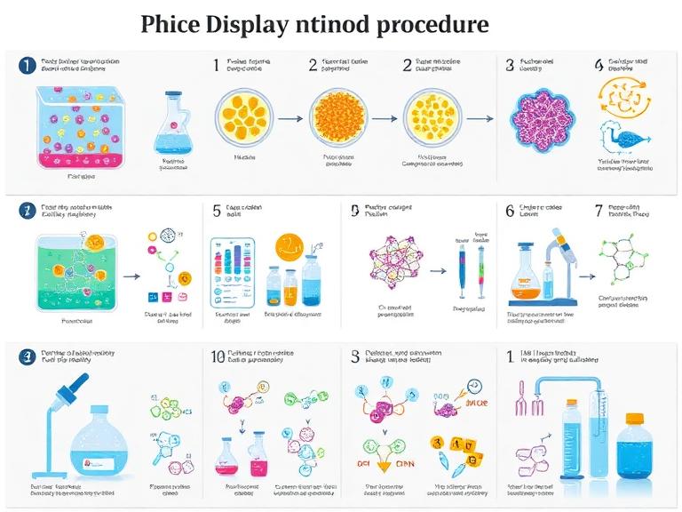
Executing a successful Phage display antibody procedure requires rigorous adherence to a multi-stage protocol. Any deviation in buffer stringency or amplification parameters can lead to the loss of rare, high-affinity clones. The process is broadly categorized into library construction, biopanning, amplification, and validation.
Stage 1: Library Construction
The foundation of the Phage display antibody procedure is the library itself. A poor library will yield poor results, regardless of how impeccable the downstream screening may be. The construction phase begins with the isolation of mRNA from B cells (harvested from naive, immunized, or synthetic sources). The mRNA is reverse-transcribed into cDNA, and the variable heavy (VH) and variable light (VL) chain domains are amplified via Polymerase Chain Reaction (PCR).
These domains are then assembled into functional fragments—most commonly Single-chain variable fragments (scFv) or antigen-binding fragments (Fab)—using overlap extension PCR. The resulting DNA repertoire is digested with restriction enzymes and ligated into a phagemid vector. Finally, the ligated vectors are electroporated into a highly competent E. coli strain (such as TG1 or XL1-Blue) to generate a library with a diversity typically ranging from 10^8 to 10^11 distinct clones.
Stage 2: Biopanning (The Core Selection Phase)
Biopanning is the critical selection phase of the Phage display antibody procedure. This process enriches the population of target-specific phages while washing away non-specific binders. A standard biopanning cycle involves four sequential actions:
Coating and Blocking: The target antigen is immobilized on a solid support, such as an immunotube or magnetic beads. The remaining binding sites on the support are then blocked using agents like BSA (Bovine Serum Albumin) or milk powder to prevent non-specific adherence.
Binding: The phage library is incubated with the immobilized antigen. Phages displaying antibodies with an affinity for the target will bind to the antigen, while non-binders remain in suspension.
Washing: This is where technical expertise is paramount. The support is washed multiple times with a buffer containing a mild detergent (like PBS-T). From our experience, adjusting the stringency of the wash buffer over successive rounds of panning is the most effective way to eliminate weak binders and isolate picomolar affinity antibodies.
Elution: The specifically bound phages are eluted from the antigen. This is typically achieved by lowering the pH using an acidic buffer (like glycine-HCl) to disrupt the antigen-antibody interaction, followed by immediate neutralization to preserve phage viability.
Stage 3: Amplification and Iteration
The eluted phages represent a tiny fraction of the original library. To proceed with the Phage display antibody procedure, these phages must be amplified. The eluted pool is used to infect fresh, exponentially growing E. coli. Helper phages (such as M13KO7 or VCSM13) are superinfected into the culture to provide the necessary structural proteins required for phage assembly and secretion. The amplified phage pool is then purified via PEG/NaCl precipitation and subjected to a subsequent round of biopanning. Typically, 3 to 5 rounds of panning are performed, with each round enriching the concentration of target-specific binders.
Stage 4: Validation and Characterization
The final stage of the Phage display antibody procedure involves isolating individual clones from the final enriched pool and validating their specificity and affinity. Individual colonies are picked and cultured in 96-well plates. Monoclonal phage ELISA (Enzyme-Linked Immunosorbent Assay) is conducted against the target antigen. Clones that exhibit a strong positive signal are sequenced to identify the unique VH and VL sequences. We recommend moving the top candidates into our Antibody Expression & Validation Platform for immediate recombinant production and advanced kinetic analysis using SPR (Surface Plasmon Resonance) or BLI (Bio-Layer Interferometry).
3. Types of Libraries Utilized in the Process
The starting point for any Phage display antibody procedure dictates the nature of the final output. At KMD Bioscience, we construct and screen various specialized libraries based on the client's ultimate therapeutic or diagnostic goals.
Immune Libraries: Generated from donors (human or animal) that have been naturally infected or intentionally immunized with the target antigen. These libraries are highly biased towards the target and often yield antibodies that have already undergone in vivo affinity maturation. We frequently utilize these in our VHH Antibody Platform to generate high-affinity alpaca or llama nanobodies.
Naive Natural Libraries: Constructed from the B cells of non-immunized donors. These massive libraries contain a vast, unbiased repertoire of antibodies and can theoretically be used to screen against any conceivable antigen, including self-antigens and toxic compounds.
Synthetic and Semi-Synthetic Libraries: These libraries are created by artificially randomizing specific regions of the antibody, primarily the Complementarity-Determining Regions (CDRs). By utilizing defined frameworks, researchers can control the biochemical properties of the resulting antibodies. Our Peptide Library Platform is highly adept at managing these complex synthetic repertoires.
4. Leveraging KMD Bioscience Platforms for Success
Managing the entirety of the Phage display antibody procedure in-house requires immense infrastructure, highly competent cell lines, and specialized bioinformatics capabilities. KMD Bioscience simplifies this process by providing an integrated suite of platforms designed to accelerate the drug discovery pipeline.
When clients initiate a project through our Phage Display Platform, they gain access to decades of accumulated biopanning expertise. Furthermore, discovering a binder is only the first step. If the initial antibody is derived from a murine or camelid source, our Antibody Humanization Platform can seamlessly transition the candidate into a humanized format suitable for clinical trials, minimizing immunogenicity risks. For completely novel targets, we integrate the Phage display antibody procedure with our Innovative Drug Discovery Platform and Single B Cell Screening Platform, ensuring that no potential therapeutic avenue is left unexplored.
5. Optimization and Troubleshooting Strategies
Even a flawlessly constructed library can yield poor results if the panning strategy is flawed. From our experience executing thousands of campaigns, we recommend paying strict attention to antigen conformation. In the Phage display antibody procedure, the antigen must be presented in a state that mimics its native biological conformation. Coating antigens directly onto polystyrene plates can sometimes cause structural denaturation, burying the true epitopes.
To circumvent this, we recommend utilizing biotinylated antigens paired with streptavidin-coated magnetic beads. This approach allows the antigen to remain in solution during the binding phase of the Phage display antibody procedure, preserving its 3D structure and significantly increasing the likelihood of isolating functionally relevant, neutralizing antibodies. Furthermore, negative selection (depletion panning) should always be employed to subtract phages that bind to the support matrix or blocking agents before exposure to the actual target.
6. Summary Table: Quick Reference Protocol
To assist researchers in navigating the workflow, we have summarized the critical phases of the Phage display antibody procedure below.
| Procedure Phase | Core Objective | Key Reagents / Equipment | KMD Bioscience Integrated Platform |
|---|---|---|---|
| Library Construction | Generate a massive repertoire of antibody genes linked to phage vectors. | RNA extraction kits, PCR, Phagemid vectors, TG1 E. coli | Custom Antibody Platform, Peptide Library Platform |
| Biopanning | Enrich target-specific clones through iterative binding, washing, and elution. | Antigen, Magnetic Beads, PBS-T, Glycine-HCl | Phage Display Platform |
| Amplification | Multiply the eluted specific phages for subsequent panning rounds. | Helper Phage (M13KO7), PEG/NaCl, Bacterial cultures | Phage Display Platform |
| Validation | Confirm specificity and determine the genetic sequence of individual clones. | Phage ELISA, Sanger Sequencing, SPR analysis | Antibody Expression & Validation Platform |
| Post-Discovery | Optimize the candidate for therapeutic viability. | Mammalian expression systems, Bioinformatics tools | Antibody Humanization Platform |
7. Frequently Asked Questions (FAQs)
How long does a standard Phage display antibody procedure take to complete?
From our experience, if starting from an existing, high-diversity naive library, the core biopanning and initial ELISA screening can be completed in approximately 4 to 6 weeks. However, if the project requires the construction of a custom immune library starting from animal immunization, the entire Phage display antibody procedure timeline will extend to 4 to 6 months.
What is the advantage of using a VHH library over a traditional scFv library?
VHH antibodies, also known as nanobodies or single-domain antibodies, are derived from the heavy-chain-only antibodies found in camelids. In the context of the Phage display antibody procedure, VHH fragments are significantly smaller, highly soluble, and exceptionally stable. They excel at recognizing hidden or concave epitopes (such as enzyme active sites) that larger scFv or Fab fragments cannot access. We heavily utilize our VHH Antibody Platform for challenging therapeutic targets.
Why is helper phage required in the Phage display antibody procedure?
Most modern libraries utilize phagemid vectors rather than true phage vectors. Phagemids carry the antibody gene fused to a coat protein but lack the full genetic machinery required to assemble a complete viral particle. Therefore, a helper phage (like M13KO7) must be introduced into the E. coli. The helper phage provides all the structural proteins and assembly enzymes required to package the phagemid DNA and secrete the hybrid phage particle, making the Phage display antibody procedure highly safe and manageable in the laboratory.
8. Academic and Industry References
For further academic exploration regarding the molecular mechanisms, historical development, and clinical applications of the Phage display antibody procedure, we recommend consulting the following authoritative sources:

- Protein Platform
- Protein Expression Services
- Protein Modification & Detection Services
- Protein Interaction Services
- Protein Purification Service
- Antibody Discovery Platform
- VHH Antibody Platform
- Phage Display Platform
- Antibody Humanization Platform
- Single B Cell Screening Platform
- Antibody Expression & Validation Platform
- Peptide Library Platform
- Custom Antibody Platform
- Innovative Drug Discovery Platform
- Product Center
- Proteins
- Antibodies
- Raw Materials Reagents
- Small Molecule Reagents
- Peptide Products
- Nanoparticle Products
- Contact Us
- Phone:400-621-6806
- Mobile:13110936064(WeC
- E-mail:[email protected]
- Address:Room 108, Building 10, No. 6 Hechang Road, Wuqing Development Zone, Tianjin City
-

WeChat
-

Service Account
-
 400-621-6806
 -
 3599163699
 -
 Monday to Friday 9:00-18:00
 -

-
 0
0
-
