


Alfa Tag-based VHH Antibody Screening System
- 2026-04-23
- 327
1.Concept of VHH Antibodies
VHH antibodies, also known as nanobodies (Nbs), are single-chain antibodies derived from the peripheral blood serum of camelids. These antibodies consist solely of heavy chains, lacking light chains as well as functional CH1 and CH4 domains. With dimensions of 2.5 nm in diameter and 4 nm in length, and a molecular weight of only 15 kDa, VHH antibodies exhibit distinct advantages over conventional antibodies.Due to their small molecular size, nanobodies demonstrate superior tissue penetration capabilities, including the ability to cross the blood-brain barrier. They can be efficiently expressed in both prokaryotic and eukaryotic systems, while maintaining high specificity and binding affinity. Additionally, they exhibit low immunogenicity in humans. These unique properties make VHH antibodies particularly valuable for applications in anticancer drug development, cardiovascular and cerebrovascular diseases, immune disorders, and other therapeutic areas.
2.What is the ALFA Tag
Specialized epitope tags are widely used for protein detection, manipulation, or purification, but their versatility is often limited. The ALFA tag is a rationally designed epitope tag that offers exceptionally broad applications in life sciences while outperforming established tags such as HA-, FLAG-, or myc-tags.
The ALFA tag features a 15-amino acid sequence that is hydrophilic, uncharged at physiological pH, and lacks residues targeted by amine-reactive immobilization agents or crosslinkers. It exhibits a strong propensity to form a stable α-helix and can spontaneously refold even after harsh chemical treatments. Due to its compact structure, the tag is physically smaller than most linear epitope tags. The ALFA tag is compatible with protein function and can be positioned at either the N-terminus or C-terminus of a protein of interest (POI), or even between two separately folded domains.
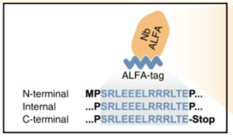
Fig. 1: Sequence of the minimized ALFA tag
3.ALFA-Tag-Specific VHH Antibody
To develop nanobodies (NbALFA) that bind the ALFA tag, we immunized alpacas and employed an in-house developed nanobody selection method to identify those specifically targeting ALFA-tagged proteins. The top-performing binder was genetically engineered with an ectopic cysteine residue to enable site-specific immobilization or fluorophore conjugation.
4.Crystal Structure of NbALFA
To better understand the interaction between NbALFA and the ALFA tag, we determined the crystal structure of NbALFA in complex with the ALFA peptide. NbALFA adopts a canonical immunoglobulin (IgG) fold, consisting of two β-sheets connected by a disulfide bond. The ALFA peptide-binding paratope extends from the N-terminal cap of the nanobody to one side formed by a five-stranded β-sheet. This binding interface is primarily composed of complementarity-determining regions (CDRs) 2 and 3, along with residues from the five-stranded β-sheet that form a hydrophobic pocket.The ALFA peptide is oriented parallel to the central axis of NbALFA. It forms an α-helical rod approximately 2 nm in length and 1.3 nm in diameter, stabilized by an intricate network of intramolecular interactions. Furthermore, the peptide establishes multiple polar and hydrophobic contacts with NbALFA.
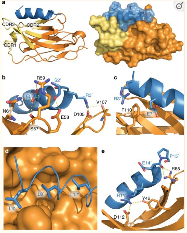
Fig. 2: Crystal structure of NbALFA
5.Applications of ALFA-tag VHH Antibodies
5.1 Detection of ALFA-Tagged Proteins by Immunofluorescence
In immunofluorescence (IF) applications using PFA-fixed mammalian cells, fluorescently labeled NbALFA specifically recognizes target proteins carrying the ALFA tag at various positions. Importantly, all tested proteins displayed their characteristic localizations (Tom70-EGFP-ALFA: mitochondrial outer membrane; ALFA-vimentin: filamentous structures; EGFP-ALFA-TM: plasma membrane), confirming that the ALFA tag is compatible with proper protein folding and correct localization.In quantitative assays, the nucleocytoplasmic distribution of EGFP tagged with ALFA at either terminus was indistinguishable from untagged EGFP. No atypical interactions with cellular membranes or organelles were observed. Thus, we conclude that the ALFA tag does not appear to impair the physiological behavior of the tagged protein of interest (POI).
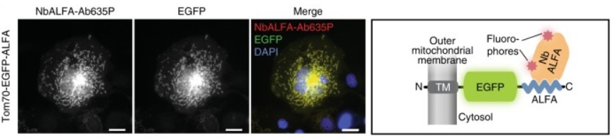
Fig. 3: Detection of ALFA-tagged proteins by immunofluorescence
5.2 Detection and Manipulation of ALFA-Tagged Proteins In Vivo
Nanobodies can be utilized to detect or manipulate target proteins in living mammalian cells. Indeed, when ALFA-tagged target proteins are co-expressed with NbALFA fused to mScarlet-I in mammalian cells, NbALFA-mScarlet-I robustly co-localizes with the ALFA-tagged target proteins, exhibiting minimal off-target signals and enabling clear visualization of ALFA-tagged structures. This demonstrates NbALFA’s ability to bind ALFA-tagged proteins in live cells. This functionality makes it highly suitable for integration with genome-editing tools such as CRISPR-Cas, allowing for the manipulation of ALFA-tagged proteins at endogenous levels in vivo.
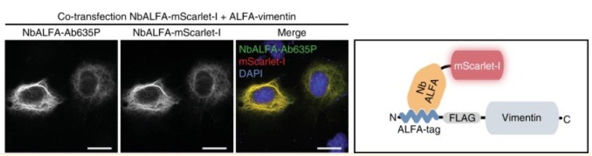
Figure 4: Detection of ALFA-tagged proteins by NbALFA.
5.3 Immunoblotting
The ALFA system was evaluated in Western blot applications, where ALFA-tagged vimentin in lysates from transfected cells could be specifically detected using IRDye800CW-labeled NbALFA. To compare the performance of the ALFA system with common epitope tags, we utilized fusion proteins containing HA-, myc-, FLAG-, and ALFA-tags. At identical concentrations of primary antibodies or nanobodies, the ALFA-tagged signals were 3–10 times stronger than those obtained with all other epitope tags. Without further optimization, NbALFA demonstrated a remarkably linear signal range spanning at least three orders of magnitude and detected target proteins at levels as low as 100 pg. Consequently, the detection limit was approximately 10-fold higher compared to all other epitope tags.
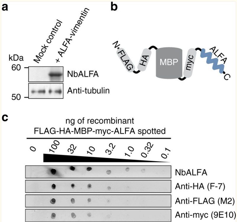
Figure 5: Detection of ALFA-tagged target proteins by fluorescent Western blotting.
6. KMD Bioscience Provides Nanobody Development Services
Compared to traditional monoclonal antibodies, nanobody production offers significant advantages. The process can be summarized as obtaining antibody genes through llama immunization and acquiring target antibody sequences via phage display. It primarily consists of four steps: llama immunization, phage library construction, antibody library screening, and antibody expression with functional validation.
6.2.1 Llama Immunization
Select age-appropriate llamas → Prepare antigens → Immunize llamas → Collect blood to measure antibody titers → PBMC cell isolation
6.2.2 Phage Library Construction
RNA extraction → cDNA synthesis by reverse transcription → PCR amplification → Vector ligation and electroporation → Bacterial library/phage library establishment
6.2.3 Antibody Library Screening
Commonly used screening methods include solid-phase screening, magnetic bead screening, and cell screening. KMD Bioscience can provide customized screening systems according to customer requirements.
6.2.4 Antibody Expression and Functional Validation
Construct mammalian expression vectors for expression → Purification via nickel column → Affinity measurement
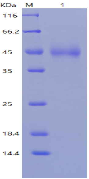
Figure 6: Expression results of Fc-VHH nanobody
To target the ALFA tag, we developed nanobodies due to their small size, monovalent nature, and robustness, which enable easy genetic modification and recombinant production in various organisms. Consequently, they can be site-specifically immobilized or quantitatively modified with fluorophores or oligonucleotides. Compared to conventional antibodies, nanobodies thus outperform in both traditional and advanced microscopy applications, as they can: (1) access more target epitopes in crowded cellular regions, (2) position fluorophores closer to the target protein, and (3) prevent protein-of-interest (POI) aggregation. Notably, NbALFA can even fold properly within eukaryotic cells, making it suitable as an intracellular antibody for detecting or manipulating ALFA-tagged proteins in vivo.
- Alfa tag-based VHH antibody screening
- VHH antibodies
- nanobodies

- Protein Platform
- Protein Expression Services
- Protein Modification & Detection Services
- Protein Interaction Services
- Protein Purification Service
- Antibody Discovery Platform
- VHH Antibody Platform
- Phage Display Platform
- Antibody Humanization Platform
- Single B Cell Screening Platform
- Antibody Expression & Validation Platform
- Peptide Library Platform
- Custom Antibody Platform
- Innovative Drug Discovery Platform
- Product Center
- Proteins
- Antibodies
- Raw Materials Reagents
- Small Molecule Reagents
- Peptide Products
- Nanoparticle Products
- Contact Us
- Phone:400-621-6806
- Mobile:13110936064(WeC
- E-mail:[email protected]
- Address:Room 108, Building 10, No. 6 Hechang Road, Wuqing Development Zone, Tianjin City
-

WeChat
-

Service Account
-
 400-621-6806
 -
 3599163699
 -
 Monday to Friday 9:00-18:00
 -

-
 0
0
-
