


Detailed Explanation of Nanobody Production Process
- 2026-05-14
- 448
1. What Are Nanobodies?
Nanobodies were first proposed and reported by Belgian scientists in Nature in 1993. Traditional antibody molecules (IgG) consist of two identical heavy chains and light chains. The light chain of an antibody molecule contains one VL (variable light) region and one CL (constant light) region, while the heavy chain consists of one VH (variable heavy) region and three CH (constant heavy) regions (CH1, CH2, and CH3).
Camels and llamas possess heavy-chain-only antibodies that lack light chains, composed of four HC (heavy chain constant) regions and two VH regions. The VHH region is referred to as a nanobody or single-domain antibody (sdAb). Nanobodies (Nb) have a crystal size of 2.5 nm, a length of 4 nm, and a molecular weight of only 15 kDa.Nanobodies are primarily used in biopharmaceutical research and development (e.g., genetic engineering drug development, ADC drug development), clinical in vitro diagnostics (e.g., colloidal gold method, ELISA, electrochemiluminescence), as well as in basic research fields such as oncology and immunology. Currently, the development of CAR-T therapeutic antibodies has garnered increasing attention from researchers.
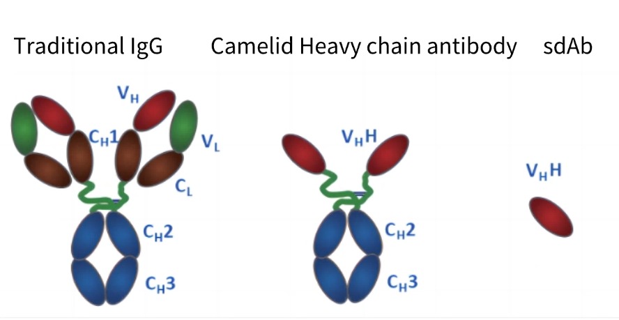
Figure 1: Structural comparison between conventional IgG antibodies and camelid nanobodies
2. Advantages and Disadvantages of Nanobodies
2.1 Advantages of Nanobodies
(1) Stability under high temperature and extreme pH conditions
(2) VHH domains can recognize epitopes that are typically inaccessible to conventional antibodies
(3) Their small molecular size facilitates rapid tissue penetration and labeling applications, including crossing the blood-brain barrier
(4) Cost-effective alternatives for large-scale production
2.2 Limitations of Nanobodies
Nanobodies (VHH antibodies) have a relatively small molecular weight of approximately 15 kDa, which is equivalent to the variable region of the heavy or light chain of a traditional IgG antibody. As a result, secondary antibodies targeting VHH antibodies are not universally applicable. To address the issue of secondary antibody recognition, the VHH can be fused with an Fc fragment for co-expression. (KMD Bioscience offers a variety of anti-llama secondary antibodies suitable for immune serum titer detection, Cat.#: SPAB01, SPAB03.)
3. Primary Applications of Nanobodies (VHH Antibodies)
Due to their unique structure and properties, nanobodies have gained significant attention in the field of CAR-T therapeutic antibody development.
CAR-T therapy is a novel precision-targeted cancer treatment, known as chimeric antigen receptor T-cell immunotherapy. Using genetic engineering techniques, activated T cells are equipped with a targeting "navigation device"—the CAR (chimeric antigen receptor)—effectively transforming them into "super soldiers" called CAR-T cells. These engineered cells utilize their CAR "targeting system" to precisely identify and eliminate tumor cells within the body.
4. KMD Bioscience can provide customers with high-quality camelid-derived nanobody development services.
KMD Bioscience offers a variety of camelid-derived nanobody development services, covering species such as alpacas, llamas, camels, and vicuñas. Leveraging our phage display technology platform, we provide comprehensive experimental processes, including antigen design, camelid immunization, library construction and screening, and functional activity validation. Additionally, we deliver highly specific and high-affinity alpaca VHH antibodies to scientists worldwide.
5. KMD Bioscience Provides a Full-Process Overview of Camelid-Derived Antibody Development for Clients
The key steps include: alpaca immunization, phage library construction, antibody library screening, and antibody expression with functional validation. The detailed workflow is as follows:
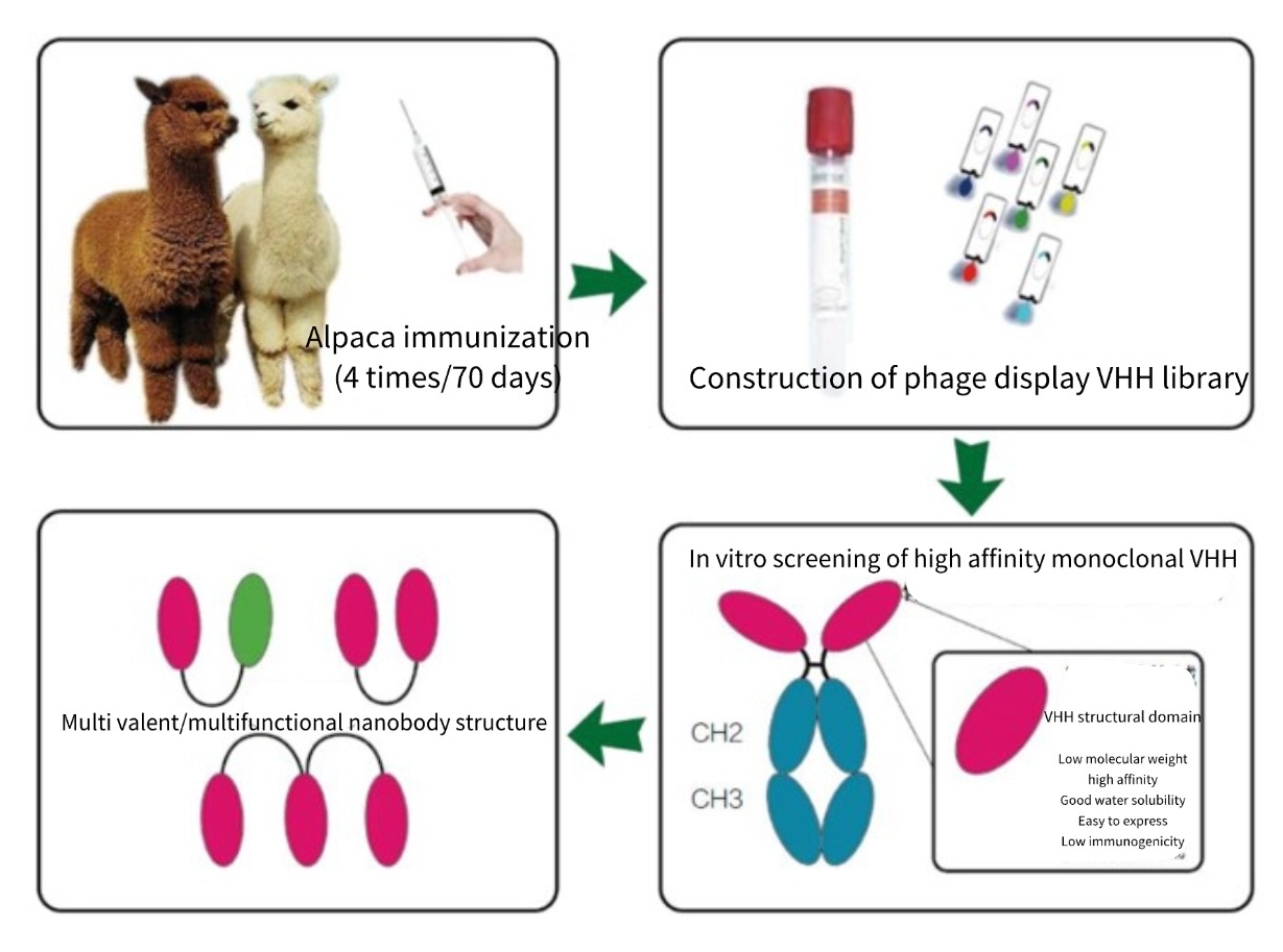
Figure 2: Development Process of Camelid-Derived Nanobodies
5.1 Llama Immunization
KMD Bioscience provides healthy adult llamas (28-36 months old) for immunization. High-quality libraries and monoclonal antibody cell lines can be obtained in approximately 16-20 weeks, along with complete experimental process records and immunization dynamic videos. Based on the customer's antigen type, KMD Bioscience will develop customized antigen preparation and immunization protocols.
5.1.1 Antigen Preparation:The customer provides the antigen. Freund’s adjuvant is used at a volume of 1 mL, and the antigen is mixed with the adjuvant at a 1:1 ratio for immunization.
5.1.2 Llama Immunization:Immunization is performed five times at 20-day intervals. The injections are administered in the llama’s neck region, and the animal is monitored for any adverse reactions for 1 hour post-immunization.
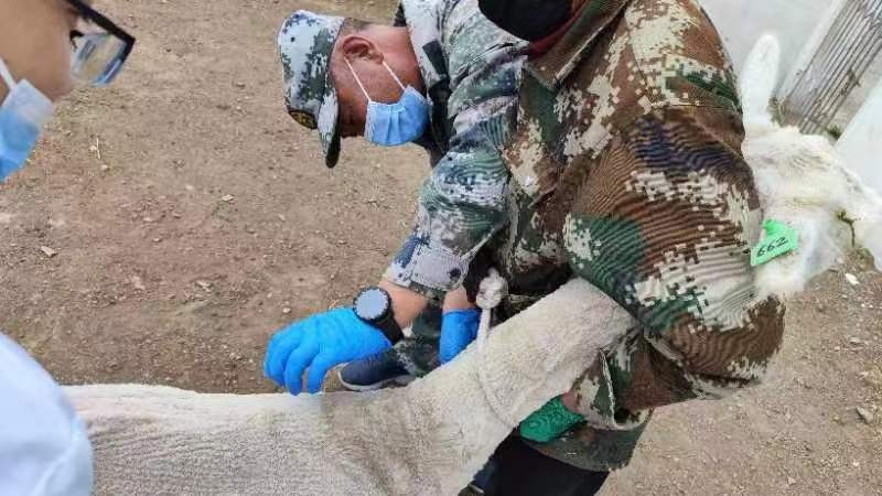
Figure 3: KMD Bioscience staff performing immunization injection on a llama.
5.1.3 Blood Collection: Blood was drawn before immunization as a negative control. One week after the third and fourth immunizations, 5 mL of blood was collected for titer determination. The serum titer was measured by ELISA, with the required titer being >10^5. KMD Bioscience provides clients with a clear immunization timeline information sheet, as shown in Table 1:
Table 1: Example of Nanobody Preparation Immunization Timeline by KMD Bioscience
| Times | Immunization date |
| The first time | November 25, 2020 |
| The second time | December 16, 2020 |
| The third time | January 6, 2021 |
| The fourth time | January 27, 2021 |
| The fifth time | February 18, 2021 |
5.1.4 PBMC Isolation: 100-120 mL of peripheral blood was collected, with a total PBMC count of 4×10^7 to 10^8 cells, all of which were shipped to the client.
5.2 Phage Library Construction
KMD Bioscience utilizes the pIII protein of M13 phage for VHH antibody (nanobody) surface display, prepares TG1 electrocompetent cells, constructs phage vectors, and performs phage library capacity determination.
5.2.1 RNA Extraction: RNA extraction was performed following the protocol of a commercial kit.
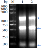
Figure 4: Electrophoresis image of RNA extraction
5.2.2 cDNA Synthesis by Reverse Transcription: cDNA was prepared following the protocol of a commercial kit.
5.2.3 PCR Amplification: Using cDNA as the template, the variable regions of heavy-chain antibody genes were amplified in two rounds of PCR.
5.2.4 Ligation and Electroporation: The PCR products were digested, ligated into a phagemid vector, and then electroporated into TG1 competent cells to construct the antibody library.
5.2.5 Bacterial Library/Phage Library Establishment: The library size was calculated, with a delivery standard of 10^8–10^9 clones.
5.3 Antibody Library Screening
The commonly used screening methods include solid-phase screening, magnetic bead screening, and cell-based screening (often employed for antibody screening targeting GPCRs and ion channel receptors).
KMD Bioscience offers customized screening systems tailored to clients' needs. Depending on specific requirements, we provide solid-phase screening, solid-phase/liquid-phase screening, and cell-based screening methods. The first two are suitable for screening target antigens such as proteins and peptides, while the third is ideal for antibody screening targeting GPCRs and ion channel receptors. Through these diverse screening approaches, we efficiently identify different types of blocking antibodies for our clients.
5.4 Antibody Expression and Functional Validation
5.4.1 Expression: A mammalian expression system was constructed for antibody production. KMD Bioscience has established a well-optimized mammalian expression platform, utilizing cell lines including but not limited to FreeStyle™ 293-F and ExpiCHO-S™, in combination with our proprietary high-expression vectors (featuring a full-length CMV promoter and an optimized secretion signal peptide sequence) for recombinant antibody expression and preparation.
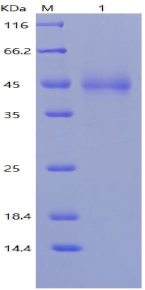
Figure 5: Expression results of Fc-VHH nanobody
5.4.2 Purification: The nanobodies were purified using a nickel column.
5.4.3 Affinity Measurement: The purified nanobodies were subjected to Biacore affinity analysis.
- Nanobody Production Process

- Protein Platform
- Protein Expression Services
- Protein Modification & Detection Services
- Protein Interaction Services
- Protein Purification Service
- Antibody Discovery Platform
- VHH Antibody Platform
- Phage Display Platform
- Antibody Humanization Platform
- Single B Cell Screening Platform
- Antibody Expression & Validation Platform
- Peptide Library Platform
- Custom Antibody Platform
- Innovative Drug Discovery Platform
- Product Center
- Proteins
- Antibodies
- Raw Materials Reagents
- Small Molecule Reagents
- Peptide Products
- Nanoparticle Products
- Contact Us
- Phone:400-621-6806
- Mobile:13110936064(WeC
- E-mail:[email protected]
- Address:Room 108, Building 10, No. 6 Hechang Road, Wuqing Development Zone, Tianjin City
-

WeChat
-

Service Account
-
 400-621-6806
 -
 3599163699
 -
 Monday to Friday 9:00-18:00
 -

-
 0
0
-
