


Identification of the Origin of Monoclonal Cells
- 2026-07-10
- 233
I. What Is Monoclonal Origin Verification
Monoclonal origin refers to the process of tracing the origin of a produced antibody to confirm that it is a highly homogeneous antibody derived from a single cell clone.
II. Purpose of Monoclonal Origin Verification
In recent years, recombinant protein therapeutics have found significant applications in the treatment of diseases, particularly cancer, and most of these therapeutics are produced through expression in mammalian CHO cells. Regulatory agencies require biopharmaceutical companies to provide evidence of the monoclonal origin of the cells used in these products; therefore, verifying the monoclonal origin of engineered cells is a critical means of ensuring the consistency of protein therapeutics.
III. Methods for Isolating Monoclonal Clones
Clonality is believed to reduce heterogeneity in cell banks. Common methods for isolating monoclonal clones include the limiting dilution method, ClonePix, flow cytometry-based fluorescence sorting, and new monoclonal selection technologies.
1. Limiting Dilution Cloning (LDC)
The limiting dilution method is a commonly used cloning technique that requires a seeding density of less than 0.5 cells per well. The advantages of this method include simple operation and minimal equipment requirements. However, it is time-consuming, inefficient, and highly labor-intensive; results can vary significantly depending on factors such as the experimenter, the laboratory environment, and experimental conditions.
2. ClonePix Method
ClonePix is a high-throughput monoclonal antibody screening system. This method requires minimal manual intervention, offers high throughput and efficiency, and provides highly accurate screening results. However, compared to the limiting dilution method, the monoclonal antibody culture medium produced by ClonePix differs more significantly from the medium used in subsequent production processes; clones generated by the limiting dilution method exhibit behavior in the microplate that more closely resembles their state during production.
3. Flow Cytometry-Based Fluorescent Cell Sorting (FACS) (FACS)
Flow cytometry-based fluorescence sorting (FACS) selects target single cells based on factors such as cell size, fluorescent dyes, and cell surface markers. The advantage of this method is a very high monoclonal formation rate; the disadvantage is that it requires optimization of conditions to ensure a one-to-one correspondence between the selected cells and the wells in the microplate. Additionally, the flow cytometer, as well as the morphology and condition of the cells, can affect the separation efficiency.
4. New Technologies for Monoclonal Cell Selection
Currently, there are various monoclonal cell screening devices available on the market, such as single-cell printing and Solentim VIPS, which can improve efficiency and reduce reliance on manual labor. At present, monoclonal cells are primarily obtained through the repeated combination and use of the above methods.
IV. Fluorescence In Situ Hybridization (FISH) for Verifying Monoclonal Origin
Fluorescence in situ hybridization (FISH) is used to verify whether cells are of a single origin; its results are visual and compelling.
Fluorescence in situ hybridization (FISH) is a technique that uses a fluorescence detection system to perform qualitative, quantitative, or relative localization analysis of DNA. Its basic principle is based on the complementarity of DNA sequences: nucleic acid probes labeled with known fluorophores, biotin, or digoxigenin are hybridized with the DNA in the sample being tested, forming detectable double-stranded nucleic acid hybrids.
V. Advantages of Fluorescence In Situ Hybridization (FISH) Technology
Fluorescence in situ hybridization (FISH) offers advantages not found in other hybridization methods:
(1) FISH uses non-radioactive labeling, making it safer
(2) FISH has a short experimental cycle and can detect multiple sequences simultaneously
(3) Due to multiple immunochemical reactions that amplify the hybridization signal, FISH technology offers sensitivity comparable to that of radioactive probes
VI. KMD Bioscience Provides High-Quality Monoclonal Antibody Origin Identification Services
KMD Bioscience has been dedicated to research in cell biology technologies for many years. We have a team of experienced scientific experts, a highly skilled laboratory team, a well-established cell culture platform, and comprehensive experimental equipment and facilities. We have built a comprehensive Fluorescence In Situ Hybridization (FISH) technical service platform and accumulated extensive experience through successful projects. Leveraging our solid technical expertise, we can provide customized end-to-end services ranging from probe design to monoclonal origin identification, striving to solve our clients’ problems in a highly efficient and cost-effective manner.
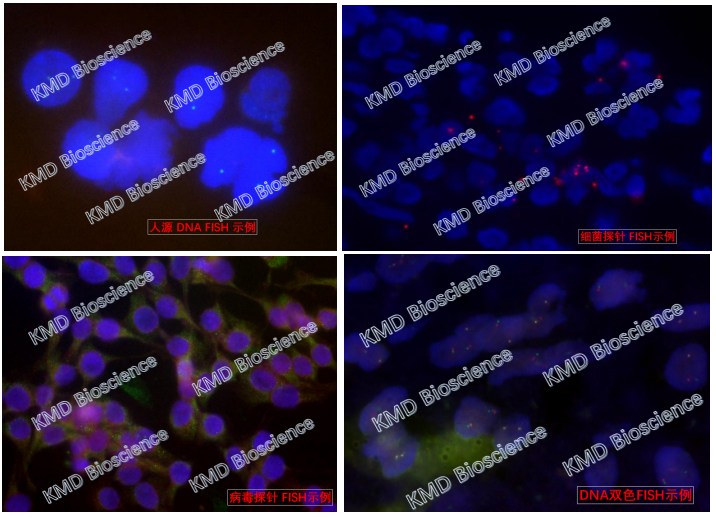
Figure 1: Photographs of Fluorescence In Situ Hybridization (FISH) on Different Samples
VII. Introduction to the Fluorescence In Situ Hybridization (FISH) Service Workflow
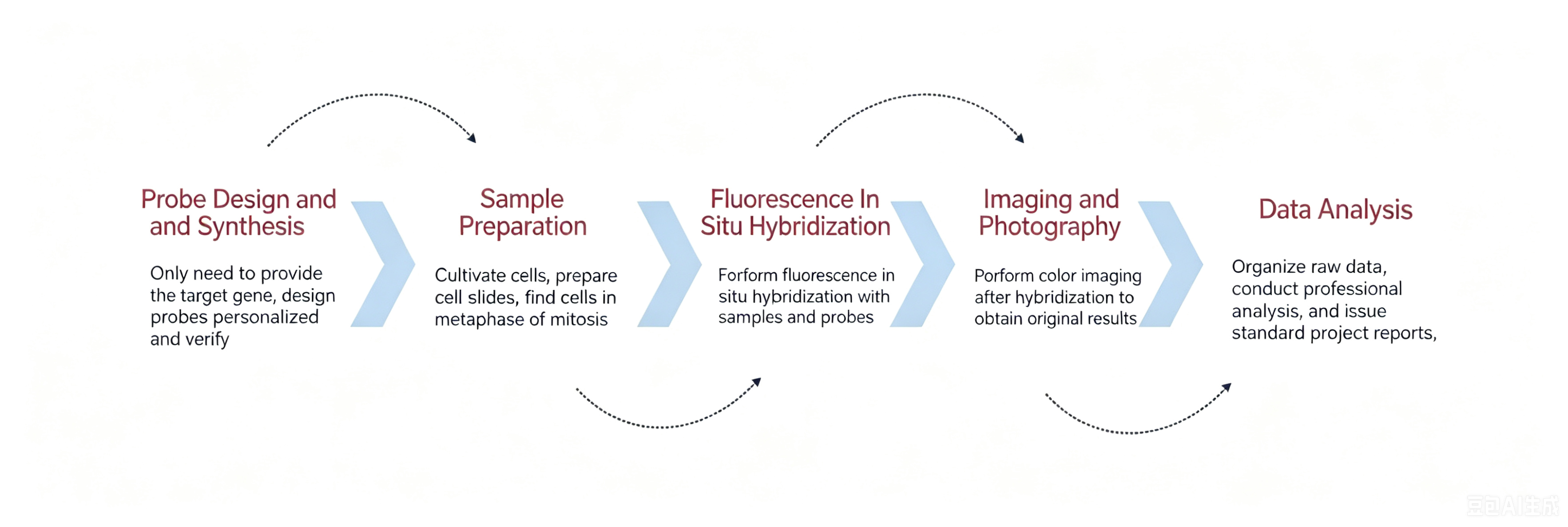
KMD Bioscience offers fluorescence in situ hybridization (FISH) services. The main technical workflow is as follows: probe design and synthesis, sample preparation, in situ hybridization experiments, imaging and photography, result analysis, and project reporting.
1. Probe Design and Synthesis
Ready-to-use probes can be hybridized directly; non-ready-to-use probes must be diluted with hybridization buffer in a 1:1:1:50 ratio, denatured at 85°C for 3 minutes, and equilibrated at 37°C for 5 minutes.
2. Sample Preparation
①. Dewaxing and rehydration: Immerse the paraffin sections in xylene to remove the paraffin twice, for 10 minutes each time;
②. Successively pass the sections through 100%, 85%, and 75% ethanol for 3 minutes each, followed by 3 minutes in PBS;
③ Add pepsin digestion solution to the slides and digest at 37°C for 15–30 minutes; wash twice with PBS for 3 minutes each time;
④ Pre-hybridization: Remove the hybridization solution from the refrigerator 30 minutes in advance to allow it to reach room temperature; add drops of the hybridization solution and incubate in a hybridization instrument at 55°C for 1 hour.
3. In Situ Hybridization Experiment
①. Remove the pre-hybridized slides, remove excess liquid, add the probe dropwise to the slides to completely cover the tissue, and incubate overnight at 37°C in a hybridization instrument;
② Wash the slides three times in 2× SSC (0.1% NP-40), 5 minutes each time;
③ Add 20 μL of DAPI-Antifade Solution, cover with a coverslip, and incubate in the dark for 20 minutes.
4. Imaging and Photography
Photograph the experimental results of the CHO cell experiment using this fluorescence in situ hybridization (FISH) technique:
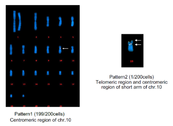
Figure 3: Monoclonal Origin Verification Experiment
5. Results Analysis and Project Report
KMD Bioscience delivers: remaining probes, slides, and samples; original hybridized and stained images; and a detailed experimental report (including the complete experimental protocol and results analysis).
KMD Bioscience has been dedicated to monoclonal origin verification for many years. Using fluorescence in situ hybridization (FISH) technology, we provide critical data to support pharmaceutical companies during the drug submission process. KMD Bioscience can analyze 100–200 cells with a short turnaround time, helping companies navigate the drug submission process.
- Monoclonal cell origin identification
- monoclonal cell isolation
- fluorescence in situ hybridization (FISH) technology
- monoclonal cell origin identification service provider

- Protein Platform
- Protein Expression Services
- Protein Modification & Detection Services
- Protein Interaction Services
- Protein Purification Service
- Antibody Discovery Platform
- VHH Antibody Platform
- Phage Display Platform
- Antibody Humanization Platform
- Single B Cell Screening Platform
- Antibody Expression & Validation Platform
- Peptide Library Platform
- Custom Antibody Platform
- Innovative Drug Discovery Platform
- Product Center
- Proteins
- Antibodies
- Raw Materials Reagents
- Small Molecule Reagents
- Peptide Products
- Nanoparticle Products
- Contact Us
- Phone:400-621-6806
- Mobile:13110936064(WeC
- E-mail:[email protected]
- Address:Room 108, Building 10, No. 6 Hechang Road, Wuqing Development Zone, Tianjin City
-

WeChat
-

Service Account
-
 400-621-6806
 -
 3599163699
 -
 Monday to Friday 9:00-18:00
 -

-
 0
0
-
