


A Guide to Fluorescence In Situ Hybridization (FISH)
- 2026-07-10
- 204
1. The Concept of Fluorescence In Situ Hybridization
Fluorescence in situ hybridization (FISH) is a non-radioactive in situ hybridization technique and a new technology that integrates molecular biology and cytogenetics. Fluorescence in situ hybridization is a technique that uses a fluorescence detection system to perform qualitative, quantitative, or relative localization analysis of DNA. Its basic principle relies on the complementarity of DNA sequences: a nucleic acid probe labeled with a known fluorophore, biotin, or digoxigenin is hybridized with the DNA in the sample being tested, forming a detectable double-stranded nucleic acid hybrid.
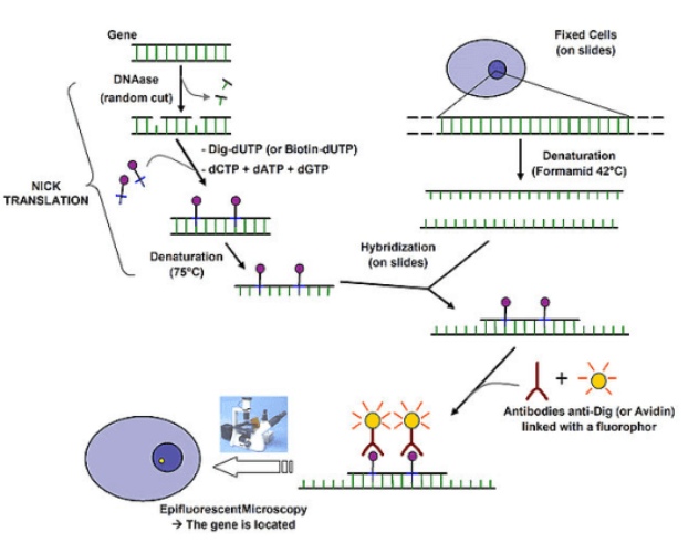
Figure 1: Schematic diagram illustrating the principle of fluorescence in situ hybridization
2. Methods for Labeling Fluorescence In Situ Hybridization Probes
There are two methods for fluorescently labeling FISH probes. The indirect labeling method involves labeling the DNA probe with biotin, while the direct labeling method involves directly conjugating a fluorophore to the probe’s nucleotides or pentose backbone. The direct labeling method involves simpler detection steps but is less sensitive than the indirect labeling method because the signal cannot be amplified.
3. Types of Fluorescence In Situ Hybridization Probes
(1) Double-stranded DNA probes: Currently widely used, simple and easy to handle, and resistant to degradation. Once successfully cloned, large quantities of labeled probes can be obtained using the same amplification and labeling procedures.
(2) Single-stranded DNA (ssDNA) probes: Stable, easier to use, more specific, resistant to RNase, with better tissue penetration, and no self-hybridization.
(3) RNA probes (ribosomal probes): Higher thermal stability, better tissue penetration, higher specificity, and less susceptibility to RNase
(4) Synthetic oligonucleotides: Cost-effective, stable, highly specific, resistant to RNase, with good tissue penetration and high reproducibility
4. Advantages of Fluorescence In Situ Hybridization (FISH) Technology
Fluorescence in situ hybridization (FISH) offers advantages not found in other hybridization methods:
(1) FISH uses non-radioactive labeling, making it safer
(2) FISH has a short experimental cycle and can detect multiple sequences simultaneously
(3) Due to multiple immunochemical reactions that amplify the hybridization signal, FISH sensitivity is comparable to that of radioactive probes
5. Applications of Fluorescence in situ Hybridization
Fluorescence in situ hybridization (FISH) technology is widely used in gene expression analysis, the detection of chromosomal structural and numerical abnormalities, rapid clinical diagnosis of cancer, and human prenatal diagnosis, among other fields, and holds significant practical value.
6. KMD Bioscience Provides High-Quality Fluorescence in situ Hybridization Services
KMD Bioscience has been dedicated to cell biology research for many years. With a team of experienced professionals in fluorescence in situ hybridization (FISH) technology, we have established a comprehensive FISH technical service platform. We offer one-stop FISH technical services ranging from probe design, sample processing, hybridization detection, gene expression studies to data analysis. We follow standardized and regulated testing procedures to ensure the accuracy of experimental results and minimize false positives and false negatives. KMD Bioscience also offers custom synthesis of a wide variety of probes, including those for most plants, animals, and microorganisms. Custom probes can be as large as 150 kb, featuring high specificity and a low error rate. Using fluorescence in situ hybridization technology, KMD Bioscience also provides monoclonal origin identification services.
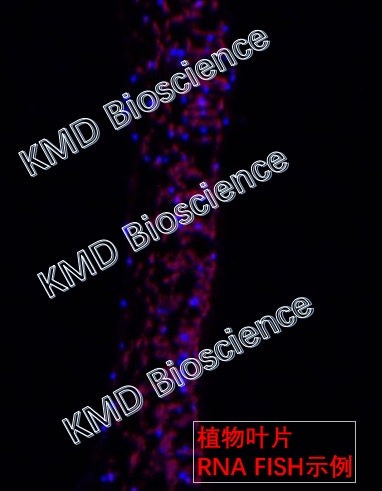
Figure 2: Example of FISH on a plant leaf
7. Overview of the Fluorescence In Situ Hybridization Service Workflow
KMD Bioscience offers fluorescence in situ hybridization (FISH) services. The main technical workflow is as follows: probe design and synthesis, sample preparation, in situ hybridization, imaging, result analysis, and project reporting.
7.1 Probe Design and Synthesis
7.1.1 Ready-to-use probes can be hybridized directly. Non-ready-to-use probes must be diluted with hybridization buffer at a ratio of 1:1:1:50, denatured at 85°C for 3 minutes, and equilibrated at 37°C for 5 minutes.
7.2 Sample Preparation
7.2.1 Dewaxing and rehydration: Immerse the paraffin sections in xylene to remove the paraffin twice, for 10 minutes each time;
7.2.2 Pass the slides sequentially through 100%, 85%, and 75% ethanol for 3 minutes each, followed by 3 minutes in PBS;
7.2.3 Add pepsin digestion solution to the slides and digest at 37°C for 15–30 minutes; wash twice with PBS for 3 minutes each;
7.2.4 Pre-hybridization: Remove the hybridization solution from the refrigerator 30 minutes in advance to allow it to reach room temperature; add the hybridization solution dropwise; incubate in a hybridization instrument at 55°C for 1 hour.
7.3 In Situ Hybridization Experiment
7.3.1 Remove the pre-hybridized slides, remove excess liquid, add the probe dropwise to the slides to completely cover the tissue, and incubate overnight at 37°C in the hybridization instrument;
7.3.2 Wash the slides three times in 2× SSC (0.1% NP-40), 5 minutes each;
7.3.3 Add 20 μL of DAPI-Antifade Solution, cover with a coverslip, and incubate in the dark for 20 minutes.
7.4 Imaging and Photography
Photographs of fluorescence in situ hybridization (FISH) results for various samples from KMD Bioscience:
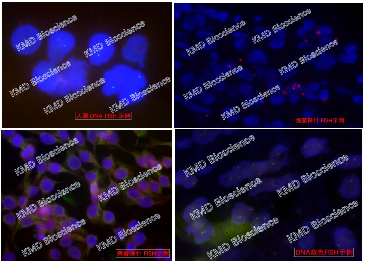
Figure 3: Photographs of Fluorescence In Situ Hybridization (FISH) on Different Samples
7.5 Results Analysis and Project Report
Deliverables from KMD Bioscience: Remaining probes, slides, and samples; original hybridized and stained images; and a detailed experimental report (including the complete experimental procedure and results analysis).
KMD Bioscience’s technical experts will provide professional, one-on-one customized solutions and testing for samples from different species, offering a full suite of customized fluorescence in situ hybridization (FISH) services—from probe design, chromosome/cell/bacterial preparation and culture to result presentation—and delivering comprehensive experimental reports and photographic images.
- Fluorescence In Situ Hybridization (FISH)
- Probe Labeling
- FISH Services

- Protein Platform
- Protein Expression Services
- Protein Modification & Detection Services
- Protein Interaction Services
- Protein Purification Service
- Antibody Discovery Platform
- VHH Antibody Platform
- Phage Display Platform
- Antibody Humanization Platform
- Single B Cell Screening Platform
- Antibody Expression & Validation Platform
- Peptide Library Platform
- Custom Antibody Platform
- Innovative Drug Discovery Platform
- Product Center
- Proteins
- Antibodies
- Raw Materials Reagents
- Small Molecule Reagents
- Peptide Products
- Nanoparticle Products
- Contact Us
- Phone:400-621-6806
- Mobile:13110936064(WeC
- E-mail:[email protected]
- Address:Room 108, Building 10, No. 6 Hechang Road, Wuqing Development Zone, Tianjin City
-

WeChat
-

Service Account
-
 400-621-6806
 -
 3599163699
 -
 Monday to Friday 9:00-18:00
 -

-
 0
0
-
