


Introduction to the Establishment of Immortalized Cell Lines
- 2026-07-10
- 328
1. What Is Cell Immortalization?
Cell immortalization refers to the process by which cells, during in vitro culture, avoid the normal aging and death associated with cell passage—due to intrinsic genetic changes or various external stimuli—thereby enabling long-term passage and indefinite division and proliferation.
2. The Significance of Cell Immortalization
(1) It provides a large supply of cell resources, addressing the challenges of limited passages and slow proliferation. It enables cells to be passaged indefinitely, and immortalized cells grow more vigorously, thereby shortening the experimental timeline.
(2) It helps researchers gain a thorough understanding of the physiological processes of cell proliferation, aging, and death.
(3) They serve as an excellent in vitro model for studying tumor cells at various stages, providing new research directions for cancer treatment and the inhibition of cancer cell proliferation.
(4) As they belong to the same genetic population, they allow for the establishment of cell banks with identical genetic traits, contributing to the stability of experimental results.
Table 1: Commonly Used Immortalized Cell Lines
Cell Name | Form | Notes |
| 3T3 | Mouse Embryonic Fibroblasts | Sturdy and easy to handle; do not touch; stop growing at high density |
| HeLa | Human epithelial cells | Cervical cancer from a human patient named Henrietta Lacks may contaminate other cell lines in culture; it is capable of growing in suspension. |
| COS | Monkey Kidney | Effective transfection; typically used as an expression system for high-level, short-term protein expression |
| 293 /293T/HEK-293T | Human Embryonic Kidney | Easy to transfect and handle; commonly used as an expression system for studying signal transduction and recombinant proteins |
| MDCK | Canine renal epithelial cells | Polarized, with distinct tops and bottoms, used for studying trafficking |
| CHO | Ovaries of Chinese Hamsters | Can be used for biochemical analysis requiring stable gene expression and high protein yield; commonly used as an expression system for studying cellular signaling and recombinant proteins |
| S2 | Fruit Fly Macrophage-Like Cells | A fruit fly cell line with desirable characteristics; highly sensitive to RNAi treatment |
| PC12 | Chromaffin cells in rat pheochromocytoma | Neuron-like cells derived from neuroendocrine adrenal tumors; in the presence of NGF, they can differentiate into neuron-like cells |
| Neuro-2a/N2a | Mouse Neuroblastoma | A model system for studying pathways involved in neuronal differentiation; can be differentiated through stimulation of cannabinoid and serotonin receptors |
| SH-SY5Y | Human Neuroblastoma Clones from Bone Marrow | Dopamine β-hydroxylase-positive, acetylcholinergic, glutamatergic, and adenosineergic; develop into clusters of neurocyte-like cells with short, thin neurites |
3. Methods of Cell Immortalization
There are generally two molecular mechanisms underlying cell immortalization: inhibition by exogenous viral proteins, or spontaneous mutations that activate proto-oncogenes or inactivate tumor suppressor genes, causing cells to bypass normal cell cycle checkpoints and blocking the cellular senescence and death pathways, thereby achieving unlimited proliferation; Another mechanism involves the activation of telomerase, which lengthens telomeres and enables sustained proliferation; oncolytic viruses such as SV40, EBV, and human papillomavirus (HPV) are examples of this.
There are various methods for cell immortalization, and different methods are suitable for different types of tissue cells. For example, HPV can mediate the immortalization of human epithelial cells, such as oral epithelial cells, epidermal cells, and keratinized epithelial cells; the SV40 large T antigen (TAg) from simian viruses is used for adherent cells, fibroblasts, and endothelial cells, while EBV is used for lymphoblastoid cells; telomerase TERT is used for mesenchymal stem cells and certain other cell types.
3.1 Telomerase TERT
Telomeres are specialized structures consisting of DNA-protein complexes located at the ends of eukaryotic chromosomes. Telomeres have important biological functions; they protect the normal structure of chromosome ends from degradation and loss, thereby playing a significant role in normal chromosome replication, apoptosis, and cellular transformation. Telomerase can synthesize new telomeres, thereby extending telomere length. Under normal circumstances, telomerase expression is low; however, under virus-mediated hTERT expression, telomerase can be normally expressed, thereby prolonging the cell’s lifespan.
3.2 SV40 T Antigen
SV40 is a eukaryotic virus. The SV40 T antigen (TAg) exerts its effect by binding to the tumor suppressor proteins Rb and p53 within tumor cells and inhibiting their function.
3.3 Oncogenes
Studies have shown that oncogenes can activate telomerase activity; therefore, the activation of oncogenes such as Myc, c-Jun, c-Ias, vsrc, Mdm2, RAS, c-fos, Bmi1, and CDK4 can promote the process of cellular immortalization.
3.4 Tumor Suppressor Genes
Inactivating tumor suppressor genes such as p53, pRb, BRCA1, and DPC4 can similarly activate telomerase, leading to cell immortalization.
4. Applications of Cell Immortalization
Immortalized cell lines are widely used as a source of biological materials for functional and molecular research and represent a potentially unlimited source for genomic DNA, expressed RNA, and protein and systems biology analyses. In addition, these cell lines can serve as in vitro models for various studies, including functional and serological studies, molecular and genetic studies, large-scale drug library screening, vaccine research, and high-throughput biological studies.
5. KMD Bioscience Provides High-Quality Cell Immortalization Services
KMD Bioscience has extensive experience in cell line immortalization and has successfully immortalized various cell types from different species (such as humans, rats, mice, and pigs), including endothelial cells, epithelial cells, fibroblasts, neurons, and more challenging cell types such as suspension cells, B cells, T cells, and macrophages. KMD Bioscience offers B-cell, T-cell, and macrophage immortalization services. Currently, the primary methods for B-cell immortalization include hybridoma cell preparation through fusion with tumor cells and EBV infection, as shown in Figure 2:
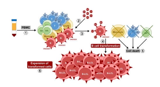
Figure 2: EBV-induced immortalization of B cells
The main methods for immortalizing T cells and macrophages are SV40 infection and EBV infection, as shown in Figure 3:
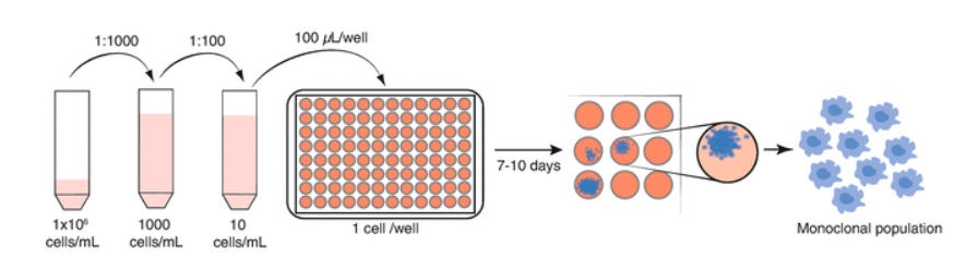
Figure 3: T/Macrophage Immortalization
6. KMD Bioscience Provides Clients with a Comprehensive Overview of the Cell Immortalization Process
Taking B-cell immortalization as an example, this section provides a detailed overview of the EBV-mediated immortalization process, which is primarily divided into three major steps: B-cell expansion and culture, EBV preparation, and B-cell preparation and EBV infection.
6.1 B-Cell Expansion and Culture
Adjust the B-cell density to (5–10) × 10⁶/L and culture in a 37°C, 5% CO₂ incubator for 5–7 days. Do not change or replenish the culture medium during this period to ensure the cell supernatant is rich in EBV.
6.2 EBV Preparation
Collect the B-cell supernatant, centrifuge it to remove cell debris, and collect the supernatant. Perform ultrafiltration of the supernatant according to the instructions for the ultrafiltration centrifuge tubes, then collect the filtrate. Divide the resulting EBV concentrate into 4 aliquots in the order of ultrafiltration, labeling them sequentially as 1, 2, 3, and 4.
6.3 Preparation of B Cells and EBV Infection
Seed the human B cells provided by the client at a density of 5 M/ml. Add 5 or 10 μl of the prepared EBV concentrate to each culture and incubate in a 37°C, 5% CO₂ incubator.
6.4 Four Weeks After EBV Infection
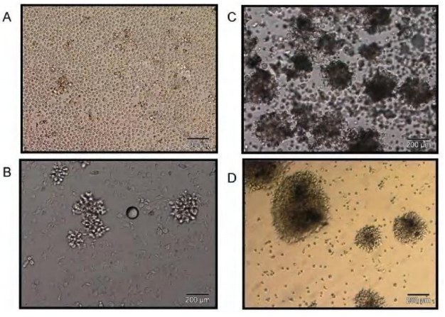
Figure 4: A (Before immortalization) – B (1 week after immortalization) – C (2 weeks after immortalization) – D (4 weeks after immortalization)
Cell immortalization is of great research significance in fields such as the study of biological growth and development mechanisms, the regulation of biological processes, and disease diagnosis and treatment. Customers need only provide isolated PBMCs—at least four tubes, each containing >10^6 cells—and leave the rest to KMD Bioscience. We possess immortalized cell lines from any species and tissue type required for your specific needs. We can establish immortalized cell lines within a short timeframe and, through quality control (QC), confirm that the cells can proliferate for 12–15 generations, delivering a product that meets your satisfaction.
- Cell Immortalization
- Cell Immortalization Services
- B-Cell Immortalization
- T-Cell Immortalization

- Protein Platform
- Protein Expression Services
- Protein Modification & Detection Services
- Protein Interaction Services
- Protein Purification Service
- Antibody Discovery Platform
- VHH Antibody Platform
- Phage Display Platform
- Antibody Humanization Platform
- Single B Cell Screening Platform
- Antibody Expression & Validation Platform
- Peptide Library Platform
- Custom Antibody Platform
- Innovative Drug Discovery Platform
- Product Center
- Proteins
- Antibodies
- Raw Materials Reagents
- Small Molecule Reagents
- Peptide Products
- Nanoparticle Products
- Contact Us
- Phone:400-621-6806
- Mobile:13110936064(WeC
- E-mail:[email protected]
- Address:Room 108, Building 10, No. 6 Hechang Road, Wuqing Development Zone, Tianjin City
-

WeChat
-

Service Account
-
 400-621-6806
 -
 3599163699
 -
 Monday to Friday 9:00-18:00
 -

-
 0
0
-
