


Designing the Perfect Antigen: Core Strategies in mRNA Immunogen Engineering
- 2026-07-21
- 209
The previous article discussed the underlying principles of mRNA-based immunisation. The very design of the antigen encoded by the mRNA serves to precisely direct the immune system to produce the antibodies we require. The ultimate aim of antigen design is to ensure that the recombinant protein is positioned correctly, so that it can present suitable epitopes to the immune system and, with sufficient yield, with sufficient yield, establish an appropriate profile.
Ⅰ. Improving the Efficiency of Transcription and Translation – Yield
(i) 5’UTR:
The most direct approach to designing a 5’UTR is to select a 5’UTR sequence from known human mRNAs that exhibit high expression efficiency in the target cell type. Even so, naturally derived 5’UTRs may contain various regulatory elements that could potentially hinder ribosomal loading under specific conditions[1]. Therefore, when designing the 5’UTR of an mRNA, a strategy should be followed that optimizes translation initiation efficiency while reducing inhibitory elements (such as stable hairpins and uORFs).
Design tools: In recent years, an increasing number of predictive models have emerged that are specifically designed to simulate the influence of the 5’UTR on protein expression and to further optimise and design 5’UTR sequences. Current mainstream methods include Random Forests (RF), Convolutional Neural Networks (CNN) and Generative Adversarial Networks (GAN), amongst others[1]. At the same time, researchers have established a number of specialised databases on 5’UTRs, which provide direct data support for the training and evaluation of these models.
(ii) 3’UTR and Poly(A) Tail:
Although the 3’UTR plays a less critical role than the 5’UTR in regulating mRNA stability and translation efficiency, there are still many factors involved. Examples include AU-rich elements (ARE) and microRNA (miRNA) binding sites.
The Poly(A) tail plays a key regulatory role in the localisation, stability and translation of mRNAs. Its length is a crucial parameter influencing the stability and translational efficiency of both endogenous mRNAs and exogenously delivered mRNAs[2]. With the development of high-throughput sequencing technologies such as TAILseq, mTAILseq, PALseq, FLAMseq, PATseq and nanopore sequencing, researchers are now able to conduct in-depth analyses of the length and sequence composition of the Poly(A) tail[3].
(iii) Codon Optimisation:
Codon optimisation is the process of selecting ideal codons for each amino acid. Synonymous codons can influence the translation efficiency and stability of mRNA[4]. One common approach involves replacing rare codons with preferred codons corresponding to highly abundant transfer RNAs (tRNAs) in order to increase the translation rate; this codon usage bias can be quantified using the codon adaptation index (CAI)[1].
Codon optimisation typically requires a CAI value optimize within the host's preference range to avoid extreme bias, during the design process, it is necessary to avoid clustering caused by repetitive sequences, which can lead to ribosomal frameshift.
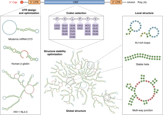
Figure 1: Flowchart of the mRNA Design Optimisation Process[1]
Ⅱ. Design of Subcellular Localisation of Antigens — Localisation
The final location of the antigen directly determines how it is ‘recognised’ by immune cells, thereby shaping the balance between humoral and cellular immunity.
(i) Secretory Type:
Secretory antigen design involves fusing a cleavable signal peptide to the N-terminus of the antigen; following maturation in the Golgi apparatus, the antigen is released into the extracellular space via secretory vesicles, thereby directing the nascent polypeptide chain to the endoplasmic reticulum (ER)[5]. This promotes the direct release of the target antigen into the culture supernatant; this strategy not only simplifies the purification process but also primarily induces a potent Th2-biased humoral immune response.
(ii) Membrane-bound Type:
Antigens are anchored to the cell membrane surface through the retention and transplantation of transmembrane domains (TMDs). Common design frameworks for membrane-fusion antigens include Type I transmembrane proteins, in which the N-terminus is located extracellularly and the C-terminus intracellularly[6]. This approach better mimics the state of natural viruses and is conducive to inducing a cellular immune response.
(iii) Cytoplasmic Type:
By removing the signal peptide and utilising the cytoplasmic anchoring provided by the N-terminal basic residues, the antigen is effectively retained in the cytoplasm. This strategy is suitable for intracellular antigen vaccines, which are presented via MHC class I molecules and strongly activate cellular immunity.
Ⅲ. Rational Design of Antigen Higher-Order Structures
Once these two modules have been completed, we obtain a protein that is highly expressed at the correct location; however, whether it is a functionally correct protein remains to be seen. More crucially, whether this antigen can effectively elicit a target antibody response during the animal immunisation process directly determines the success or failure of subsequent antibody discovery work. Whether the ultimate goal is to obtain polyclonal antibodies for diagnostic purposes or to prepare monoclonal antibodies via phage display or hybridoma technology, the conformational integrity of the antigen, the accessibility of its epitopes and the degree of polymerisation are all core prerequisites. Therefore, the rational design of the antigen’s higher-order structure must be tailored to the specific immunological strategy and antibody screening approach.
(i) Conformation Design:
Using PDB or AlphaFold structures, Optimizing and enhancing thermal stability by introducing disulfide bonds or hydrophobic cores, thereby ensuring that the protein maintains its trimeric conformation at 37 °C; this is because only by retaining its native trimeric conformation can the correct antigenic epitopes be exposed, thereby inducing effective functional antibodies.
(ii) Epitope Focus:
Alanine scanning can be used to identify immunodominant epitopes, and point mutations can be introduced to disrupt their binding affinity with antibodies, thereby eliminating immunodominant but non-protective epitopes. Glycosylation masking can also be employed for this purpose; this involves inserting an N-X-S/T motif into the region adjacent to the non-neutralising epitope, utilising the steric hindrance of the glycan to mask the region and thereby redirect the immune response towards conservative neutralising epitopes.
(iii) Presentation Format:
①Ferritin Nanocages: An antigen monomer is fused to the N-terminus of ferritin, positioning key epitopes on the surface of the nanoparticles to enable strong binding to B-cell receptors.
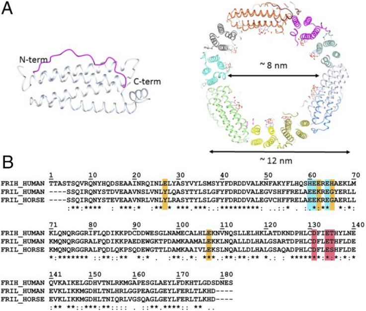
Figure 2: Schematic Diagram of the Structure of Ferritin
②Virus-like Particles: By utilising the envelope protein as a scaffold and chimerising the target epitope into the exposed loop region on the particle surface, these particles can be preferentially captured by lymph nodes due to their size.
Ⅳ. Tools and Methods for Verifying Effectiveness
(i) Rapid Preliminary Screening for Expression and Localisation:
①Protein Immunoblotting (Western blot, WB): By analysing whole-cell lysates and supernatants, this technique allows rapid confirmation of whether the molecular weight is correct, whether the protein is expressed in full, and whether there is any degradation.
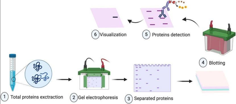
Figure 3: Schematic diagram of the WB process
②Immunofluorescence: Co-staining with specific labels and organelle markers to accurately determine protein localisation.
(ii) Verification of Conformation and Epitope Accessibility:
① Enzyme-Linked Immunosorbent Assay (ELISA): The experiment is validated by using a linear-epitope antibody as a reference and a conformation-specific antibody as the test reagent. If the signal ratio of the conformation-specific antibody to the linear-epitope antibody exceeds a predefined threshold, the antigen is considered to have the correct conformation. If the signal ratio of the conformation-specific antibody to the linear-epitope antibody exceeds a predefined threshold, this indicates that the conformation is correct.
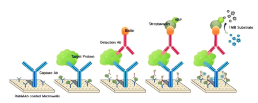
Figure 4: Schematic Diagram Illustrating the Principle of the ELISA method
②Fluorescence-Activated Cell Sorting (FACS): To assess the exposure of epitopes in the native conformation of membrane-bound antigens, flow cytometry is used to detect the binding capacity of neutralising antibodies. If the experimental results show that the fluorescence intensity exhibits a dose-dependent saturation curve as the antibody concentration increases, this confirms that the target epitope is fully exposed and accessible on the surface of live cells.
This article systematically outlines a comprehensive strategy for mRNA immunogen engineering: from 5′UTR and codon optimisation to Poly(A) tail design, to enhance antigen yield; regulating the direction of the immune response through secretory, membrane-bound or cytoplasmic localisation; and subsequently ensuring correct epitope presentation through conformational locking, epitope focusing and nanoparticle polymerisation. Supplemented by validation methods such as ELISA and FACS, this approach provides a practical design framework for the efficient induction of antibody production, ultimately aiming to optimise germinal centre responses and achieve broad-spectrum, long-lasting immunity.
In terms of antigen production, KMD Bioscience offers bespoke recombinant protein expression services using a variety of expression systems, including E. coli, yeast, insect cells and mammalian cells, covering a wide range of receptors, cytokines and viral antigens; in terms of conformation validation, the company provides one-stop antibody preparation services ranging from gene sequence analysis to antibody purification, including humanised antibodies and nanobodies.
From antigen design to protein production, and from antibody development to cell line construction—with its one-stop technical platform, KMD Bioscience helps you turn your blueprint for the ‘perfect antigen’ into a tangible, usable research tool in the laboratory.
Once we have designed mRNA capable of displaying the ‘perfect state’ using the strategies outlined above, our ultimate goal is to establish a highly efficient, long-lasting and broad-spectrum antibody response. How can this goal be achieved? How can the germinal centre response within the body be optimised? Can mRNA be used directly to encode therapeutic antibodies? The final instalment will provide comprehensive answers to these questions.
References
[1]Jin L, Zhou Y, Zhang S, Chen SJ. mRNA vaccine sequence and structure design and optimization: Advances and challenges. J Biol Chem. 2025;301(1):108015. doi:10.1016/j.jbc.2024.108015
[2]Spiewla T, Czubak K, Pilch Z, et al. PolyA tail segmentation improves the stability of the template DNA and increases the translatability of in vitro transcribed mRNA. Nucleic Acids Res. 2026;54(2):gkaf1412. doi:10.1093/nar/gkaf1412
[3]Passmore LA, Coller J. Roles of mRNA poly(A) tails in regulation of eukaryotic gene expression. Nat Rev Mol Cell Biol. 2022;23(2):93-106. doi:10.1038/s41580-021-00417-y
[4]Li Y, Wang F, Yang J, Han Z, Chen L, Jiang W, Zhou H, Li T, Tang Z, Deng J, He X, Zha G, Hu Z, Hu Y, Wu L, Zhan C, Sun C, He Y, Xie Z. Deep generative optimization of mRNA codon sequences for enhanced mRNA translation and therapeutic efficacy. Nat Commun. 2025 Nov 12;16(1):9957. doi: 10.1038/s41467-025-64894-x. PMID: 41224770; PMCID: PMC12612108.
[5]Sato S, Minagawa N, Hirata Y, et al. Evaluating Signal Peptide Efficiency for Extracellular Protein Secretion for mRNA Vaccine Design. Biol Pharm Bull. 2025;48(5):706-712. doi:10.1248/bpb.b25-00155
[6]Sueda S, Tsuruga R, Hirakawa T, Fujii S. Cell surface display of a protein based on a tail-anchored membrane protein. Biochem Biophys Res Commun. 2025;761:151738. doi:10.1016/j.bbrc.2025.151738
FAQs
Ⅰ. The article mentions that, when designing 5’UTRs, naturally derived UTRs may contain elements that hinder ribosome loading. Specifically, which types of regulatory elements are considered ‘superfluous’ and need to be removed?
There are two main types to focus on: firstly, upstream open reading frames, which competitively bind to the scanning ribosome, thereby reducing the translation initiation efficiency of the primary open reading frame; and secondly, regions with overly stable secondary structures—particularly the neck-loop structures near the 5’ UTR—which physically obstruct the scanning of the 43S pre-initiation complex. Furthermore, certain cell-type-specific inhibitory regulatory motifs must also be specifically excluded depending on the target cell line.
Ⅱ. Regarding the length of the poly(A) tail, the article mentions that ‘length is an important parameter’. In practical mRNA design, is there an optimal range for the poly(A) length?
The prevailing view is that 100–120 adenosines (A) is the optimal length range. A poly(A) tail that is too short (<30 nt="">200 nt) may actually induce translation inhibition and complicate plasmid construction and IVT (in vitro transcription). However, the optimal length may vary slightly depending on cell type and delivery method; it is recommended to refer to empirical data for similar antigens in the target system when designing the construct.
Ⅲ. In codon optimisation, a CAI value of at least 0.85 is a common benchmark. If the CAI value for a given antigen sequence is already high, is it still necessary to consider the secondary structure of the mRNA?
This is absolutely essential. The CAI measures the degree of alignment between codon usage and host tRNA abundance, but it does not directly reflect the translatability of mRNA. Even if the CAI is high, if the mRNA forms a highly stable hairpin structure around the initiation codon AUG, the ribosome will still be unable to bind effectively and initiate scanning, and translation efficiency will be significantly reduced. Therefore, the CAI and the free energy of the mRNA’s secondary structure must be optimised in tandem; neither can be neglected.
Ⅳ. The section on ‘conformational design’ in the article mentions that introducing disulphide bonds increases the Tm by 5–10 °C. Why is an increase of this magnitude in the Tm biologically significant?
The human core temperature is approximately 37 °C, whilst the temperature environment encountered by recombinant antigens within the body may be higher. The Tm value represents the midpoint temperature at which a protein transitions from its native folded state to an unfolded state. Raising the Tm value from 40 °C to above 45 °C means that the proportion of the protein in an unfolded state at 37 °C decreases exponentially. This enables the antigen to be continuously presented to immune cells in the correct conformation during its exposure window of several hours to several days within the body, rather than rapidly transforming into a non-functional aggregated state.
Ⅴ. Both ferritin nanocages and virus-like particles (VLPs) are used for polymeric display; what are the fundamental differences in their design approaches?
The key difference between the two lies in their presentation logic. Ferritin nanocages are ‘synthetically engineered scaffolds’, in which antigen monomers are attached to the N- or C-terminus of ferritin subunits via genetic fusion or chemical conjugation; upon assembly, the antigens are ‘suspended’ on the surface of 24-mer nanoparticles. The advantages of this approach are its well-defined structure, uniform size, and independence from virus-derived components. VLPs, on the other hand, utilise a ‘naturally derived scaffold’, whereby viral structural proteins self-assemble to form particles, with the target epitope being genetically engineered to be inserted into the exposed loop regions on the surface of the capsid protein. The advantage of VLPs lies in their retention of the viral particle’s natural immunostimulatory properties; however, the scaffold itself may trigger an anti-vector immune response, and the vector suppression effect must be taken into account during repeated immunisations.
- Immunological strategies
- animal immunization
- antibody discovery

- Protein Platform
- Protein Expression Services
- Protein Modification & Detection Services
- Protein Interaction Services
- Protein Purification Service
- Antibody Discovery Platform
- VHH Antibody Platform
- Phage Display Platform
- Antibody Humanization Platform
- Single B Cell Screening Platform
- Antibody Expression & Validation Platform
- Peptide Library Platform
- Custom Antibody Platform
- Innovative Drug Discovery Platform
- Product Center
- Proteins
- Antibodies
- Raw Materials Reagents
- Small Molecule Reagents
- Peptide Products
- Nanoparticle Products
- Contact Us
- Phone:400-621-6806
- Mobile:13110936064(WeC
- E-mail:[email protected]
- Address:Room 108, Building 10, No. 6 Hechang Road, Wuqing Development Zone, Tianjin City
-

WeChat
-

Service Account
-
 400-621-6806
 -
 3599163699
 -
 Monday to Friday 9:00-18:00
 -

-
 0
0
-
