


The Underlying Logic of mRNA Immunisation: From Innate Recognition to the Initiation of the Adaptive Response
- 2026-07-06
- 152
The 2023 Nobel Prize in Physiology or Medicine was awarded to Drs Katalin Karikó and Drew Weissman in recognition of their discoveries regarding nucleoside modifications, which enabled the development of effective mRNA vaccines against COVID-19[1]. This award has also brought the mechanism of action of messenger RNA (mRNA) to the forefront of public attention. This naturally raises the question: when an mRNA vaccine is administered, what changes occur within the body?

Figure 1: Drs Katalin Karikó and Drew Weissman, Winners of the 2023 Nobel Prize in Physiology or Medicine[2]
I. Inherent Immune Recognition of mRNA
(i) The PRR Recognition Mechanism of mRNA
mRNA can induce innate immunity, which serves as the body’s first line of defence against non-self substances. Pathogen-associated molecular patterns (PAMPs) on mRNA can be recognised by pattern recognition receptors (PRRs) on the cell surface. The binding of the ligand-receptor complex triggers signal transduction into the cell, which in turn initiates a cascade of signalling pathways[3].
(ii) RNA-Sensing Receptor
One of the major RNA-sensing PRRs is the Toll-like receptors (TLRs). Humans possess ten TLRs (TLR1–10), nearly half of which recognise nucleic acid ligands. TLR3, TLR7/8 and TLR9 are located intracellularly and respond to double-stranded RNA (dsRNA), single-stranded RNA (ssRNA) and deoxyribonucleic acid (DNA), respectively [4]. On the other hand, intracellular RNA is recognised by a specific family of proteins known as RIG-I-like receptors (RLRs), which include Retinoic Acid-inducible Gene I (RIG-I) and melanoma differentiation-associated gene 5 (MDA-5); these proteins are capable of detecting viral RNA.
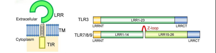
Figure 2: Schematic Diagram of the TLR Structure[4]
In summary, TLRs can recognise RNA, whilst RLR acts as an alarm system for abnormal RNA.
(iii) The Central Dilemma
The current dilemma is that the more easily mRNA is detected by the body, the more intense the body’s inflammatory response becomes; if this response is too intense, it prevents cells from translating the mRNA normally. Therefore, a solution must be found that both activates the immune system and preserves the mRNA.There are currently two relatively well-established methods:
①Replace the Uridine (U) bases in an RNA sequence with naturally occurring pseudouridine (Ψ)[5] or N1-methylpseudouridine (m1ψ)[6].
②Encapsulation Using Lipid Nanoparticles (LNPs): Unencapsulated mRNA degrades very readily within the body; LNP-encapsulated mRNA, however, benefits from the protective shell provided by the LNP, which slows down the rate of degradation. Furthermore, the ionisable lipids within the LNP carry a positive charge in the acidic environment of the endosome, helping the mRNA to escape from the endosome into the cytoplasm, thereby enabling the efficient translation of the antigen protein[7]. Furthermore, certain LNP components can activate the innate immune response and may exhibit some adjuvant effects in vaccine applications, providing activation signals to the immune system.
II. The Bridge from Innate to Adaptive Immunity: Antigen Presentation and Initial T-Cell Activation
Once mRNA enters the body, it is translated by ribosomes to produce antigenic proteins; these antigens must be presented to the immune system in order to be effective.
Adaptive immune responses require the involvement of antigen processing and presentation pathways. These pathways enable glycoproteins encoded by the Major Histocompatibility Complex (MHC) to be loaded with their corresponding ligands[8]. Only MHC molecules bearing ligands, when expressed on the surface of antigen-presenting cells, can activate T cells; the T cells then carry out effector functions such as cytotoxicity, providing help to B cells, and producing cytokines[8].
The presence of MHC Class I and Class II molecules determines the antigen-presenting function on the surface of most nucleated cells, respectively. The former is primarily responsible for presenting intracellular antigenic peptides to CD8⁺ cytotoxic T cells, thereby initiating the killing of target cells in the event of infection or malignancy; the latter, on the other hand, is a function performed specifically by certain antigen-presenting cells, which digest and absorb antigens, break them down into peptide fragments, and then present these to CD4⁺ T cells, thereby modulating the subsequent immune response[9].
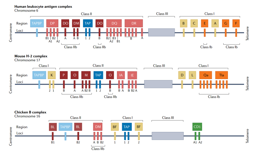
Figure 3: Simplified Diagram of the MHC in Humans, Mice and Chickens[8]
Ordinary cells can present antigens via MHC molecules, but their ‘calls’ are not loud enough during this process; they require the assistance of specific ‘informants’—DCs—to do their job.
DCs are the most efficient antigen-presenting cells and are responsible for initiating the primary immune response. Localised at various sentinel sites throughout the body, they are capable of efficiently capturing and processing antigens. Subsequently, DCs migrate to lymphoid organs, where they initiate and regulate T-cell and B-cell responses by expressing co-stimulatory molecules and secreting bioactive molecules[10]. Furthermore, DCs not only activate lymphocytes to induce immune responses, but also suppress the onset of autoimmune reactions by inducing T-cell tolerance to self-antigens[10].
III. The Initiation of Adaptive Immunity: Follicular Helper T cells and the Prelude to the Germinal Centre
Follicular helper T cells (Tfh cells) are cells that specifically assist B cells. Tfh cells have two key characteristics: selective localisation and direct physical interaction with B cells, enabling them to provide assistance throughout the entire B-cell response[11]. This includes assisting B cells in maturing within the germinal centre, driving the production of high-affinity antibodies by B cells, and promoting the formation of long-lived memory B cells. Most high-affinity antibody responses induced by protein antigens depend on Tfh cell help; in the absence of T cell help, B cells are likely to die within 24 hours of antigen recognition.
With the assistance of Tfh cells, activated B cells enter a specialised structure known as the germinal centre. Within the germinal centre, B cells undergo a ruthless process of ‘survival of the fittest’. The germinal centre is a microanatomical site where B-cell clones expand and antibody affinity matures. Here, B cells undergo the processes of immunoglobulin somatic diversification and affinity-driven selection—a Darwinian evolutionary process that ultimately yields high-affinity antibodies, which are crucial for effective humoral immunity. Ultimately, the surviving B cells differentiate into antibody-secreting cells or memory B cells. The result of this entire process is that the immune system does not simply recognise any antibody, but rather precisely identifies and selectively targets pathogens using high-affinity antibodies with exceptional binding capacity.
The aforementioned germinal center reactions ultimately give rise to long-lived plasma cells and memory B cells, which together constitute the humoral immune memory and continuously produce populations of antibodies directed against different epitopes. The production of monoclonal and polyclonal antibodies, as we commonly refer to them, depends on this reaction. These two types of antibodies serve not only as indicators for evaluating the immunogenicity of mRNA vaccines in experiments, but also as the primary sources for antibody development.
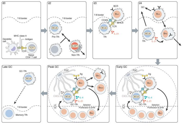
Figure 4: Kinetics of Tfh and B-cell Responses[12]
This article systematically outlines the complete chain of events by which mRNA triggers an immune response: the innate immune system recognises mRNA via PRRs such as TLRs and RLRs; nucleoside modification and LNP delivery resolve the conflict between immunogenicity and translational efficiency; antigens are presented by MHC, DCs activate T cells, which in turn guide B cells into the germinal centre via Tfh cells, where they undergo high-frequency somatic mutation and affinity selection, ultimately differentiating into plasma cells secreting high-affinity antibodies and memory B cells, thereby laying the immunological foundation for the discovery of precise antibodies.
In the context of innate immune recognition, KMD Bioscience offers a range of primary antibody products targeting various innate immune receptors.In the areas of antigen expression and presentation, KMD Bioscience supplies recombinant protein products that can be used as standards for ELISA assays or as positive controls for Western blot analyses,Furthermore, KMD Bioscience’s antibody product range covers a wide variety of high-demand targets and can serve as a reference tool for validating antigen-antibody binding activity.Furthermore, KMD Bioscience’s phage display technology platform offers antibody screening services using antibodies derived from immunised animals or from libraries, enabling the efficient in vitro enrichment and affinity maturation of antibody candidates.Furthermore, our range of secondary antibodies for serum antibody titre testing fully supports research requirements throughout the entire process, from the assessment of immune responses to the validation of antibody function.
Having understood how the immune system is ‘awakened’ by mRNA and how Tfh-B cell collaboration is initiated, the next key question is: how can we design the mRNA-encoded antigen itself to precisely guide the immune system to produce the antibodies we require? The next article will explore strategies for immunogen engineering in greater depth.
References
[1]https://www.cell.com/immunity/collections/mRNA-vaccines?utm_campaign=Immunity&utm_content=268990791&utm_medium=social&utm_source=twitter&hss_channel=tw-295993800
[2]https://www.nobelprize.org/prizes/medicine/2023/summary/
[3]Wang Y, Zhang Z, Luo J, Han X, Wei Y, Wei X. mRNA vaccine: a potential therapeutic strategy. Mol Cancer. 2021;20(1):33. Published 2021 Feb 16. doi:10.1186/s12943-021-01311-z
[4]Shimizu T. RNA recognition in toll-like receptor signaling. Curr Opin Struct Biol. 2024;88:102913. doi:10.1016/j.sbi.2024.102913
[5]Karikó K, Muramatsu H, Welsh FA, et al. Incorporation of pseudouridine into mRNA yields superior nonimmunogenic vector with increased translational capacity and biological stability. Mol Ther. 2008;16(11):1833-1840. doi:10.1038/mt.2008.200
[6]Andries O, Mc Cafferty S, De Smedt SC, Weiss R, Sanders NN, Kitada T. N(1)-methylpseudouridine-incorporated mRNA outperforms pseudouridine-incorporated mRNA by providing enhanced protein expression and reduced immunogenicity in mammalian cell lines and mice. J Control Release. 2015;217:337-344. doi:10.1016/j.jconrel.2015.08.051
[7]Jiang S, Lu Z. mRNA-LNP vaccines: rational design, delivery optimization, and clinical translation. J Mater Chem B. 2025;13(48):15447-15467. Published 2025 Dec 10. doi:10.1039/d5tb01972a
[8]Pishesha N, Harmand TJ, Ploegh HL. A guide to antigen processing and presentation. Nat Rev Immunol. 2022;22(12):751-764. doi:10.1038/s41577-022-00707-2
[9]Wu Y, Zhang N, Hashimoto K, Xia C, Dijkstra JM. Structural Comparison Between MHC Classes I and II; in Evolution, a Class-II-Like Molecule Probably Came First. Front Immunol. 2021;12:621153. Published 2021 Jun 14. doi:10.3389/fimmu.2021.621153
[10]Yao V, Platell C, Hall JC. Dendritic cells. ANZ J Surg. 2002;72(7):501-506. doi:10.1046/j.1445-2197.2002.02450.x
[11]Crotty S. Follicular helper CD4 T cells (TFH). Annu Rev Immunol. 2011;29:621-663. doi:10.1146/annurev-immunol-031210-101400
[12]Crotty S. T Follicular Helper Cell Biology: A Decade of Discovery and Diseases. Immunity. 2019 May 21;50(5):1132-1148. doi: 10.1016/j.immuni.2019.04.011. PMID: 31117010; PMCID: PMC6532429.
FAQs
Ⅰ.What is the most fundamental difference between mRNA vaccines and traditional vaccines?
Traditional vaccines (such as inactivated vaccines) introduce a whole, dead virus into the body, allowing the immune system to recognise the virus’s entire structure. mRNA vaccines, on the other hand, introduce a ‘blueprint’—that is, one that encodes only a key component of the virus—which the body’s cells then produce themselves and display on their surface for the immune system to recognise.
Ⅱ.Why are Tfh cells crucial for the production of high-quality antibodies?
Tfh cells are the dedicated ‘mentors’ of B cells; without their assistance, most B cells would die within 24 hours of recognising an antigen. Through signals such as the secretion of IL-21 and the expression of CD40L, Tfh cells help B cells enter the germinal centre, where they undergo somatic hypermutation and affinity sorting, ultimately differentiating into plasma cells that secrete high-affinity antibodies and long-lived memory B cells. mRNA vaccines are particularly effective at activating Tfh cells, which is one of the key reasons for their ability to elicit a potent antibody response.
Ⅲ.How do antibodies induced by mRNA vaccines differ from those induced by conventional vaccines?
Antibodies induced by conventional vaccines (such as inactivated vaccines) primarily target a variety of antigens on the viral surface, including some non-neutralising epitopes; consequently, the specificity of these antibodies is relatively broad.
In contrast, mRNA vaccines use immunogenic design strategies to precisely encode specific antigens (such as the RBD region of the spike protein), thereby focusing the immune response on key neutralising epitopes. Furthermore, the mRNA-LNP platform naturally favours a Th1-type immune response, inducing predominantly IgG2a antibodies, which typically exhibit stronger neutralising activity than the IgG1 antibodies induced by conventional vaccines.
Research also indicates that mRNA vaccines are particularly effective at activating Tfh cells, thereby driving more efficient germinal centre responses and affinity maturation; the resulting high-affinity antibodies are superior to those produced by conventional vaccines in terms of both quality and persistence.
Ⅳ.What happens between the immune response induced by mRNA vaccines and the production of monoclonal antibodies?
Following administration of an mRNA vaccine, the antigen is expressed in the body and presented by dendritic cells to T cells; Tfh cells guide B cells into the germinal centre, where they undergo high-frequency somatic mutation and affinity selection, ultimately differentiating into antibody-secreting cells and memory B cells.
B cells isolated from the spleen or peripheral blood of these immunised animals can be used to produce monoclonal antibodies via two pathways:
(ⅰ)Hybridoma technology—fusing B cells with myeloma cells and screening for clones capable of stably secreting the target antibody;
(ⅱ)Phage display technology—amplifying the antibody gene pool from B cells, constructing a phage display library, and obtaining high-affinity antibodies through in vitro screening.
The advantage of the mRNA immunisation strategy lies in the fact that the B-cell repertoire activated by antigens in their natural conformation, as expressed in vivo, exhibits greater epitope diversity, thereby providing a higher-quality ‘source pool’ for the subsequent screening of monoclonal antibodies.
Ⅴ.What are the unique advantages of mRNA-based immunological strategies in antibody discovery?
Traditional antibody discovery workflows typically involve immunising animals with recombinant proteins; however, it is difficult to obtain the native conformation of membrane proteins or conformationally unstable antigens through recombinant expression. The mRNA-LNP immunisation strategy overcomes this bottleneck by injecting mRNA encoding the target antigen directly into animals, thereby utilising host cells to translate and present the antigen protein in situ with its native conformation and post-translational modifications.
This approach not only eliminates the cumbersome steps involved in protein expression and purification, but also induces high-quality polyclonal and monoclonal antibodies directed against native epitopes. For example, in the production of antibodies against the GPCR protein PAR4, mRNA-LNP immunisation successfully induced functional antibodies that specifically recognise the native conformation.
- monoclonal antibodies
- mRNA Immunisation

- Protein Platform
- Protein Expression Services
- Protein Modification & Detection Services
- Protein Interaction Services
- Protein Purification Service
- Antibody Discovery Platform
- VHH Antibody Platform
- Phage Display Platform
- Antibody Humanization Platform
- Single B Cell Screening Platform
- Antibody Expression & Validation Platform
- Peptide Library Platform
- Custom Antibody Platform
- Innovative Drug Discovery Platform
- Product Center
- Proteins
- Antibodies
- Raw Materials Reagents
- Small Molecule Reagents
- Peptide Products
- Nanoparticle Products
- Contact Us
- Phone:400-621-6806
- Mobile:13110936064(WeC
- E-mail:[email protected]
- Address:Room 108, Building 10, No. 6 Hechang Road, Wuqing Development Zone, Tianjin City
-

WeChat
-

Service Account
-
 400-621-6806
 -
 3599163699
 -
 Monday to Friday 9:00-18:00
 -

-
 0
0
-
