


Principles and Applications of Monoclonal Antibody Preparation
- 2026-07-10
- 678
1. Basic Concepts of Monoclonal Antibodies
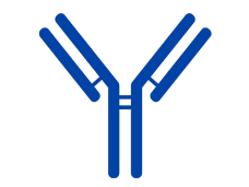
An antibody (Ab) is a Y-shaped globular protein secreted by effector B cells (effector lymphocytes) and used by the body to defend against foreign substances such as viruses, bacteria, and other antigens. Antibodies are found only in the blood of vertebrates and on the surface of B lymphocytes.As the name suggests, a monoclonal antibody (mAb) refers to immunoglobulins secreted by the progeny B cells derived from a single parent B cell. These antibodies exhibit high uniformity, recognizing the same antigenic epitope.The production of monoclonal antibodies typically employs hybridoma technology. For instance, in the preparation of mouse monoclonal antibodies, the immortal characteristics of myeloma cells are utilized to enable B cells to continuously produce antibodies in vitro. The fusion of these two cell types results in an antibody-producing hybridoma.
2. Principle of Hybridoma Monoclonal Antibody Preparation
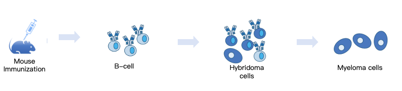
The preparation of hybridomas involves the acquisition and cultivation of two types of cells: myeloma cells and antibody-secreting cells. The antibody-secreting cells commonly used are B lymphocytes, though under certain conditions, other cell types such as peripheral blood mononuclear cells may also be employed. B lymphocytes cannot sustain antibody production under in vitro culture conditions and thus require immortalization, whereas myeloma cells are inherently capable of indefinite proliferation in vitro. The fusion of these two cell types results in a hybrid cell that retains both the antibody-secreting ability of effector B cells and the immortal growth characteristics of myeloma cells.For murine hybridoma preparation, the myeloma cell lines SP2/0 and NS0 are typically used, whereas hybridomas from other species are generally produced through heterologous fusion of these myeloma cells with splenocytes. The schematic principle of hybridoma preparation is as follows:
3. Basic Process of Monoclonal Antibody Preparation
3.1 Antigen Preparation
Antigens come in various types, including small molecule peptides, compounds, proteins, and more. According to immunological theory, when the molecular weight of an antigen is less than 5 kDa, its immunogenicity is insufficient to stimulate an immune response in the body, meaning it cannot elicit the production of spleen cells or other lymphocytes that secrete antigen-specific antibodies. Such antigens are also known as haptens. Before animal immunization, these antigens need to be modified—for example, through carrier conjugation (e.g., BSA, KLH, etc.)—to become complete antigens.
3.2 Animal Immunization
Typically, 6-8-week-old female Balb/c mice are selected for immunization. Each mouse is administered an intramuscular injection at a dose of 50–100 μg/0.5 mL per immunization. The immunization is repeated 4–6 times, and serum is collected before each subsequent immunization. The antibody titer in the serum is then measured by ELISA.
3.3 Cell Fusion
When the serum antibody titer in mice reaches the expected level (~10⁴ ELISA titer), blood is collected, and the mice are euthanized and sterilized. The spleen is then harvested, minced, and pressed through a mesh screen to prepare a single-cell suspension. Pre-recovered myeloma cells are mixed with mouse splenocytes at a specific ratio, and fusion between lymphocytes and myeloma cells is induced using PEG or electrofusion to generate hybridoma cells.
3.4 Selective Culture
Selective culture utilizes the characteristics of hybridoma cells. Properly fused myeloma cells, which encode hypoxanthine-guanine phosphoribosyltransferase (HGPRT), can survive in HAT selective medium, whereas myeloma-myeloma cell fusions and B cell-B cell fusions fail to survive under these selective culture conditions.
3.5 Hybridoma Cell Cloning and Subcloning
After screening with HAT selective medium, hybridoma cells tend to aggregate into clusters. These clustered cells are dispersed using the limiting dilution method and then transferred into 96-well plates for clonal cultivation. The antibody titer, specificity, affinity, and antigen epitope recognition in the cell culture supernatant are assessed via ELISA. Positive clones are selected and subjected to repeated subcloning following the same procedure, ultimately yielding hybridoma cell lines derived from a single B cell. This step is critical in the entire hybridoma preparation process, as the screening strategy and methodology play a pivotal role in obtaining the desired monoclonal antibody hybridomas.
3.6 Large-Scale Monoclonal Antibody Production
Large-scale monoclonal antibody (mAb) production primarily involves two methods: ascites production and in vitro cultivation.
3.6.1 Ascites Production Method:Syngeneic Balb/c mice are pretreated intraperitoneally with liquid paraffin or pristane for 1–2 weeks. Subsequently, hybridoma cells are injected into the peritoneal cavity. The hybridoma cells proliferate within the mouse abdomen and secrete monoclonal antibodies. After approximately 1–2 weeks, abdominal swelling becomes visible. Ascitic fluid is then extracted via syringe puncture, yielding a high concentration of monoclonal antibodies.
3.6.2 In Vitro Cultivation Method:The in vitro cultivation method typically involves large-scale hybridoma cell culture using cell factories. The cell culture supernatant is collected for antibody purification. The advantage of this method is that it produces animal-free monoclonal antibodies, while the disadvantage is the relatively low antibody concentration in the supernatant, necessitating large-scale production and supernatant concentration.
4. Applications of Monoclonal Antibody Production
① Medical Diagnosis and Treatment: Monoclonal antibodies are used in medical diagnosis to detect specific proteins, pathogens, or other biomolecules. In therapeutics, they are employed to treat cancers, autoimmune diseases, infectious diseases, and more. For example, certain monoclonal antibodies in cancer therapy are designed to target tumor cells, such as Rituximab for treating non-Hodgkin's lymphoma.
② Biological Research: Monoclonal antibodies serve as tools in laboratory research to study the expression, localization, and function of specific proteins in biological systems. They are used in experimental techniques such as Western blotting, flow cytometry, and immunohistochemistry.
③ Drug Development: In the drug development process, monoclonal antibodies can act as biological targets for candidate drugs. They are also used as drug carriers—for instance, by attaching drugs to monoclonal antibodies to achieve targeted delivery to specific cells.
④ Diagnostic Reagents: Monoclonal antibodies are widely used in the preparation of medical diagnostic reagents, such as ELISA (enzyme-linked immunosorbent assay) and immunochromatography. They can detect biomarkers in serum to aid in disease diagnosis.
⑤ Immunotherapy: Some monoclonal antibodies are designed to enhance immune system functions, such as by activating T cells or inhibiting immune checkpoints to boost anti-tumor immune responses.
⑥ Vaccine Production: Monoclonal antibodies can be used as raw materials in vaccine production, such as for quality control in viral vaccine manufacturing.
- Monoclonal antibody preparation
- mouse monoclonal antibody production
- hybridoma technology
- hybridoma monoclonal antibody preparation

- Protein Platform
- Protein Expression Services
- Protein Modification & Detection Services
- Protein Interaction Services
- Protein Purification Service
- Antibody Discovery Platform
- VHH Antibody Platform
- Phage Display Platform
- Antibody Humanization Platform
- Single B Cell Screening Platform
- Antibody Expression & Validation Platform
- Peptide Library Platform
- Custom Antibody Platform
- Innovative Drug Discovery Platform
- Product Center
- Proteins
- Antibodies
- Raw Materials Reagents
- Small Molecule Reagents
- Peptide Products
- Nanoparticle Products
- Contact Us
- Phone:400-621-6806
- Mobile:13110936064(WeC
- E-mail:[email protected]
- Address:Room 108, Building 10, No. 6 Hechang Road, Wuqing Development Zone, Tianjin City
-

WeChat
-

Service Account
-
 400-621-6806
 -
 3599163699
 -
 Monday to Friday 9:00-18:00
 -

-
 0
0
-
