


Flow Cytometry Antibody Color Schemes: Five Classic Targets in the CD Series
- 2026-09-15
- 14
Flow cytometry, as a powerful tool for single-cell, multiparameter analysis, has been widely applied in numerous research fields such as immunology, oncology, and hematology. One of the keys to the success of flow cytometry experiments lies in the precise selection of antibody targets and the appropriate combination of fluorescent dyes. As core members of cell surface markers, the CD series molecules are central to various flow cytometry applications, including immunophenotyping, cell activation assessment, and tumor microenvironment analysis.
Today, we will systematically examine the five major targets—CD3, CD28, CD31, CD45, and CD326—from the dual perspectives of molecular biological function and flow cytometry applications, and provide you with practical guidance on dye selection.
I. CD3—The Identity Marker of T Cells
(i) Molecular Overview
CD3 is a key component of the T-cell receptor (TCR) complex. The TCR itself lacks significant intracellular domains and instead transmits signals by binding to CD3 molecules[1]. CD3 consists of CD3εγ, CD3εδ, and CD3ζζ dimers; these CD3 molecules contain intracellular signaling domains capable of coupling antigen recognition signals to downstream signaling pathways. The TCR-CD3 complex is a multichain structure responsible for antigen recognition in T cells[2].
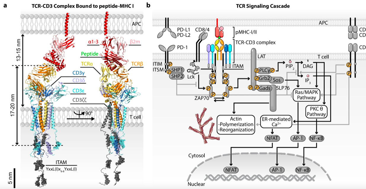
Figure 1: Schematic Diagram of the TCR-CD3 Complex Structure and Signaling Pathway[3]
(ii) Flow-Based Use Cases
Ø A classic marker of T cells in general, expressed on all T cells and NKT cells
Ø A core marker for identifying T cell populations in immunophenotyping
Ø When used in combination with CD4/CD8, it can further distinguish T cell subsets
II. CD28—T-Cell Co-stimulatory Receptor
(i) Molecular Overview
CD28 is a transmembrane glycoprotein receptor on the surface of T cells that belongs to the immunoglobulin superfamily. Full activation of T cells requires two signals: the primary signal mediated by TCR/CD3, and the costimulatory signal mediated by CD28[4]. CD28 binds to its ligands CD80 (B7-1) or CD86 (B7-2) to transduce the most important costimulatory signal[4].
CD28 is widely recognized as the primary co-stimulatory pathway for initial T-cell activation, and the CD28/B7 pathway plays a central role in anti-pathogen immune responses, autoimmune diseases, and graft rejection[5].
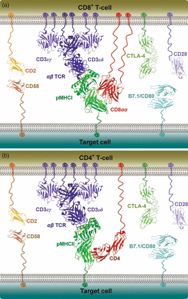
Figure 2: Schematic Diagram of T-cell–APC Immunosynapse Molecules[6]
(ii) Flow-Based Use Cases
Ø Assess the activation status and function of T cells
Ø When used in combination with CD3 detection, provide a comprehensive analysis of the dual signaling pathways involved in T cell activation
Ø Investigate the cross-regulation between immune checkpoints and co-stimulatory signals
III. CD31 (PECAM-1) — A Marker for Endothelial Cells and Platelets
(i) Molecular Overview
CD31, also known as platelet-endothelial cell adhesion molecule-1 (PECAM-1), is a member of the immunoglobulin superfamily. It is expressed on all cells within the vascular compartment—endothelial cells (primarily concentrated at intercellular junctions), most leukocyte subtypes, and platelets[7].
CD31 not only possesses adhesion properties but also serves as a potent signaling molecule that plays multiple roles in vascular biology, including angiogenesis, platelet function, thrombosis, the mechanical sensing of fluid shear stress by endothelial cells, and the regulation of various stages of leukocyte migration across the venous wall[7].
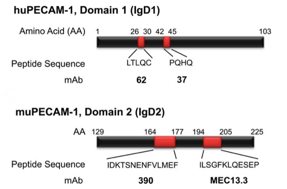
Figure 3: Schematic Diagram of the PECAM-1/CD31 Protein Domains[8]
(ii) Flow-Based Use Cases
Ø Endothelial cell identification and angiogenesis studies
Ø Platelet function analysis
Ø Assessment of vascular density in the tumor microenvironment
Ø Analysis of selected T/B cell subsets and myeloid cells
IV. CD45—A Common Antigen of White Blood Cells
(i) Molecular Overview
CD45, also known as the common leukocyte antigen, is the predominant tyrosine phosphatase in hematopoietic cells[9]. It is expressed on the surface of all nucleated hematopoietic cells (except red blood cells and platelets). CD45 regulates various signaling molecules through dephosphorylation and plays a crucial role in the regulation of T-cell receptor-associated kinases and cytokine receptor signaling.
The extracellular domain of CD45 exhibits considerable variability due to alternative splicing and glycosylation, which enables anti-CD45 antibodies to distinguish between cell populations at different stages of maturation and function[9]. Polymorphic variations in human CD45 have been associated with autoimmune and infectious diseases, establishing CD45 as an important immunoregulatory factor[10].
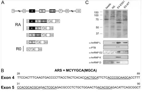
Figure 4: Schematic Diagram of CD45 Alternative Splicing and Isoforms[11]
(ii) Flow-Based Use Cases
Ø CD45/SSC gating strategy—the most classic method for white blood cell identification in flow cytometry
Ø Comprehensive immunophenotyping of hematopoietic cell populations
Ø Immunophenotypic analysis of leukemia and lymphoma
Ø Chimerism monitoring after bone marrow transplantation (CD45.1/CD45.2 allele-specific labeling)
V. CCD326 (EpCAM) — Epithelial Cell and Tumor MarkerD
(i) Molecular Overview
CD326, also known as epithelial cell adhesion molecule (EpCAM), is a type I transmembrane glycoprotein with a molecular weight of approximately 40 kDa. Originally identified as a major surface antigen in human colon cancer, it mediates epithelial-specific cell-cell adhesion[12]. Recent studies have shown that EpCAM’s functions extend far beyond those of an adhesion molecule—it is also involved in cell signaling, migration, proliferation, and differentiation[12].
(ii) Flow-Based Use Cases
Ø Circulating Tumor Cell (CTC) Detection—Defined as CD326⁺/CD45⁻ Cells
Ø Diagnosis and Prognostic Assessment of Epithelial Tumors[12]
Ø Identification and Isolation of Tumor Stem Cells
Ø Detection of Tumor Cells in Malignant Effusions[12]
VI. Principles of Antibody Color Coding
Now that you understand the functions and application scenarios of each marker, how can you effectively combine them into a multicolor flow cytometry panel? Here are a few key principles to keep in mind:
(i) The Principle of Combining Strong and Weak Elements
Pair weakly expressed antigens with strongly fluorescent dyes, and strongly expressed antigens with weakly fluorescent dyes. Fluorophore brightness reference: PE and APC are high-brightness; FITC and PerCP-Cy5.5 are medium or low-brightness.
(ii) Fluorescence Interference Mitigation
Weakly expressed antigens should be placed in a non-interfering channel, while strongly expressed antigens can be placed in channels where spillover may occur. For example, CD45 is highly expressed in all white blood cells and can be paired with a Violet-series fluorophore to avoid interference with key channels at 488 nm or 633 nm.
(iii) Considerations Regarding Antigen Expression Relationships
Mutually exclusive antigens (such as CD3 and CD19) allow for a certain degree of fluorescence bleed; for co-expressed antigens (such as CD3 and CD28), fluorescent combinations with minimal spectral overlap should be selected.
(iv) Instrument Configuration Matching
Before selecting a color scheme, be sure to familiarize yourself with the flow cytometer’s laser and filter configuration to ensure that the selected fluorophores can be properly excited and detected by the instrument.
Item Number | Products | Type | Specifications |
YR1072 | Research-Grade Reference Monoclonal Antibodies,Mouse IgG2a | 1mg/5mg | |
YR1618 | Research-Grade Reference Monoclonal Antibodies,Human IgG4 | 1mg/5mg | |
YR1153 | Research-Grade Reference Monoclonal Antibodies,Fab-G1 Kappa | 100ug/1mg | |
YR1154 | Research-Grade Reference Monoclonal Antibodies | 100ug/1mg |
KMD Bioscience is committed to providing high-quality target-related products to research institutions. The company offers high-quality target products such as CD28 antibodies and EpCAM/CD326 antibodies, as well as monoclonal antibodies labeled with FITC, HRP, and other markers for use in flow cytometry. The company’s antibody functional validation services include flow cytometry (FC) analysis to ensure the efficacy, stability, and biological activity of the antibodies in relevant applications.
FAQs
Q1: Why can CD3 serve as an identity marker for T cells? How does it differ from other T-cell markers?
CD3 is a key component of the T-cell receptor (TCR) complex, consisting of CD3εγ, CD3εδ, and CD3ζζ dimers. The TCR itself lacks a significant intracellular domain and must bind to CD3 molecules to transmit signals. Since CD3 is expressed on all T cells and NKT cells but not on B cells or NK cells (except NKT cells), it is widely used as a classic marker for T cells in general. In contrast, CD4 and CD8 mark the helper and cytotoxic T-cell subsets, respectively, while CD3 serves as the “master switch” for identifying the entire T-cell population—in immunophenotyping, CD3 is typically used first to identify T cells, followed by further subsetting using CD4/CD8.
Q2: Why is CD28 necessary for T-cell activation? Isn’t CD3 signaling alone sufficient?
No, it is not. Full T-cell activation requires two signals: first, the primary signal mediated by TCR/CD3 (antigen recognition signal); and second, the costimulatory signal mediated by CD28. CD28 binds to its ligands CD80 (B7-1) or CD86 (B7-2) to transduce the most critical costimulatory signal. If only CD3 signaling is present without CD28 costimulation, T cells enter a state of anergy and are unable to proliferate effectively or perform their effector functions. Therefore, in flow cytometry experiments, CD3 and CD28 are often detected together to comprehensively assess the activation status of T cells.
Q3: What does the CD45/SSC gating strategy mean? Why is it so widely used?
CD45 is a surface marker found on all nucleated hematopoietic cells (except red blood cells and platelets). SSC (side scatter) reflects a cell’s granularity and complexity. By plotting the fluorescence intensity of CD45 (y-axis) against SSC (x-axis) to create a two-dimensional scatter plot, it is possible to clearly distinguish populations such as lymphocytes (CD45⁺/SSCˡᵒ), monocytes (CD45⁺/SSCᵐᵉᵈ), and granulocytes (CD45⁺/SSCʰⁱ) in peripheral blood. This method is considered a classic because it is simple, rapid, and does not require complex antibody combinations. It effectively excludes interference from debris and red blood cells while accurately identifying leukocyte populations, making it the most commonly used gating starting point in clinical flow cytometric immunophenotyping.
Q4: What are the fundamental differences between CD31 and CD326 in flow cytometry applications? For which research areas are they respectively suitable?
The fundamental difference between the two lies in their completely distinct expression profiles and biological functions.
CD31 (PECAM-1) is expressed on all cells within the vascular compartment—endothelial cells (primarily concentrated at intercellular junctions), most leukocyte subtypes, and platelets. It is primarily used for: endothelial cell identification and angiogenesis research, platelet function analysis, and assessment of vascular density in the tumor microenvironment.
CD326 (EpCAM), on the other hand, is specifically expressed on epithelial cells and epithelial-derived tumors. It is primarily used for: circulating tumor cell (CTC) detection (defined as CD326⁺/CD45⁻ cells), diagnosis and prognostic assessment of epithelial-derived tumors, and detection of tumor cells in malignant effusions, among others.
In short: CD31 targets blood vessels and the endothelium, while CD326 targets epithelium and tumors.
Q5: In multicolor flow cytometry, how exactly is the “strong-weak pairing principle” applied? Can you provide an example?
The core of the strong-weak pairing principle is to pair weakly expressed antigens with strong fluorescent dyes, and strongly expressed antigens with weak fluorescent dyes. This is because antigens with weak expression produce inherently low signals, requiring high-intensity fluorophores (such as PE or APC) to distinguish them from background noise; conversely, antigens with strong expression generate sufficient signals and can be detected using medium- or low-intensity fluorophores (such as FITC or PerCP-Cy5.5), thereby “reserving” the high-intensity fluorophores for the weakly expressed targets that need them most.
For example: In a panel containing CD3 and CD45, CD45 is highly expressed in all white blood cells, while a specific target of interest is weakly expressed. In this case, high-intensity fluorophores such as PE or APC should be allocated to the weakly expressed target, while FITC or V450 should be allocated to CD45. This approach ensures the detection rate of the weakly expressed target while preventing the strong antigen from occupying the valuable high-intensity channel.
[1]Kuhns MS, Davis MM, Garcia KC. Deconstructing the form and function of the TCR/CD3 complex. Immunity. 2006 Feb;24(2):133-9. doi: 10.1016/j.immuni.2006.01.006. PMID: 16473826.
[2]Feito MJ, Jiménez-Periañez A, Ojeda G, Sánchez A, Portolés P, Rojo JM. The TCR/CD3 complex: molecular interactions in a changing structure. Arch Immunol Ther Exp (Warsz). 2002;50(4):263-72. PMID: 12371622.
[3]Travaglino S, Jeon Y, Kim Y, Zhu C, Choi HK. Mechanotransduction through T cell receptors: consensus, controversies and future outlooks. Exp Mol Med. 2026 Mar;58(2):319-335. doi: 10.1038/s12276-026-01639-w. Epub 2026 Feb 5. PMID: 41639425; PMCID: PMC12992811.
[4]Matsumoto A, Dobashi H, Ohnishi H, Tanaka T, Kubota Y, Kitanaka A, Ishida H, Tokuda M, Waki M, Kubo A, Ishida T. Tyrosine phosphorylation of a novel 100-kDa protein coupled to CD28 in resting human T cells is enhanced by a signal through TCR/CD3 complex. Microbiol Immunol. 2003;47(1):63-9. doi: 10.1111/j.1348-0421.2003.tb02787.x. PMID: 12636255.
[5]Bour-Jordan H, Blueston JA. CD28 function: a balance of costimulatory and regulatory signals. J Clin Immunol. 2002 Jan;22(1):1-7. doi: 10.1023/a:1014256417651. PMID: 11958588.
[6]Bridgeman JS, Sewell AK, Miles JJ, Price DA, Cole DK. Structural and biophysical determinants of αβ T-cell antigen recognition. Immunology. 2012 Jan;135(1):9-18. doi: 10.1111/j.1365-2567.2011.03515.x. PMID: 22044041; PMCID: PMC3246648.
[7]Woodfin A, Voisin MB, Nourshargh S. PECAM-1: a multi-functional molecule in inflammation and vascular biology. Arterioscler Thromb Vasc Biol. 2007 Dec;27(12):2514-23. doi: 10.1161/ATVBAHA.107.151456. Epub 2007 Sep 13. PMID: 17872453.
[8]Chacko AM, Nayak M, Greineder CF, Delisser HM, Muzykantov VR. Collaborative enhancement of antibody binding to distinct PECAM-1 epitopes modulates endothelial targeting. PLoS One. 2012;7(4):e34958. doi: 10.1371/journal.pone.0034958. Epub 2012 Apr 13. PMID: 22514693; PMCID: PMC3325922.
[9]Poppema S, Lai R, Visser L, Yan XJ. CD45 (leucocyte common antigen) expression in T and B lymphocyte subsets. Leuk Lymphoma. 1996 Jan;20(3-4):217-22. doi: 10.3109/10428199609051610. PMID: 8624459.
[10]Tchilian EZ, Beverley PC. Altered CD45 expression and disease. Trends Immunol. 2006 Mar;27(3):146-53. doi: 10.1016/j.it.2006.01.001. Epub 2006 Jan 18. PMID: 16423560.
[11]Motta-Mena LB, Smith SA, Mallory MJ, Jackson J, Wang J, Lynch KW. A disease-associated polymorphism alters splicing of the human CD45 phosphatase gene by disrupting combinatorial repression by heterogeneous nuclear ribonucleoproteins (hnRNPs). J Biol Chem. 2011 Jun 3;286(22):20043-53. doi: 10.1074/jbc.M111.218727. Epub 2011 Apr 20. PMID: 21507955; PMCID: PMC3103377.
[12]Patriarca C, Macchi RM, Marschner AK, Mellstedt H. Epithelial cell adhesion molecule expression (CD326) in cancer: a short review. Cancer Treat Rev. 2012 Feb;38(1):68-75. doi: 10.1016/j.ctrv.2011.04.002. Epub 2011 May 14. PMID: 21576002.
- Flow cytometry antibodies
- CD series flow cytometry antibodies
- Immunophenotyping
- CD3\CD28\CD31\CD45\CD326

- Protein Platform
- Protein Expression Services
- Protein Modification & Detection Services
- Protein Interaction Services
- Protein Purification Service
- Antibody Discovery Platform
- VHH Antibody Platform
- Phage Display Platform
- Antibody Humanization Platform
- Single B Cell Screening Platform
- Antibody Expression & Validation Platform
- Peptide Library Platform
- Custom Antibody Platform
- Innovative Drug Discovery Platform
- Product Center
- Proteins
- Antibodies
- Raw Materials Reagents
- Small Molecule Reagents
- Peptide Products
- Nanoparticle Products
- Contact Us
- Phone:400-621-6806
- Mobile:13110936064(WeC
- E-mail:[email protected]
- Address:Room 108, Building 10, No. 6 Hechang Road, Wuqing Development Zone, Tianjin City
-

WeChat
-

Service Account
-
 400-621-6806
 -
 3599163699
 -
 Monday to Friday 9:00-18:00
 -

-
 0
0
-
