


A Brief Overview of the Virus Isolation and Purification Process
- 2026-07-09
- 359
1. A Brief Introduction to Viruses
Viruses contain an RNA or DNA genome surrounded by a protective protein shell encoded by the virus itself. The primary function of a virus is to deliver its DNA or RNA genome into a host cell so that the host cell can express (transcribe and translate) the genome. The viral genome is typically packaged within a symmetrical protein capsid along with associated basic proteins. Nucleic acid-associated proteins, known as nucleoproteins, form the nucleocapsid together with the genome.
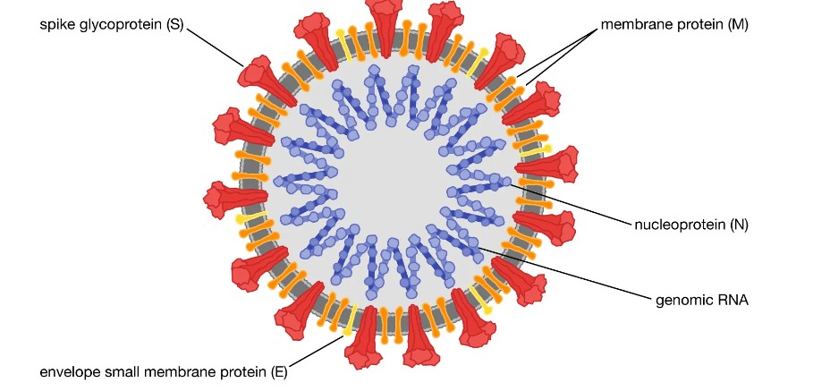
Figure 1: Structure of the SARS-CoV-2 virus
2. Advantages and Disadvantages of Virus Purification Methods
2.1 Ultracentrifugation
A common method for virus concentration and purification involves a combination of virus precipitation and ultracentrifugation, followed by buffer exchange via dialysis or ultrafiltration. This method is well-suited for robust viruses with icosahedral symmetry, but it is not suitable for polymorphic viruses with varying particle sizes, and these viruses typically lose their integrity during ultracentrifugation.
2.2 Chromatography
Chromatography is another method used for virus purification. It can effectively remove contaminants while maintaining particle integrity, thereby ensuring good virus recovery rates. Ion-exchange chromatography on a whole-column system can be used to purify polymorphic viruses. The advantage is that the purification and concentration steps are combined, and particle integrity is maintained due to low shear forces. The disadvantage is that it requires stringent elution conditions and often necessitates an additional buffer exchange step. The use of size-exclusion resins is an alternative method that allows for the purification of polymorphic viruses in their native buffer. However, the viruses may still degrade during loading onto the chromatography column.
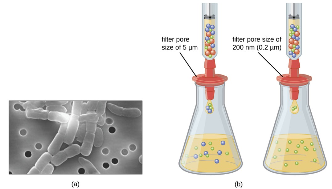
Figure 2: Membrane filters can be used to remove cells or viruses from a solution
3. Virus Isolation and Culture
Vero-E6 and MK-2 (ATCC) cells were maintained in modified Eagle’s medium supplemented with 10% fetal bovine serum and 1× penicillin-streptomycin at 37°C in the presence of 5% CO₂. Virus culture is initiated in standard screw-cap culture tubes (16 × 125 mm; Thermo Fisher Scientific). When cells reach 80% to 90% confluence, inoculate with 500 μl of virus solution containing 33 μl of sample and 2× penicillin-streptomycin solution, and incubate at 37°C for 1 hour. Subsequently, add 5 mL of viral culture medium—composed of MEM, 2% fetal bovine serum, and 1× penicillin-streptomycin solution—to the tube. Maintain the cells in a 37°C incubator and observe for cytopathic effects daily. Starting two days after the initial inoculation, perform RT-PCR analysis using RNA extracted from the culture supernatant every two days to verify the presence of the target virus.
4. Sequencing
The target viral genome was isolated from the viral-containing supernatant using a viral RNA extraction kit (Macherey-Nagel, NucleoSpin RNA Virus). As described previously, viral RNA was transcribed into cDNA in a 10 µL reaction using Superscript IV (Thermo Scientific, Waltham, MA, USA) and random hexamers (Thermo Scientific). Multiplex primers for whole-virus genome sequencing were designed using Primalscheme. The primers were designed to amplify approximately 450 bp overlapping fragments via PCR. Truncated Illumina TruSeq adapter sequences were added to the 5’ ends of the primers. The resulting PCR products were purified using MagSi-NGSPREP Plus magnetic beads (Magtivio) according to the manufacturer’s protocol. Perform paired-end sequencing of the purified PCR products on an Illumina MiSeq sequencer using the v3 600-cycle kit.
5. Virus Purification/Concentration
The virus stock collected in Step 3 was pre-purified at 15 °C at 10,000×g for 20 minutes and used for the further purification/concentration methods described below.
5.1 Virus Particle Formation
For precipitation, 9.5 mL of the pre-purified virus-containing supernatant was spread over a 1.5 mL cushion of 20% (w/v) sucrose buffer containing 20 mM HEPES and 155 mM NaCl, pH 7.0 (HN), and centrifuged for 2 hours at 100,000×g at 4°C. Discard the supernatant, wash the visible precipitate with HN to remove residual sucrose, and resuspend it in 200 µL of HN overnight at 4 °C.
5.2. Purification of the Target Virus Using Size-Exclusion Resin
Wash the Capto Core 700 size-exclusion chromatography resin six times with 10 volumes of HN buffer. Add a 1.2 mL aliquot of Capto Core (bed volume) to 12 mL of pre-clarified supernatant and centrifuge at 4°C for 20 minutes. Precipitate the resin by centrifuging at 800×g for 3 minutes at 4°C, and collect the supernatant containing the virus. Repeat this process using a fresh aliquot of Capto Core resin. Concentrate the purified virus to a final volume of 150 µL using a 4 mL Amicon Ultra ultrafiltration device with a 100 kD MWCO.
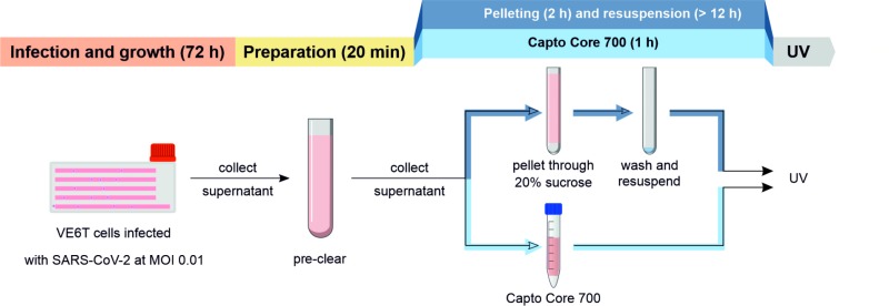
Figure 3: Main Steps in Virus Isolation and Purification
6. Mass Spectrometry
After desalting the peptide samples using a C18 Macrospin column (Nest Group), the desalted samples were analyzed using an Evosep One liquid chromatography system coupled with a CaptiveSpray nano-electrospray ion source and a hybrid capture-ion-flow quadrupole TOF mass spectrometer (Bruker timsTOF Pro).
7. Cryo-Electron Microscopy and Tomography
For cryo-EM, a Leica EM GP plunger was used to vitrify viral samples onto a glow-discharge Quantifoil electron microscopy grid coated with a porous carbon film (R1.2/1.3, mesh 300) at 22 °C, 85% humidity, and a 3-second imprinting time.
The isolation and purification of viruses is essentially the isolation and purification of proteins. KMD Bioscience offers high-quality natural protein purification services, with a variety of protein purification systems (affinity chromatography, ion exchange, molecular sieves, hydrophobic chromatography, HPLC, etc.) to choose from. Our advanced AKTA purification system and pre-packed purification columns in various specifications enable the purification of recombinant or natural proteins from diverse sources. With a purification cycle of just 2–3 weeks, we can deliver high-purity proteins and related experimental data to you.
- Virus Isolation
- Virus Purification

- Protein Platform
- Protein Expression Services
- Protein Modification & Detection Services
- Protein Interaction Services
- Protein Purification Service
- Antibody Discovery Platform
- VHH Antibody Platform
- Phage Display Platform
- Antibody Humanization Platform
- Single B Cell Screening Platform
- Antibody Expression & Validation Platform
- Peptide Library Platform
- Custom Antibody Platform
- Innovative Drug Discovery Platform
- Product Center
- Proteins
- Antibodies
- Raw Materials Reagents
- Small Molecule Reagents
- Peptide Products
- Nanoparticle Products
- Contact Us
- Phone:400-621-6806
- Mobile:13110936064(WeC
- E-mail:[email protected]
- Address:Room 108, Building 10, No. 6 Hechang Road, Wuqing Development Zone, Tianjin City
-

WeChat
-

Service Account
-
 400-621-6806
 -
 3599163699
 -
 Monday to Friday 9:00-18:00
 -

-
 0
0
-
