


A Brief Discussion on Natural Protein Purification Services
- 2026-07-09
- 293
1. Concepts of Natural Protein Purification
The separation and purification of proteins are widely used in biochemical research and applications and constitute an important technical procedure. A typical eukaryotic cell can contain thousands of different proteins; to study a specific protein, it must first be purified from other proteins and non-protein molecules.
KMD Bioscience can provide purification services for both labeled and unlabeled recombinant proteins, as well as the purification and separation of native proteins from tissue fluids, saliva, intestinal fluids, animal or human serum, and even certain animal tissues.
2. Methods for Purifying Native Proteins
Depending on the characteristics of the protein, there are generally five purification methods: affinity chromatography, ion exchange, molecular sieve chromatography, hydrophobic chromatography, and HPLC. We use the advanced AKTA purification system and a variety of pre-packed purification columns to purify recombinant or native proteins from different sources.
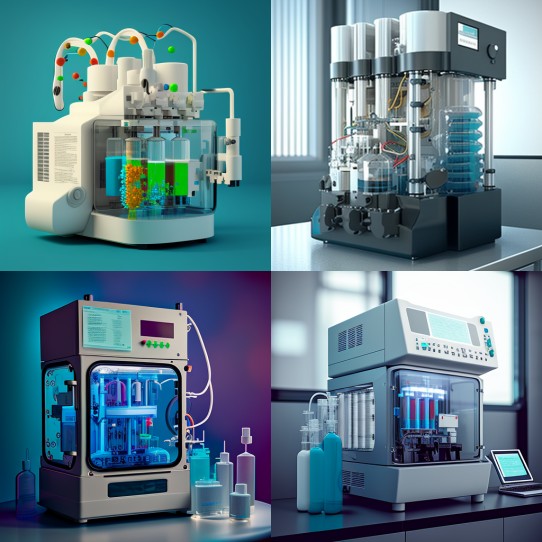
Figure 1: AKTA Purifier
3. Separation and Purification of Various Natural Proteins
3.1 Separation and Purification of Natural Macromolecular Proteins
Based on the physicochemical properties of the target protein to be purified as specified by the client, molecular sieves with appropriate pore sizes are selected for separation and purification. Then, depending on the pH of the sample buffer, suitable anion and cation exchange columns as well as HPLC are selected to identify and collect the corresponding elution peaks.
3.2 Separation and Purification of Natural Small-Molecule Proteins
First, macromolecular impurities are removed through multi-stage separation and filtration using molecular sieves. Next, small-molecule compounds are purified using macroporous resins or reverse-phase resins, followed by further identification of the small-molecule proteins via HPLC and MS.
3.3 Separation and Purification of Mixed Unknown Protein Samples
A tailored protocol is developed based on the characteristics of the target protein to be separated, and the separated products are finally confirmed through various methods, including mass spectrometry and bioactivity assays.
4. Steps for the Purification of Natural Proteins
The general procedure for the separation and purification of natural proteins can be divided into three steps: pretreatment, crude separation, and fine separation.
4.1 Pretreatment
To separate and purify a specific protein, the protein must first be released from its original tissue or cells in a dissolved state while maintaining its natural state and preserving its biological activity. Therefore, connective tissue and adipose tissue should first be removed from animal materials. Then, depending on the specific circumstances, an appropriate method should be selected to disrupt the tissues and cells. Since plant tissues and cells possess cell walls composed of substances such as cellulose, hemicellulose, and pectin, the desired result is generally achieved by grinding them with quartz sand or glass powder in combination with a suitable extraction solution, or by treating them with cellulase.
4.2 Rough Separation
Once the protein extract (which may sometimes be contaminated with nucleic acids, polysaccharides, and the like) is obtained, methods such as salt precipitation, isoelectric point precipitation, and fractional separation using organic solvents are generally employed. These methods are characterized by their simplicity and high throughput; they can remove a large amount of impurities while simultaneously concentrating the protein solution.
4.3 Fine Separation
After rough separation, the protein solution is typically of smaller volume, with most impurity proteins already removed. Methods such as hydrophobic chromatography, ion-exchange chromatography, molecular sieve separation, and affinity chromatography are used for further fine separation.
4.3.1 Pre-purification Testing and Pilot-Scale Purification
First, SDS-PAGE is performed to assess the protein profile. Subsequently, an ion-exchange chromatography protocol is developed by optimizing conditions (buffer, resin, elution conditions, salt gradient, etc.).
(1) The bacterial culture is centrifuged and filtered. The Akta Purifier instrument is used in conjunction with a G25 desalting column for loading and elution to achieve purification.
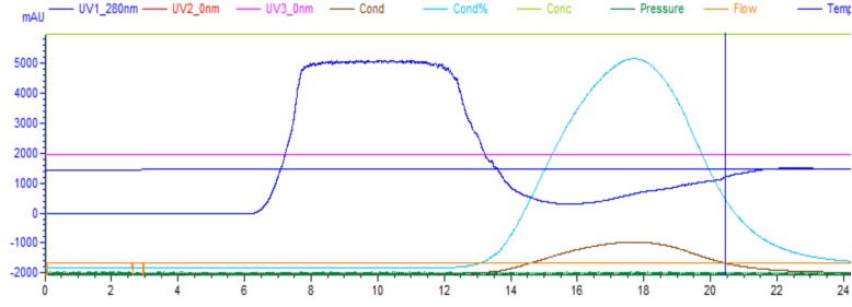
Figure 2: Purification chromatogram of the desalted sample
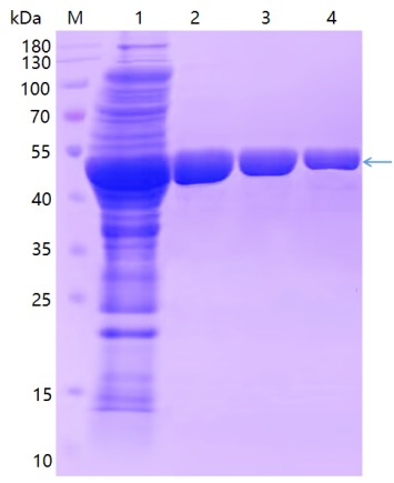
Figure 3: SDS-PAGE gel of the desalted sample
(2) The sample, after desalting on G25, was subjected to SPFF (agarose gel) column chromatography. After centrifugation through the membrane, the sample was purified through the steps of equilibration, loading, and elution.
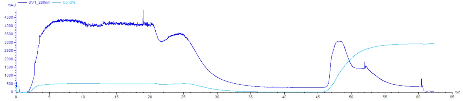
Figure 4: SPFF Separation and Purification Chromatogram
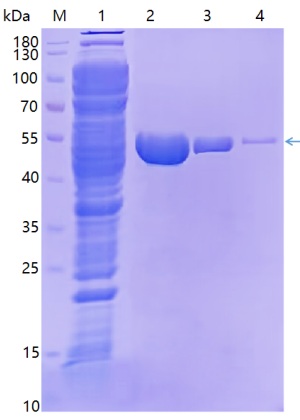
Figure 5: SDS-PAGE gel of the SPFF-purified sample
4.3.2 Protein Purification and Assay
Once the pilot-scale conditions for protein purification have been optimized, large-scale purification can be scaled up based on these conditions. Concentration can be determined using SDS-PAGE (for purity verification), UV absorption, the BCA method, and other techniques.
5. Delivery
KMD Bioscience can provide you with the finished protein, a purification report, and image data.
- Natural Protein Purification
- Recombinant Proteins
- Protein Purification
- Purification Methods

- Protein Platform
- Protein Expression Services
- Protein Modification & Detection Services
- Protein Interaction Services
- Protein Purification Service
- Antibody Discovery Platform
- VHH Antibody Platform
- Phage Display Platform
- Antibody Humanization Platform
- Single B Cell Screening Platform
- Antibody Expression & Validation Platform
- Peptide Library Platform
- Custom Antibody Platform
- Innovative Drug Discovery Platform
- Product Center
- Proteins
- Antibodies
- Raw Materials Reagents
- Small Molecule Reagents
- Peptide Products
- Nanoparticle Products
- Contact Us
- Phone:400-621-6806
- Mobile:13110936064(WeC
- E-mail:[email protected]
- Address:Room 108, Building 10, No. 6 Hechang Road, Wuqing Development Zone, Tianjin City
-

WeChat
-

Service Account
-
 400-621-6806
 -
 3599163699
 -
 Monday to Friday 9:00-18:00
 -

-
 0
0
-
