


Vector Construction Techniques
- 2026-07-09
- 198
1. What is vector construction?
Vector construction is one of the common methods used in molecular biology research. This technique involves inserting an exogenous DNA fragment into an artificially constructed plasmid, with the goal of delivering the ligated DNA molecule into host cells for replication and amplification in subsequent scientific experiments.
2. What are the common methods of vector construction?
Currently, common vector construction techniques include overlapping PCR, traditional restriction enzyme digestion and ligation, and homologous recombination. The traditional restriction enzyme digestion and ligation method involves digesting both the plasmid and the DNA containing the target fragment with the same restriction enzymes to obtain linear DNA with either sticky ends or blunt ends. The digestion products are purified, and then DNA ligase is used to join the fragment to the vector, resulting in a recombinant plasmid. The recombinant plasmid is then transformed into competent cells for subsequent screening and amplification. Homologous recombination cloning technology uses homologous recombination enzymes to recombine two linearized vectors and an insert fragment—both of which have identical terminal sequences—into a recombinant plasmid, which is similarly transformed into competent cells for cell-based panning and amplification.
3. What are the commonly used vector construction expression systems?
Prokaryotic expression vectors: The E. coli expression system requires that the vector contain an SD sequence, a strong promoter with regulatory sequences flanking it, a correct reading frame between the promoter and the exogenous gene, and a transcription termination site downstream of the exogenous gene. The most commonly used vectors are the pET series and the pGEX series. KMD Bioscience achieves highly efficient expression of recombinant proteins through codon optimization, modification of expression vector elements, and the selection of fusion tags and host diversity.
Mammalian cell expression vectors: These contain regulatory elements such as prokaryotic sequences, promoters, enhancers, selection marker genes, terminators, and polyadenylation signals. The primary vector choices are the pcDNA3.1 series and the pcDNA4 series. The high-expression vectors designed by KMD Bioscience enable the company to provide clients with mammalian cell expression and purification services for recombinant proteins at high expression levels.
Yeast Plasmid Vectors: These consist of selection markers (e.g., the nutritional deficiency screening marker HIS3 and the antibiotic screening marker chloramphenicol) and regulatory sequences (including promoters, ARS, 2μM plasmids, and yeast centromeric regions). The primary vector choices are the pPIC9 series and the pPIC3.5 series. KMD Bioscience can simultaneously provide clients with yeast fermentation services for protein production in various scales, combined with a protein purification platform, to obtain high-quality recombinant proteins in a short period of time.
Insect expression vectors: Baculovirus expression vectors use Sf9 and Sf21 cell lines as well as the silkworm as expression hosts. Exogenous genes are cloned into a transfer vector, which is then co-transfected with viral DNA into cells. Recombinant viruses are obtained through homologous recombination and cell-based panning. The primary vector choices are the pFastBac1 series and the pFastBacHT series. Based on the codon preferences of Sf9, Sf21, Hi-5, and S2 insect cells, KMD Bioscience provides free codon optimization to effectively increase the expression levels of recombinant proteins.
4. Basic Workflow for Vector Construction?
4.1 Restriction Enzyme Digestion and Ligation Method
(1) Vector Selection:
Select an appropriate vector based on the purpose of constructing the recombinant plasmid, and analyze the restriction sites present in both the target fragment and the vector.
(2) Primer Design:
Primers should bind specifically to the target fragment and enable its amplification. They should be approximately 18–25 bp in length, with a GC content between 40% and 60%, and should include two suitable restriction sites.
(3) PCR Amplification:
Using the subject’s DNA or cDNA as a template, amplify the target fragment via PCR, then detect and recover the target gene band using 1% agarose gel electrophoresis.
(4) Restriction Enzyme Digestion of the Vector and Target Fragment:
Perform a double-digestion of the target fragment and the plasmid vector using the appropriate restriction enzymes. The entire sample addition process is performed on ice. Samples are mixed thoroughly with a pipette, the bottom of the tube is gently tapped to remove air bubbles, and the sample is then centrifuged to the bottom of the tube using a palm-held centrifuge. Finally, digestion is carried out in a 37°C water bath for 1.5 hours. After the reaction is complete, the products are subjected to 1% agarose gel electrophoresis; if the band positions are correct, the products are recovered.
(5) Ligation and Transformation:
Use T4 DNA ligase to ligate the recovered products from the restriction digestion. This enzyme exhibits high efficiency in ligating sticky ends at 16°C; ligation overnight is sufficient for complete ligation.
(6) Isolation of Single Clones and Plasmid Verification:
Bacterial Suspension PCR: Transfer a single clone to a 1.5 mL Eppendorf tube containing the appropriate antibiotic and incubate for 4–5 h. Add the components according to the PCR protocol used for amplifying the target fragment, using 1 µL of the bacterial suspension as the template DNA. Include a negative control with water as the template and a positive control with the amplified target fragment. After the reaction is complete, run all samples on a 1% agarose gel and make a preliminary assessment of whether ligation was successful based on the intensity and position of the bands.
Restriction digestion verification: Using a double-restriction digestion product of the target fragment as the positive control and a blank double-restriction digestion product as the negative control, subject the post-reaction samples to 1% agarose gel electrophoresis. If a sample group exhibits two bands at the same positions as those in the positive and negative controls, this further confirms the successful ligation of the recombinant plasmid.
Sequencing verification: Send the sample to a sequencing company and verify the results through alignment.
4.2 Homologous Recombination Method
(1) Vector linearization: Use restriction enzyme digestion or reverse PCR amplification to linearize the vector by selecting an appropriate cloning site.
(2) Preparation of the Insert Fragment: Introduce homologous sequences matching both ends of the linearized vector into the 5’ ends of the forward and reverse amplification primers for the insert fragment. This ensures that the 5’ and 3’ termini of the amplified insert fragment carry homologous sequences (15–20 bp, excluding restriction sites) that correspond to the respective ends of the linearized cloning vector.
(3) Recombination reaction: Recover the products from agarose gel electrophoresis and calculate the required amounts of linearized vector and insert DNA for the recombination reaction based on the measured concentrations.
(4) Transform the competent cells.
(5) Identification of recombination products: Perform PCR on bacterial cultures and sequence the products.
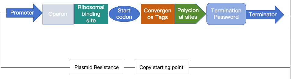
Figure 1: Schematic diagram of an expression plasmid
5. How does the homologous recombination method compare to the traditional restriction enzyme digestion method?
The homologous recombination method is less dependent on restriction sites, takes less time to construct, and involves a simpler process that saves a lot of effort; however, it has a slightly higher false-positive rate.
The traditional restriction enzyme digestion method has been in use for a long time and is technically mature, but it relies on the selection of restriction sites. It has low digestion and ligation efficiencies, and the construction process is relatively cumbersome.
6. Applications of vector construction technology?
(1) Constructing cloning vectors for amplifying the target gene;
(2) Constructing expression vectors for expressing the target gene;
(3) Constructing gene-editing vectors for gene editing;
(4) Constructing viral packaging vectors for cell transfection.
KMD Bioscience offers a wide range of vector construction services, including standard protein expression vector construction (E. coli, mammalian, yeast, and insect systems), as well as vector element replacement and modification. With a professional and dedicated approach, we provide clients with precise consulting services, testing technologies, and research support.
- Vector construction technology
- prokaryotic expression vectors
- mammalian expression vectors
- yeast expression vectors

- Protein Platform
- Protein Expression Services
- Protein Modification & Detection Services
- Protein Interaction Services
- Protein Purification Service
- Antibody Discovery Platform
- VHH Antibody Platform
- Phage Display Platform
- Antibody Humanization Platform
- Single B Cell Screening Platform
- Antibody Expression & Validation Platform
- Peptide Library Platform
- Custom Antibody Platform
- Innovative Drug Discovery Platform
- Product Center
- Proteins
- Antibodies
- Raw Materials Reagents
- Small Molecule Reagents
- Peptide Products
- Nanoparticle Products
- Contact Us
- Phone:400-621-6806
- Mobile:13110936064(WeC
- E-mail:[email protected]
- Address:Room 108, Building 10, No. 6 Hechang Road, Wuqing Development Zone, Tianjin City
-

WeChat
-

Service Account
-
 400-621-6806
 -
 3599163699
 -
 Monday to Friday 9:00-18:00
 -

-
 0
0
-
