


Experimental Procedure for Inserting a Target Gene into a Yeast Strain
- 2026-07-09
- 191
I. Preparation of Competent Cells
1. Inoculate a single colony of the yeast recipient strain onto a YPD agar plate and incubate at 30°C for 2 days;
2. Pick a single colony from the plate and inoculate it into 10 mL of YPD liquid medium; incubate overnight on a shaking incubator at 30°C;
3. After overnight incubation, inoculate 100 ml of YPD medium with approximately 1% of the culture and incubate on a shaking incubator until the OD reaches 1.2–1.5;
4. Centrifuge at 4°C and 5000 rpm for 5 minutes to collect the cell pellet, then resuspend the cells in 100 ml of pre-chilled sterile water;
5. Centrifuge at 4°C and 5000 rpm for 10 minutes to collect the precipitated cells, then resuspend the cells in 100 mL of pre-chilled sterile water;
6. Centrifuge again at 4°C and 5000 rpm for 10 minutes to collect the precipitated cells, then resuspend the cells in 100 mL of pre-chilled sterile water;
7. Wash once with 20 mL of 1 mol/L sorbitol;
8. Dissolve the cells in 200 μL of 1 mol/L pre-chilled sorbitol without adding glycerol, and store at -80°C for several hours to allow for transformation.
II. Electroporation Plating
1. Prepare 80 μL of yeast competent cells and mix with 1–5 μg of linearized plasmid (pre-chilled on ice for 15 min). Quickly transfer the mixture to a 0.2 cm electroporation cup (pre-chilled and sterilized on ice) and perform electroporation;
2. Immediately after electroporation, add 1 mL of sorbitol and plate the mixture (plating may also be performed after 1 hour of incubation on a shaking incubator);
3. Identify the colonies after 3–4 days of growth on MD medium and 4–5 days on ROB medium.
Precautions for Electroporation:
1. The linearized plasmid should be in a concentration of 1–5 µg; higher purity is preferable, and sufficient quantity must be ensured. If you are unable to find positive transformants, consider whether this factor is the cause;
2. When collecting the competent cell suspension, ensure the OD value is between 1.2 and 1.5. You may dilute the suspension by different factors to determine if there is a linear relationship. If the suspension is turbid but the OD value is not high, it may be because the dilution factor was insufficient;
3. Storage of competent cells: If competent cells have been prepared but other steps are not yet complete, the time they spend on ice can affect transformation efficiency. Therefore, the general principle remains: prepare and use immediately. Also, aliquot them into portions sufficient for a single use; once removed, do not return them to the original container to avoid contamination from repeated pipetting;
4. Cleaning of electroporation cups: First, wash and dry them thoroughly, then soak them in 75% ethanol. Before use, UV-sterilize them in a laminar flow hood. Reusing them may also have a certain impact on the experiment;
5. Electroporation parameters: Experiment with voltage and electroporation time by appropriately increasing the voltage or extending the duration. Perform the electroporation process on ice.
III. Isolating Monoclonal Antibodies
Determination of Mut+ and Muts Phenotypes:
After the transformants have grown on the plates for a period of time, screen for Mut+ and Muts. Isolate monoclonal clones by streaking or plating them onto MM and MD media (first plate onto MM plates, then onto MD plates, changing the toothpick for each clone), and incubate at 30°C for 2 days. Observe the colonies: those that grow on MD medium but do not grow or grow very little on MM medium are the Muts phenotype; the rest are the Mut+ phenotype.
IV. Methods for PCR Identification of Recombinants
Template Preparation:
1. When colonies on the plate have grown to the point where they are visible to the naked eye (approximately 12 hours);
2. Prepare all components of the PCR reaction mixture except for the template, and aliquot them. It is recommended to use the detection-specific primers provided in the kit, or one primer from the vector and one gene-specific primer (this approach allows for the determination of the orientation of non-directional clones);
3. Pick up a colony with a sterile toothpick, swab it against the inside of a PCR tube, and transfer it to a sterile 1.5-mL centrifuge tube; label both the PCR tube and the 1.5-mL centrifuge tube;
4. Perform PCR amplification and 1% agarose gel electrophoresis.
Yeast Genome Extraction Method:
1. Inoculate recombinant and empty plasmid transformants into 5 mL of YPDZ medium; use GS115 cells in YPD medium as a control. Incubate at 30°C for 16–18 hours;
2. Centrifuge at 1,500 g for 5–10 minutes at room temperature to collect the cells;
3. Resuspend in 100 μL TE buffer (pH 7.0), add 300 μL EDTA (pH 8.0), 0.07 M Tris-HCl, 3 μL ethanol, and 1 μL Lyticase, then incubate in a 37°C water bath for 30 minutes;
4. Add 200 μL of saturated phenol and 200 μL of chloroform, mix well, centrifuge for 30 seconds, and collect the upper aqueous phase;
5. Add twice the volume of anhydrous ethanol and 1/10 of the volume of NaAC, and let stand at –20°C for 30 minutes;
6. Centrifuge at 10,000 g for 20 min, discard the supernatant; wash the pellet once with 75% ethanol;
7. After drying, dissolve in 15 μL of TE or H₂O, and store at -20°C for later use.
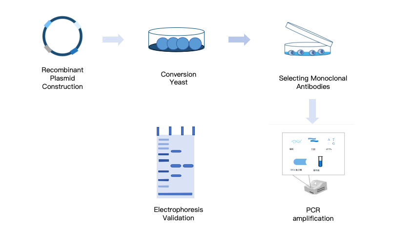
Figure 1: Experimental Procedure for Inserting the Target Gene into a Yeast Strain
V. KMD Bioscience Offers Recombinant Protein Yeast Expression Services
KMD Bioscience has been dedicated to recombinant protein expression and purification services for many years. We offer comprehensive recombinant protein yeast expression and purification services, including the pPICZaA, pGAPZaA, and pPIC9K expression vectors, as well as strains such as X33, GS115, and Saccharomyces cerevisiae.
- Eukaryotic expression
- recombinant protein yeast expression system
- yeast expression
- protein purification

- Protein Platform
- Protein Expression Services
- Protein Modification & Detection Services
- Protein Interaction Services
- Protein Purification Service
- Antibody Discovery Platform
- VHH Antibody Platform
- Phage Display Platform
- Antibody Humanization Platform
- Single B Cell Screening Platform
- Antibody Expression & Validation Platform
- Peptide Library Platform
- Custom Antibody Platform
- Innovative Drug Discovery Platform
- Product Center
- Proteins
- Antibodies
- Raw Materials Reagents
- Small Molecule Reagents
- Peptide Products
- Nanoparticle Products
- Contact Us
- Phone:400-621-6806
- Mobile:13110936064(WeC
- E-mail:[email protected]
- Address:Room 108, Building 10, No. 6 Hechang Road, Wuqing Development Zone, Tianjin City
-

WeChat
-

Service Account
-
 400-621-6806
 -
 3599163699
 -
 Monday to Friday 9:00-18:00
 -

-
 0
0
-
