


Culture of Mammalian Adherent Cells
- 2026-07-10
- 290
I. Principle of Cell Adhesion
Cells secrete an extracellular matrix that can adhere to and anchor onto the surfaces of supporting materials (such as the bottom of culture flasks or petri dishes). Cell adhesion primarily occurs through the expression of various adhesion factors on the cell membrane, enabling binding to these surfaces and exogenous matrices. Therefore, whether a cell can firmly attach to the surface depends not only on the cell's own adhesion capacity (determined by the types and quantities of adhesion molecules it expresses and the extracellular or exogenous matrix molecules it secretes) but also on the specific surface structure of the culture dish material.
II. Culture of Adherent Cells
1.Cell Thawing:
(1) Turn on the UV sterilization of the biosafety cabinet in advance for 30 minutes.
(2) Preheat the water bath to 37°C and warm the culture medium.
(3) Retrieve the cryopreserved cells from liquid nitrogen and quickly thaw them in the 37°C water bath following the principle of "slow freezing, rapid thawing." This process should ideally be completed within 2 minutes to maintain maximum cell viability. After thawing, centrifuge the cells at 1000 rpm for 3-5 minutes. Carefully discard the supernatant, resuspend the cell pellet in pre-warmed complete medium, and transfer the mixture to an appropriate culture dish. Finally, place the dish in a cell culture incubator for further cultivation.
2.Passaging of Adherent Cells:
(1) Before passaging, observe the cell status under a microscope to ensure they are in good condition.
(2) Check the cell confluence to determine if it has reached 70%-90%, as both low and high confluence levels are unfavorable for cell growth.
(3) During passaging, first discard the culture medium and gently wash the cells 2-3 times with sterile phosphate-buffered saline (PBS, pH 7.4), taking care to avoid cell detachment.
(4) After washing, add 0.25% trypsin digestion solution to dissociate the cells. Monitor the digestion process under the microscope until the cells detach and round up into individual spheres. Then, add complete medium to terminate digestion (prolonged digestion may harm cell viability, while insufficient digestion may lead to incomplete detachment).
(5) After resuspending the detached cells, split them into new culture dishes at a ratio of 1:3 to 1:5, add sufficient complete medium, and continue expanding the culture.
3.Cryopreservation of Adherent Cells:
(1) Discard the culture medium and gently wash the cells 2-3 times with sterile phosphate-buffered saline (PBS, pH 7.4), taking care to avoid cell detachment.
(2) Add 0.25% trypsin digestion solution to the washed cells and observe under a microscope until the cells detach and round up. Then, terminate digestion by adding complete medium.
(3) Transfer the detached cells into cryovials at the following ratio: 60% fetal bovine serum (FBS) + 30% cell suspension + 10% dimethyl sulfoxide (DMSO).
(4) Label the cryovials with the cell line name, freezing date, passage number, and other relevant information. Place the vials in a -80°C freezer for 12 hours before transferring them to liquid nitrogen for long-term storage.
4.Transfection of Adherent Cells:
(1) One day before transfection, seed the cells into a culture dish with complete medium and incubate for 24 hours until the cell confluency reaches 60%-80%.
(2) Before transfection, replace the complete medium with antibiotic-free complete medium to avoid interference with transfection efficiency.
(3) Use an appropriate amount of serum-free medium to dilute the plasmid and Lipo3000 transfection reagent. Then, add P3000 to the diluted plasmid and mix thoroughly. Combine this mixture with the diluted Lipo3000, gently pipette to mix, and incubate at room temperature for 20 minutes. After incubation, add the plasmid-P3000-Lipo3000 mixture to the cell culture dish, mix gently, and return the dish to the incubator for 6 hours. Finally, replace the medium with complete medium containing antibiotics.
III. Common Adherent Cell Lines
| Cell name | CHO-K1 cells | 293 cells | vero cells |
| Form | Epidermal cells | Epithelial cells | Epithelial cells |
| Culture medium | Ham's F12K (containing 2 mM L-glutamine, 1.5 g/L NaHCO3), 10% fetal bovine serum | MEM (containing 2 mM L-glutamine, Earle's BSS, 1.5 g/L NaHCO₃, 0.1 mM non-essential amino acids, 1.0 mM sodium pyruvate), 10% horse serum (heat-inactivated) | MEM (containing 2 mM L-glutamine, Earle's BSS, 1.5 g/L NaHCO₃, 0.1 mM non-essential amino acids, 1.0 mM sodium pyruvate), 10% fetal bovine serum |
| Passage | Aspirate the culture medium. Rinse once with 0.25% (w/v) Trypsin-0.53 mM EDTA to remove residual serum (serum inhibits trypsin activity). Add 2–3 mL of Trypsin-EDTA and incubate at 37°C for approximately 5 minutes, or until the cells detach completely. Add 6–8 mL of fresh culture medium and gently dissociate the cells by pipetting. Transfer an appropriate volume of the cell suspension to a new culture vessel. (A 1:4 to 1:8 split ratio is recommended. Change the medium 1–2 times during subculture.) | Aspirate the culture medium. Rinse once with 0.25% (w/v) Trypsin-0.53 mM EDTA to remove serum (serum inhibits trypsin activity). Add 2-3 mL of Trypsin-EDTA and place in a 37°C incubator for approximately 5 minutes, until the cells detach completely. Add 6-8 mL of culture medium and disperse the cells by pipetting. Transfer an appropriate amount of cells to a new culture vessel. (A subcultivation ratio of 1:2 to 1:4 is recommended, and the medium should be changed every 2-3 days during subculture.) | Aspirate the culture medium. Rinse once with 0.25% (w/v) Trypsin-0.53 mM EDTA to remove serum (serum inhibits trypsin activity). Add 2-3 mL of Trypsin-EDTA and place in a 37°C incubator for approximately 5 minutes, until the cells detach completely. Add 6-8 mL of culture medium and disperse the cells by pipetting. Transfer an appropriate amount of cells to a new culture vessel. (A subcultivation ratio of 1:2 to 1:4 is recommended, and the medium should be changed every 2-3 days during subculture.) |
| Cryopreservation | Freeze in liquid nitrogen using 95% culture medium + 5% DMSO. | Freeze in liquid nitrogen using 95% culture medium + 5% DMSO. | Freeze in liquid nitrogen using 95% culture medium + 5% DMSO. |
IV. Applications of Adherent Cell Culture
1.Cell Biology Research: Adherent cell culture can provide valuable information for cytological studies, such as cell division, differentiation, and transformation.
2.Disease Modeling: By culturing adherent cells from patients, researchers can establish cell models related to certain diseases, such as cancer.
3.Drug Screening: The efficacy and toxicity of drugs can be tested using adherent cell cultures, aiding in the identification of potential therapeutic compounds.
4.Gene Editing: Adherent cell culture plays a crucial role in supporting gene-editing technologies like CRISPR-Cas9.
V. Adherent Cell Growth Observation
Cells in closed culture exhibited slow proliferation and strong refractivity. Even after 120 hours of cultivation, the 3% group still failed to form a confluent monolayer.
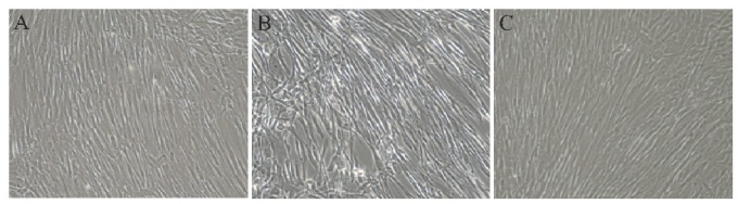
A: Cells in the 2% closed group;B: Cells in the 3% closed group;C: Cells in the 3% open group
KMD Bioscience and our experienced technicians have years of expertise in cell culture. We offer a range of experimental services, including cell transfection, proliferation, apoptosis, co-culture, and stable cell line screening. We develop comprehensive cell culture solutions to provide high-quality services for our clients.
- Adherent cell culture
- Adherent cell thawing/recovery
- Adherent cell passaging
- Adherent cell cryopreservation/freezing

- Protein Platform
- Protein Expression Services
- Protein Modification & Detection Services
- Protein Interaction Services
- Protein Purification Service
- Antibody Discovery Platform
- VHH Antibody Platform
- Phage Display Platform
- Antibody Humanization Platform
- Single B Cell Screening Platform
- Antibody Expression & Validation Platform
- Peptide Library Platform
- Custom Antibody Platform
- Innovative Drug Discovery Platform
- Product Center
- Proteins
- Antibodies
- Raw Materials Reagents
- Small Molecule Reagents
- Peptide Products
- Nanoparticle Products
- Contact Us
- Phone:400-621-6806
- Mobile:13110936064(WeC
- E-mail:[email protected]
- Address:Room 108, Building 10, No. 6 Hechang Road, Wuqing Development Zone, Tianjin City
-

WeChat
-

Service Account
-
 400-621-6806
 -
 3599163699
 -
 Monday to Friday 9:00-18:00
 -

-
 0
0
-
