


How Much Do You Know About SDS-PAGE?
- 2026-07-10
- 283
SDS-PAGE (Sodium Dodecyl Sulfate-Polyacrylamide Gel Electrophoresis) is a discontinuous electrophoretic system developed by Ulrich K. Laemmli, commonly used for separating proteins with molecular weights ranging from 5 to 250 kDa. The combined use of sodium dodecyl sulfate (SDS, also known as sodium lauryl sulfate) and polyacrylamide gel eliminates the effects of protein structure and charge, enabling protein separation based solely on molecular weight differences. In experiments involving expression and purification of foreign proteins in E. coli, SDS-PAGE is indispensable and is routinely employed to analyze protein expression characteristics (including target protein expression levels, expression patterns, purity, etc.).
1.Principle of SDS-PAGE
The principle of SDS-PAGE is that when placed in an electric field, charged molecules migrate toward the electrode of the opposite charge. The separation of charged molecules depends on their relative mobility.
Due to lower resistance during electrophoresis, smaller molecules migrate faster. The structure and charge of proteins also influence their migration speed. Sodium dodecyl sulfate (SDS) and polyacrylamide eliminate the effects of protein structure and charge, allowing proteins to be separated based on the length of their polypeptide chains.
2. The Role of SDS in SDS-Page
SDS is a detergent in the SDS-PAGE sample buffer. It breaks intra- and intermolecular hydrogen bonds, disrupting the secondary and tertiary structures of protein molecules. Meanwhile, DTT (or β-mercaptoethanol) is a strong reducing agent that cleaves disulfide bonds between cysteine residues.
3. Sample processing methods
Based on the purpose of sample separation, there are primarily three treatment methods: reducing SDS treatment, non-reducing SDS treatment, and reducing SDS treatment with alkylation.
Ø Restore SDS treatment
The loading buffer contains SDS and DTT (or β-mercaptoethanol), which denature proteins by disrupting their conformation and neutralizing their charges. This results in the formation of SDS-protein complexes that separate during electrophoresis based solely on molecular weight. This is the standard treatment method for most electrophoresis procedures.
The sample is diluted to an appropriate concentration, mixed with loading buffer, centrifuged, boiled for 5 minutes, and centrifuged again before loading.
Ø Reducing SDS treatment with alkylatio
The alkylation effect of iodoacetamide can permanently and stably protect the SH groups, thereby yielding narrower bands. Additionally, iodoacetamide can also capture excess DTT, preventing streaking during silver staining.
Add 10 μL of 20% iodoacetamide to 100 μL of sample buffer and incubate at room temperature for 30 min.
Ø Non-reducing SDS treatment
When only SDS is added to the sample without the reducing agent DTT, the disulfide bonds remain unbroken. As a result, the proteins are not completely unfolded and retain some quaternary structure. This method is suitable for physiological fluids, serum, urea, and similar samples.
Typically, the sample is only boiled in 1% SDS for 3 minutes and should not be used for molecular weight determination.
4. Dyeing method
After electrophoresis is complete, all proteins are separated by size and can then be analyzed using other methods, such as protein staining. Common staining techniques include Coomassie Brilliant Blue (the most widely used and easiest to apply), silver staining (the most sensitive), total protein staining, Amido Black 10B staining, and Fast Green FCF staining. During gel electrophoresis, molecular weight markers (Markers) are typically loaded alongside the samples to estimate protein sizes. The figure on the left shows Coomassie Brilliant Blue staining results, while the one on the right displays silver staining results.
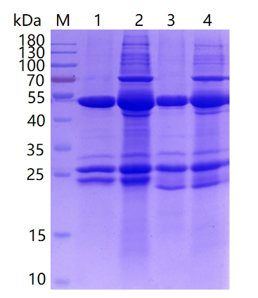
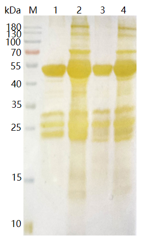
Coomassie Brilliant Blue staining results The result of Coomassie Brilliant Silver Staining
5. Common Issues and Solutions in SDS-PAGE Electrophoresis
| Problem | Reason | Solution |
| The adhesive strip shows upward curling on both sides with a depression in the middle. | The middle part of the gel solidifies unevenly, which mostly occurs in thicker gels. | Allow it to fully solidify before proceeding with subsequent experiments. |
| The adhesive strip exhibits downward edges and bulging in the center. | Incomplete removal of air bubbles at the bottom gap between the two plates, often seen in protein vertical electrophoresis tanks. | Add an appropriate amount of buffer between the two plates to remove air bubbles. |
| The bands show tailing. | Poor sample dissolution or excessively high concentration of the separating gel. | Centrifuge before loading; choose a suitable sample buffer and add an appropriate amount of sample solubilizer; if the electrophoresis buffer is too old, prepare a fresh solution; reduce the gel concentration. |
| The bands display texture phenomena. | The sample consists of insoluble particles. | Centrifuge before loading; add an appropriate amount of sample solubilizer. |
| Bromophenol blue fails to function as an indicator (e.g., bromophenol blue has run off the bottom of the gel, but the proteins have not yet migrated down). | The concentration of the buffer and the separating gel. | Replace with a buffer of the correct pH; reduce the concentration of the separating gel. |
| The protein bands are excessively wide. | Overloading of samples or leakage. | Appropriately reduce the sample loading volume. |
| The electrophoresis voltage is very high, but the current is very low. | This phenomenon is relatively common among beginners. For example, the voltage may exceed 50V, but the current remains below 5mA. The primary cause is incorrect assembly of the electrophoresis tank, which prevents the formation of a current pathway. Specific reasons include:1.The inner and outer tanks are installed in reverse;2.Insufficient buffer solution in the outer tank;3.Failure to remove the insulator at the bottom of the electrophoresis tank (such as the rubber mat used for gel casting). | Assemble the electrophoresis tank correctly. |
- SDS-PAGE;SDSPAGE;Protein gel electrophoresis;Polyacrylamide gel electrophoresis (PAGE)

- Protein Platform
- Protein Expression Services
- Protein Modification & Detection Services
- Protein Interaction Services
- Protein Purification Service
- Antibody Discovery Platform
- VHH Antibody Platform
- Phage Display Platform
- Antibody Humanization Platform
- Single B Cell Screening Platform
- Antibody Expression & Validation Platform
- Peptide Library Platform
- Custom Antibody Platform
- Innovative Drug Discovery Platform
- Product Center
- Proteins
- Antibodies
- Raw Materials Reagents
- Small Molecule Reagents
- Peptide Products
- Nanoparticle Products
- Contact Us
- Phone:400-621-6806
- Mobile:13110936064(WeC
- E-mail:[email protected]
- Address:Room 108, Building 10, No. 6 Hechang Road, Wuqing Development Zone, Tianjin City
-

WeChat
-

Service Account
-
 400-621-6806
 -
 3599163699
 -
 Monday to Friday 9:00-18:00
 -

-
 0
0
-
