


Principles and distinctions of common PCR techniques: PCR, qPCR, RT-PCR, and RT-qPCR
- 2026-05-21
- 230
As technology has evolved, various PCR techniques developed for different purposes have been widely applied in laboratories. Among them, the most common include Polymerase Chain Reaction (PCR), quantitative real-time PCR (qPCR), reverse transcription PCR (RT-PCR), and quantitative real-time reverse transcription PCR (RT-qPCR). Although these four techniques are easily confused in nomenclature, they differ fundamentally in principles and applications.
I. PCR: The Classic Method for DNA Amplification
PCR is the most widely used foundational technique. Its principle involves the use of a heat-stable DNA polymerase, artificially synthesized primers, and four deoxynucleotide triphosphates (dNTPs) in a temperature cycle of denaturation, annealing, and extension to achieve exponential amplification of a specific DNA fragment. After 30 to 40 cycles, the target DNA fragment can be amplified millions of times.
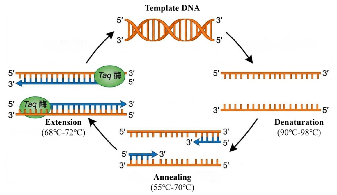
Figure 1: PCR Workflow
Following PCR, the amplified fragments are typically analyzed qualitatively or semi-quantitatively using agarose gel electrophoresis. This method is straightforward, cost-effective, and the amplified products can be directly used for subsequent cloning experiments.
II. RT-PCR: The Bridge from RNA to DNA
RT-PCR is a technique that combines RNA reverse transcription with PCR amplification. Its core principle lies in using reverse transcriptase to synthesize complementary DNA (cDNA) from an RNA template, followed by conventional PCR amplification using this cDNA as the template.
The advent of RT-PCR overcame the limitation of basic PCR, which cannot directly amplify RNA. In practice, RT-PCR can be performed using either a one-step or a two-step method.
• One-step method: Reverse transcription and PCR amplification are carried out sequentially in a single tube, simplifying the procedure and reducing the risk of contamination.
• Two-step method: RNA is first reverse transcribed into cDNA, and then an aliquot of the product is used for PCR. This setup facilitates the detection of multiple genes from the same RNA sample.
RT-PCR is crucial in recombinant protein production research. When researchers aim to construct expression vectors by obtaining a gene of interest from tissues or cells, they first isolate total RNA, obtain the coding region cDNA via RT-PCR, and then clone it into an expression plasmid. This approach is particularly suitable for eukaryotic genes, as it avoids the interference caused by introns when amplifying from genomic DNA. This technique can also be used to detect the transcription levels of specific mRNAs, indirectly reflecting the transcriptional efficiency of the target gene.
III. qPCR: Precise Quantification of DNA
qPCR builds upon conventional PCR by incorporating a fluorescence detection mechanism. Its defining feature is the real-time collection of fluorescence signals during the amplification process, enabling quantitative analysis of the starting template by monitoring the amount of amplified product in each cycle.
The detection chemistries for qPCR are primarily divided into two categories: one is the SYBR Green I fluorescent dye method, where the dye emits fluorescence upon binding to double-stranded DNA, with signal intensity proportional to the amount of double-stranded DNA; the other is the TaqMan probe method, which utilizes a sequence-specific probe labeled with a fluorophore and a quencher. Fluorescence is released when the probe is hydrolyzed by Taq polymerase, enabling sequence-specific detection.
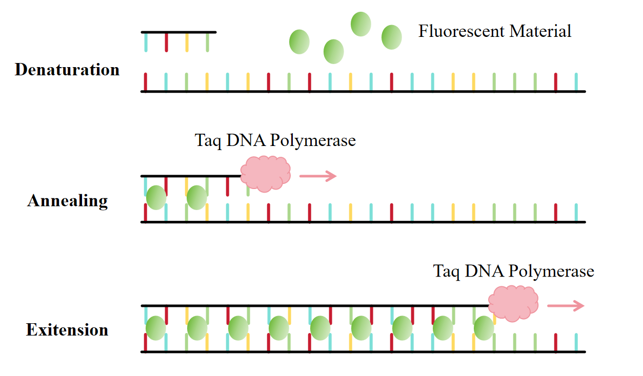
Figure 2: SYBR Green I Fluorescent Dye Method
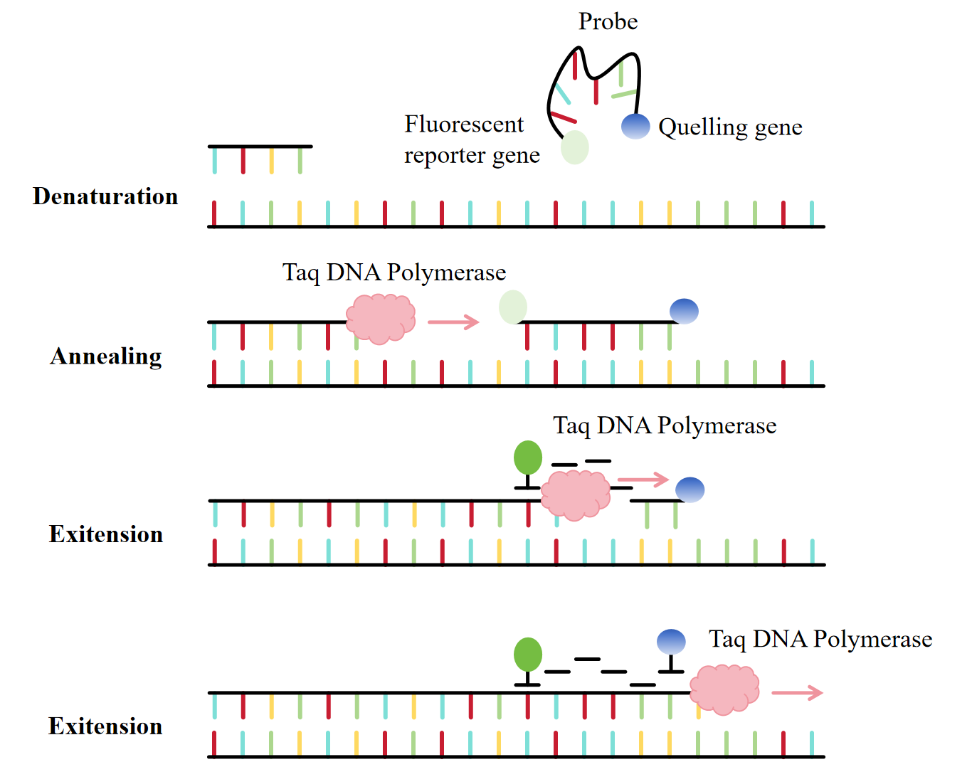
Figure 3: TaqMan Probe Method
Unlike basic PCR, qPCR does not require electrophoresis; amplification occurs in a closed tube, which not only reduces contamination but also allows for reaction quality control through amplification curves and melting curves. The absolute copy number or relative expression level of a target nucleic acid in a sample can be accurately calculated using a standard curve.
qPCR is a common method for detecting changes in transcription levels in studies of protein expression regulation. Researchers can analyze the impact on downstream protein expression by comparing the changes in mRNA levels of a target gene before and after drug treatment or gene editing.
IV. RT-qPCR: The Ultimate Solution for RNA Quantification
Quantitative real-time reverse transcription PCR (RT-qPCR) organically combines reverse transcription with fluorescence quantitative technology and represents the most sensitive and reliable method for RNA quantification available today. The workflow involves first extracting total RNA or mRNA from a sample, synthesizing cDNA through reverse transcription, and then using that cDNA as a template for qPCR amplification and real-time detection.
RT-qPCR combines the RNA detection capability of RT-PCR with the precise quantification feature of qPCR. This technology offers high sensitivity, capable of detecting RNA molecules at extremely low copy numbers, making it suitable for gene expression analysis, RNA virus quantification, and detection of minimal residual disease.
In biopharmaceutical research and development, RT-qPCR is used to detect the transcription levels in engineered cell lines, providing crucial data for screening high-expression clones.
In recombinant protein process development, RT-qPCR can monitor the dynamic changes in the mRNA of the target gene under different culture conditions, aiding in the optimization of induction conditions and harvest timing to improve recombinant protein production efficiency.
V. Comparison and Selection of the Four Techniques
Technique | Template Type | Detection Method | Primary Applications |
PCR | DNA | End-point electrophoresis | Gene cloning, DNA fragment acquisition, qualitative pathogen detection |
RT-PCR | RNA | End-point electrophoresis | Gene cloning (obtaining cDNA from RNA), qualitative RNA virus detection |
qPCR | DNA/cDNA | Real-time fluorescence quantification | Absolute DNA quantification, gene copy number analysis, SNP detection |
RT-qPCR | RNA | Real-time fluorescence quantification | Quantitative gene expression analysis, RNA viral load determination |
Although PCR, RT-PCR, qPCR, and RT-qPCR have similar names, they each have distinct strengths and complementary roles regarding template compatibility, detection methods, and application scenarios. Basic PCR established the foundation for nucleic acid amplification, RT-PCR built the bridge for converting RNA to DNA, qPCR enabled precise DNA quantification, and RT-qPCR combines the advantages of both, becoming the gold standard for RNA quantification.
Leveraging extensive project experience, KMD Bioscience can flexibly select and integrate these four technologies to cover the entire experimental workflow, from nucleic acid extraction and qualitative detection to precise quantification. Additionally, we offer services such as Protein Expression, Gene Library Customization, and Probe Customization, empowering researchers to explore the essence of life phenomena more efficiently and accurately.
[1] Hussain SM, Sharif A, Bashir F, Ali S, Javid A, Hussain AI, Ghafoor A, Alshehri MA, Naeem A, Naeem E, Amjad M. Polymerase Chain Reaction: A Toolbox for Molecular Discovery. Mol Biotechnol. 2026 Feb;68(2):409-421.
[2] Hampton TH, Taub L, Ferreria-Fukutani K, Stanton BA, MacKenzie TA. Analyzing qPCR data: Better practices to facilitate rigor and reproducibility. Biochem Biophys Rep. 2025 Nov 19;44:102356.
[3] Li Z, Yang X, Yang B, Yang J, Tao C, Zhang D, Yamaguchi Y. A comprehensive review of methodological and technological advancement in PCR during the last 15 years. Biotechnol Adv. 2025 Dec;85:108719.
Q1. What are the key differences among the four PCR-related techniques?
The core differences among the four PCR-related techniques lie in template compatibility, detection methods, and functional roles. Basic PCR uses DNA as a template and relies on end-point electrophoresis for detection, with the primary function of achieving exponential amplification of a target DNA fragment to meet fundamental nucleic acid acquisition needs. RT-PCR uses RNA as a template, which is first converted into cDNA via reverse transcription before amplification; it also uses end-point electrophoresis for detection and specifically addresses the challenge of amplifying RNA directly. qPCR is compatible with DNA or cDNA templates and incorporates real-time fluorescence detection, allowing precise quantification of the starting template without the need for electrophoresis. RT-qPCR integrates reverse transcription with real-time quantitative technology, uses RNA as a template, and combines the capability for RNA detection with the advantage of precise quantification, making it the core method for RNA quantitative analysis. Each of the four techniques has its own focus, catering to different experimental requirements.
Q2. What are the operational and applicational differences between the one-step and two-step methods of RT-PCR?
Differences in operational workflow: The one-step method performs reverse transcription and PCR amplification sequentially within a single reaction tube, eliminating the need for sample transfer between steps. This streamlined operation significantly reduces the risk of contamination during sample handling. The two-step method, in contrast, is carried out in two separate phases: RNA is first converted into cDNA using reverse transcriptase, and then an aliquot of the resulting cDNA product is used as a template for PCR amplification. While this process is more involved, it offers greater flexibility in controlling the reaction conditions for each step.
Differences in application scenarios: The one-step method is suitable for routine detection where operational efficiency is critical and sample volumes are high, particularly for rapid qualitative detection of RNA viruses. The two-step method, by preserving the cDNA product, facilitates the detection of multiple genes from the same RNA sample. This makes it more appropriate for research experiments requiring multi-gene analysis, such as the acquisition and validation of target genes in recombinant protein production.
Q3. What are the characteristics of the two fluorescence detection chemistries used in qPCR?
The two fluorescence detection chemistries used in qPCR differ significantly in specificity, operational complexity, and suitable applications.
The SYBR Green I fluorescent dye method is straightforward to perform and cost-effective. Its core principle involves the dye binding non-specifically to double-stranded DNA, emitting fluorescence proportional to the amount of double-stranded DNA, which allows rapid detection of amplification products. However, because the dye lacks sequence specificity, it may bind to non-specific amplification products, potentially leading to false-positive results. Quality control via melting curve analysis is therefore essential.
The TaqMan probe method offers higher sequence specificity. It utilizes a sequence-specific probe labeled with a fluorophore and a quencher. Fluorescence is released only when the probe hybridizes to the target sequence and is hydrolyzed by Taq polymerase. This effectively prevents interference from non-specific amplification, resulting in higher detection accuracy. However, the cost of custom probe synthesis is higher, and the procedure is relatively more complex, making this method suitable for experiments with stringent requirements for detection specificity.
Q4. How should one select the appropriate PCR-related technique based on the experimental objective?
If the experimental objective is to obtain specific DNA fragments, perform gene cloning, or conduct qualitative pathogen detection without the need for quantification, basic PCR is suitable. It is simple to perform and cost-effective, meeting the needs for fundamental DNA amplification.
If the experimental sample is RNA and the aim is to obtain cDNA for gene cloning or qualitative RNA virus detection without precise quantification, RT-PCR is the appropriate choice. The one-step or two-step method can be selected flexibly based on the sample detection requirements.
If the experiment requires precise quantification of DNA or cDNA, such as for gene copy number analysis or SNP detection, qPCR should be chosen. The fluorescence detection method can be selected based on the required specificity of the assay.
If the experimental sample is RNA and precise quantification of the target RNA is required, such as for gene expression analysis or RNA viral load determination, RT-qPCR is the recommended method. Its sensitivity and reliability are well-suited to meet these experimental demands.
Q5. How do the four PCR-related techniques complement each other in practical applications?
Complementarity between fundamental amplification and conversion: Basic PCR serves as the foundational technology for nucleic acid amplification, enabling the rapid generation of large quantities of DNA fragments to provide materials for subsequent experiments. RT-PCR, on the other hand, establishes the bridge for converting RNA to DNA, transforming RNA samples into amplifiable cDNA and overcoming the limitation of basic PCR, which cannot directly process RNA samples. Together, these techniques fulfill the amplification needs for different types of nucleic acids.
Complementarity between qualitative and quantitative analysis: RT-PCR and basic PCR are primarily used for qualitative nucleic acid detection, allowing rapid determination of the presence or absence of a target nucleic acid. qPCR and RT-qPCR focus on precise quantification, enabling the quantitative analysis of DNA or RNA. Combining these approaches facilitates a complete experimental workflow from qualitative screening to quantitative validation. For instance, one might first use RT-PCR for qualitative detection of an RNA virus and then employ RT-qPCR for quantitative determination of the viral load.
Complementarity between research and applications: These four techniques cover the entire experimental workflow from nucleic acid extraction and qualitative detection to precise quantification. In research, they can be used for gene cloning, protein expression regulation studies, and more. In practical applications, they are employed in scenarios such as pathogen detection and drug development. Their flexible combination can meet experimental needs across various fields, enhancing experimental efficiency and accuracy.
- PCR
- qPCR
- RT-PCR
- RT-qPCR

- Protein Platform
- Protein Expression Services
- Protein Modification & Detection Services
- Protein Interaction Services
- Protein Purification Service
- Antibody Discovery Platform
- VHH Antibody Platform
- Phage Display Platform
- Antibody Humanization Platform
- Single B Cell Screening Platform
- Antibody Expression & Validation Platform
- Peptide Library Platform
- Custom Antibody Platform
- Innovative Drug Discovery Platform
- Product Center
- Proteins
- Antibodies
- Raw Materials Reagents
- Small Molecule Reagents
- Peptide Products
- Nanoparticle Products
- Contact Us
- Phone:400-621-6806
- Mobile:13110936064(WeC
- E-mail:[email protected]
- Address:Room 108, Building 10, No. 6 Hechang Road, Wuqing Development Zone, Tianjin City
-

WeChat
-

Service Account
-
 400-621-6806
 -
 3599163699
 -
 Monday to Friday 9:00-18:00
 -

-
 0
0
-
