


mRNA Immunofluorescence Assay
- 2026-07-10
- 245
1. The Concept of Immunofluorescence
Immunofluorescence (IF) is an important immunochemical technique used to detect and localize various antigens in different types of tissues from various cell preparations.
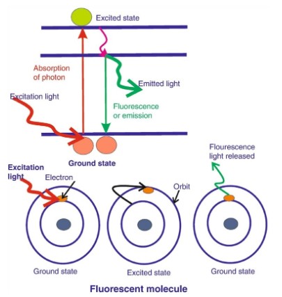
Figure 1: Principles of Immunofluorescence
2. Types of Immunofluorescence
Depending on the scope of the experiment or the specific antibodies used, there are currently three methods: the direct method, the indirect method, and the complement-fixation technique.
2.1 Direct Method
Primary or direct immunofluorescence uses a single antibody chemically conjugated to a fluorophore. The antibody recognizes the target molecule and binds to it, and the fluorophore it carries can be detected under a microscope. Because the antibody is directly conjugated to the fluorophore, this technique offers several advantages over the indirect method described below. It reduces the number of steps in the staining process, making it faster, and can minimize background signal by avoiding issues such as antibody cross-reactivity or nonspecific binding. However, due to the limited number of fluorescent molecules that can bind to a single primary antibody, direct immunofluorescence is less sensitive than indirect immunofluorescence.
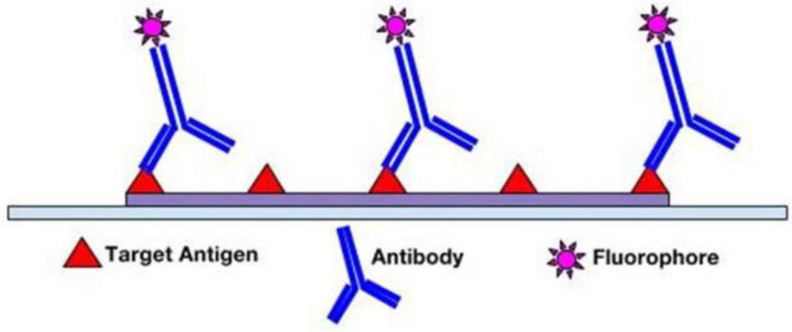
Figure 2: Direct Immunofluorescence
2.2 Indirect Method
Secondary or indirect immunofluorescence uses two antibodies: an unlabeled primary antibody that specifically binds to the target molecule, and a secondary antibody carrying a fluorophore that recognizes and binds to the primary antibody. Multiple secondary antibodies can bind to a single primary antibody. This amplifies the signal by increasing the number of fluorescent molecules per antigen. This approach is more complex and time-consuming than the direct method described above, but it offers greater flexibility because a variety of different secondary antibodies and detection techniques can be used with a given primary antibody.
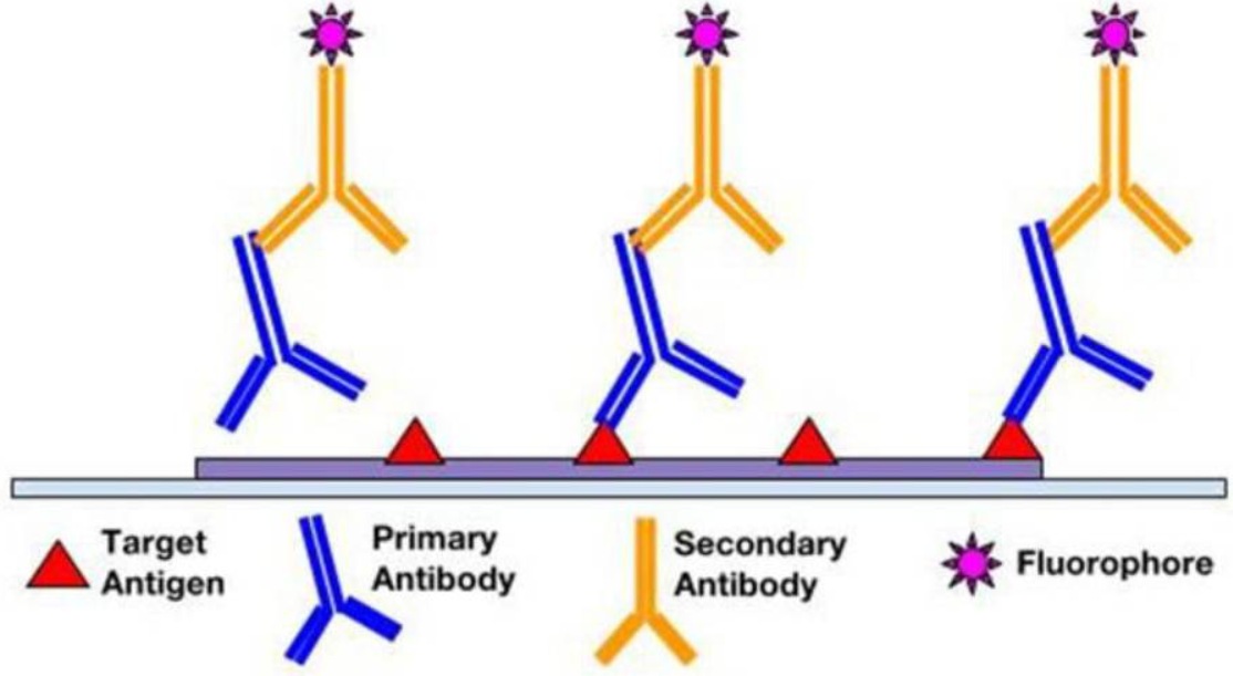
Figure 3: Indirect Immunofluorescence
2.3 Complement-Binding Assay
The complement-binding IIF technique is a highly sensitive three-step serological assay. Using this method, very small amounts of circulating antibodies can be detected due to their high affinity for complement. FITC-labeled anti-complement can be used to detect complement. This technique is more sensitive than conventional IIF because amplification is achieved by binding more than one complement molecule to each immunoglobulin (Ig), which are then visualized.
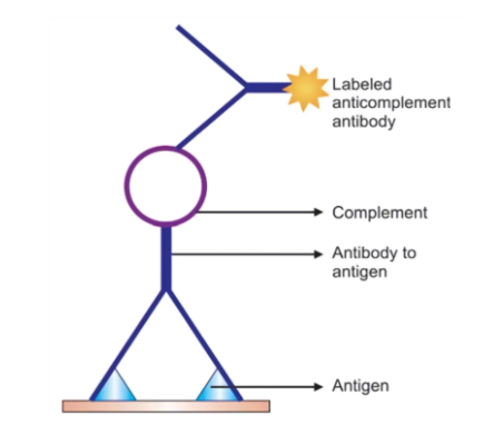
Figure 4: Complement-binding technology
3. Advantages and Disadvantages of Immunofluorescence
3.1 Advantages
(1) Simple and reproducible
(2) Short procedure time of 1–3 hours
(3) High sensitivity
(4) Significant prognostic value, particularly for cases of herpes
3.2 Disadvantages
(1) Autofluorescence
(2) Fluorescence overlap
(3) Nonspecific fluorescence
(4) Occasional polymerization or interference with the kinetics and expression levels of target molecules
4. Applications of Immunofluorescence
Immunofluorescence is widely used in the study of autoimmune diseases; defining antigen-antibody interactions at the subcellular level; identifying viral, protozoal, bacterial, and parasitic antigens; and identifying small cell surface structures, such as receptors on lymphocytes.
5. KMD Bioscience Provides High-Quality Immunofluorescence Technical Services
KMD Bioscience provides high-quality immunofluorescence technical services and technical support. The main steps of the immunofluorescence technique are as follows: specimen preparation, fixation, permeabilization, blocking, primary antibody incubation, secondary antibody incubation, and fluorescence detection.
5.1 Sample Preparation
5.1.1 To enhance cell adhesion, treat the coverslips with a 1:10 dilution of poly-L-lysine solution at room temperature for 5 minutes.
5.1.2 Seed the cells at an appropriate dilution and allow them to grow until they reach the desired confluence (~70%).
5.1.3 Briefly rinse the cells in PBS.
5.2 Fixation
5.2.1 Aspirate the PBS and cover the cells with PBS containing formaldehyde (work in a fume hood). Allow the cells to fix at room temperature for 15 minutes.
5.2.2 Aspirate the fixative and rinse three times with PBS, for 5 minutes each time.
5.3 Permeabilization (Optional)
Fixation with methanol and acetone produces permeabilized cell preparations. For polyformaldehyde-fixed preparations, treat with Triton X-100 for 5 minutes or with methanol for 5 minutes.
5.3.1 Methanol Permeabilization Procedure: After formaldehyde fixation, cover the cells with ice-cold 100% methanol (use enough to completely cover the cells to a depth of 3–5 mm; do not allow the cells to dry), and incubate the cells in methanol at 4°C for 10 minutes.
5.3.2 Rinse in PBS for 5 minutes.
5.4 Blocking
5.4.1 Block the samples for 1 hour in PBS containing 5% normal serum (e.g., normal goat serum, normal donkey serum) and Triton X-100. (Recommended blocking buffers may vary between 1–2% bovine serum albumin, fetal bovine serum albumin, or skim milk powder in TBS (PBS) with or without 0.2% Tween 20 and sodium azide.)
Note: Unless otherwise specified, all incubations should be performed at room temperature to prevent drying and to protect the fluorescent dyes from light exposure.
5.5 Primary Antibody Incubation
5.5.1 Dilute the primary antibody in PBS with Triton. Volume: 50–100 μL per section; 25–50 μL per coverslip, well, or孔 (48- or 96-well plate).
5.5.2 Aspirate the blocking solution and apply the diluted primary antibody.
5.5.3 Incubate overnight at 4°C with gentle shaking or agitation.
5.5.4 Wash three times with PBS containing 0.1% Triton X-100, for 5 minutes each time.
5.6 Secondary Antibody Incubation
5.6.1 Incubate with a fluorescently labeled secondary antibody diluted in PBS containing 0.1% Triton X-100 for 1–2 hours at room temperature in the dark. (From this point onward, the slides must be stored in the dark.)
5.6.2 Wash in PBS containing 0.1% Triton X-100.
5.7 Fluorescence Detection
5.7.1 Mount the coverslips.
5.7.2 Examine immediately under a fluorescence microscope or store flat in the dark at 4°C.
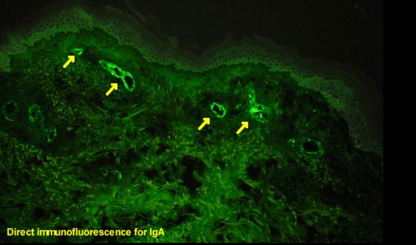
Figure 5: Human skin tissue sections prepared for direct immunofluorescence
KMD Bioscience is a trusted provider of standard and customized IHC and IF services. It employs outstanding scientists with over 10 years of experience in IHC and IF staining, and a team of seasoned experts provides technical support to resolve various IHC and IF technical issues, ensuring the quality and speed of its services.
- mRNA Immunofluorescence Assay
- mRNA
- Immunofluorescence Assay
- Immunofluorescence

- Protein Platform
- Protein Expression Services
- Protein Modification & Detection Services
- Protein Interaction Services
- Protein Purification Service
- Antibody Discovery Platform
- VHH Antibody Platform
- Phage Display Platform
- Antibody Humanization Platform
- Single B Cell Screening Platform
- Antibody Expression & Validation Platform
- Peptide Library Platform
- Custom Antibody Platform
- Innovative Drug Discovery Platform
- Product Center
- Proteins
- Antibodies
- Raw Materials Reagents
- Small Molecule Reagents
- Peptide Products
- Nanoparticle Products
- Contact Us
- Phone:400-621-6806
- Mobile:13110936064(WeC
- E-mail:[email protected]
- Address:Room 108, Building 10, No. 6 Hechang Road, Wuqing Development Zone, Tianjin City
-

WeChat
-

Service Account
-
 400-621-6806
 -
 3599163699
 -
 Monday to Friday 9:00-18:00
 -

-
 0
0
-
