


Yeast Surface Display Technology: A Comprehensive Solution from Enzyme Engineering to Antibody Discovery
- 2026-09-24
- 93
In the course of enzyme research, have you encountered the following issues: high costs associated with the expression, isolation, and purification of target enzymes; rapid inactivation of free enzymes under high temperatures, extreme pH conditions, or in organic solvents, making it difficult to achieve the required stability; enzymes being lost with the product stream after each reaction batch, preventing their recovery and reuse; low screening efficiency when dealing with massive mutagenic yeast display libraries; intermediate products rely on slow diffusion to transfer between enzyme active sites, resulting in a significant decrease in reaction rates.
Although traditional immobilization methods have alleviated these problems to some extent, they still have certain limitations: fundamental issues such as operational complexity, high costs, significant loss of enzyme activity, and resistance to mass transfer remain unresolved[1].
Consequently, the Yeast Surface Display (YSD) technology was developed. Compared to traditional intracellular or extracellular expression systems, the surface display system eliminates the need for enzyme purification, overcomes substrate transport limitations, and demonstrates enhanced activity, stability, and selectivity[2]. Furthermore, yeast display for antibody discovery has emerged as a highly promising alternative to phage display, particularly for the screening of antibody fragments that require proper folding and glycosylation.
What problems can YSD help you solve? When is YSD the right choice? How can you successfully turn your ideas into results? This article will break it all down step by step from a problem-solving perspective.
I. Problems Yeast Surface Display Can Solve
(i) Reduced Purification Costs
Ø Challenge: The expression and purification of the target enzyme require multiple steps, including cell lysis, centrifugation, and chromatography, which is time-consuming, and each step results in a loss of enzyme activity.
Ø Yeast Surface Display technology Solution: YSD technology anchors enzymes to the surface of yeast cells as they are expressed, eliminating the need for separate purification and immobilization steps. This method eliminates the need to isolate, purify, and chemically immobilize proteins on a carrier, resulting in a faster, simpler, and more cost-effective process.
(ii) Improved Stability
Ø Challenge: Free enzymes have poor stability under industrial production conditions (high temperatures, extreme pH levels, and organic solvents), making it difficult to meet the requirements for large-scale applications.
Ø Yeast Surface Display technology Solution:The yeast surface display system significantly enhances enzyme thermal stability and pH tolerance by anchoring enzymes to the natural protective matrix of the cell wall. The glycosylation modifications of the yeast cell wall further improve thermal stability and resistance to degradation, which is particularly beneficial for maintaining the long-term activity of antibodies in yeast-based antibody discovery.
(iii) Reusability
Ø Challenge:Free enzymes are for single-use only and are expensive; although traditional immobilized enzymes can be reused, their preparation is complex and they suffer significant loss of enzymatic activity.
Ø Yeast Surface Display technology Solution:Yeast cells containing enzymes can be separated from the reaction mixture by simple centrifugation and reused multiple times. The reusability of whole-cell catalysts significantly reduces the cost of enzymes per reaction.
(iv) Improved Efficiency
Ø Challenge:Conventional screening methods are inefficient and are powerless in the face of mutation libraries containing millions of variants.
Ø Yeast Surface Display technology Solution:Since the target enzyme is displayed on the cell surface, its activity or binding capacity can be monitored using fluorescently labeled substrates, antibodies, or products, thereby enabling high-throughput sorting of yeast cells expressing different enzyme mutants via flow cytometry (FACS).
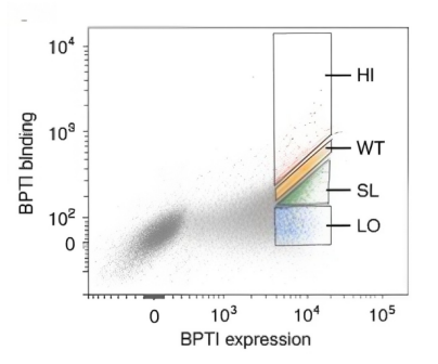
Figure 1: Flow Cytometry Sorting Diagram[3]
II. Yeast Surface Display technology Use Cases
Suitable Scenarios
Ø Directed evolution of enzymes: It is necessary to screen for highly stable variants from a vast database.
Ø Whole-cell catalyst construction: Reduces purification costs by using enzymes directly in catalytic reactions.
Ø Multienzyme-linked reaction: Requires the synergistic catalysis of multiple enzymatic reactions.
Ø Stability Modification of Industrial Enzymes: It is necessary to improve the thermal stability and pH tolerance of the enzymes.
Ø Engineering of soluble proteins: target proteins that are hydrolytic enzymes and fold correctly on the yeast cell surface
Situations Where It Is Not Appropriate
Ø Enzymes requiring an intracellular environment: Yeast surface display technology is expressed on an oxidative extracellular surface and is not suitable for enzymes requiring a reducing environment.
Ø Enzymes that use insoluble macromolecules as substrates: The substrates have difficulty effectively binding to the enzymes displayed on the cell surface.
Ø Membrane proteins: They require an intact cell membrane structure to function.
Ø Ultra-high-molecular-weight proteins: The transport capacity of the yeast secretory pathway is limited, so the display efficiency may be relatively low.
III. The Complete Yeast Surface Display technology Process
(i) Design of Anchor Proteins and Fusion Strategies
The choice of anchor protein directly affects display efficiency and the activity of the target protein. Currently, commonly used anchor proteins include Aga1p and Aga2p, which are suitable for most protein displays.
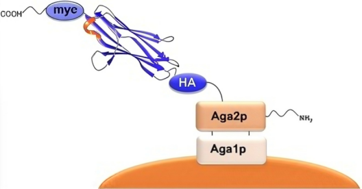
Figure 2: View of the Yeast Surface[4]
(ii) Gene Fusion and Vector Construction
The method for designing fusion proteins involves fusing the gene encoding the target enzyme with the gene encoding a anchor protein and cloning the construct into a suitable yeast expression vector. Key decision points include: the strength and inducibility of the promoter, the secretion efficiency of the signal peptide, and the length and flexibility of the linker peptide; all of these factors influence the final display outcome[1].
(iii) Yeast Transformation and Validation
To verify whether the target enzyme has been successfully displayed on the yeast surface, immunofluorescence labeling combined with flow cytometry can be used to determine the positive display rate. At the same time, enzyme activity assays are required to confirm that the displayed enzyme retains its native conformation and catalytic function.
(iv) Functional Screening
Ø Magnetic bead sorting: This method can be used for initial screening to sort 109–1010 cells; however, it has low resolution and is used for coarse screening to eliminate negative cells.
Ø Flow cytometry: This method enables precise screening with extremely high resolution, capable of distinguishing cell populations based on different fluorescence intensities, with a throughput of approximately 107 cells per hour.
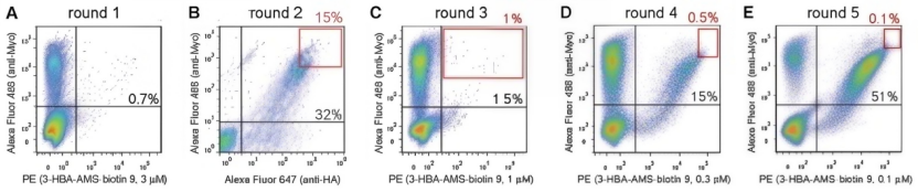
Figure 3: Results of Magnetic Bead Preliminary Screening and Flow Cytometry-Based Refined Screening[5]
(v) Clone Identification and Functional Validation
To determine whether the clones screened by FACS with low-positive rates truly exhibit the expected performance, the selected clones must undergo sequencing for identification (to confirm the mutation sequence) and solubility expression validation (to confirm the enzyme’s performance under actual application conditions).
IV. How KMD Bioscience Can Accelerate Your Research
KMD Bioscience offers a proven yeast surface display technology platform and yeast expression system services. Combined with its high-throughput FACS screening capabilities, the company is committed to providing researchers with comprehensive solutions ranging from technical consulting to experimental implementation.
Yeast surface display technology has undergone nearly three decades of development. It has evolved from a simple antibody screening tool into a comprehensive technological platform in the field of enzyme engineering that integrates expression, immobilization, and screening.
However, the maturation of technology does not mean that every researcher needs to build an entire platform from scratch. Let the experts handle the expert work—by entrusting platform-related tasks such as anchor protein screening, display vector optimization, and FACS sorting to KMD Bioscience, you can focus more of your energy on core scientific questions.
FAQs
Q1: The capacity of a phage display library typically reaches 1011, while that of a yeast display library generally ranges from 107 to 10⁹. How does this difference in order of magnitude affect actual screening? Could some rare clones be missed?
For a theoretical library with a theoretical diversity of 10¹¹, the YSD libraries ranging from 10⁷ to 10⁹ are indeed unable to provide complete coverage.
Actual Impact:
Ø Different screening strategies: Phage display relies on repeated “screening” rounds to enrich variants and can accommodate larger library sizes. YSD, on the other hand, is typically combined with FACS for screening; although the library size per run is smaller, the high sensitivity and quantitative capabilities of FACS allow for the efficient identification of high-affinity or high-activity variants even in smaller libraries.
Ø Definition of “rare clones”: If “rare” refers to the top 0.01% of clones in terms of function, then provided the library is constructed to a high standard, a library size of 10⁷–10⁹ is typically sufficient to cover them. For ultra-large libraries (e.g., 10¹¹), enrichment is usually achieved through multiple rounds of selection, whereas YSD places greater emphasis on the quality and resolution of the first round of screening.
Q2: In the section on “Anchor Protein and Fusion Strategy Design,” are there any anchor proteins other than Aga1p/Aga2p to choose from?
In addition to the classic Aga1-Aga2 system, there are other anchor protein strategies, such as the Sed1p and Pir protein families. The Tianjin Institute of Industrial Biotechnology, Chinese Academy of Sciences, recently developed a new system called CEN-Display. Its 28_YI anchoring module achieved an efficiency of up to 71.3% when displaying α-galactosidase, representing an approximately 63.5% increase in displayed enzyme activity compared to the traditional Aga1-Aga2 system.
Q3: “The length and flexibility of the linker peptide affect the display efficiency”—so in actual design, what is the appropriate number of amino acids for the linker peptide? Are there any general sequence recommendations?
Ø Design Principles: The linker peptide must be sufficiently long and flexible to ensure that the target protein protrudes from the glycoprotein layer of the yeast cell wall, folds correctly, and comes into contact with the substrate or antigen. At the same time, the linker peptide should not be so long as to cause protein instability or proteolytic degradation.
Ø Length Guidelines: There is no fixed number of amino acids; the length typically needs to be optimized based on the structure of the target protein. Some studies have optimized enzyme activity by inserting linker peptides of varying lengths.
Ø General Sequences: The most commonly used flexible linker peptides are (GGGGS)n or (EAAAK)n. Trials typically start with n ranging from 1 to 3 (i.e., 5 to 15 amino acids).
Ø Improved Strategy: Research has led to the development of a Pir-tag peptide segment measuring only 4.5 kDa, which promotes surface display with an efficiency comparable to that of the full-length Hsp150 anchor protein. This suggests that a carefully designed short peptide may be more effective than a long, haphazardly constructed linker peptide. The optimal approach often requires experimental screening and optimization.
Q4: The article mentions that “the transport capacity of the yeast secretory pathway is limited.” For proteins with what molecular weight (e.g., > how many kDa) might low display efficiency become an issue? Are there any quantitative or semi-quantitative reference thresholds?
The transport capacity of the yeast secretory pathway is indeed a limiting factor in the application of YSD. Although there is no strictly defined molecular weight cutoff, proteins with molecular weights exceeding 100–150 kDa or complex proteins with multiple subunits generally become significantly more difficult to express efficiently in yeast. This is typically manifested as low expression efficiency, low protein yield, or proteins that are not properly folded and cannot function correctly. This limitation is not absolute; for example, nanobodies (approximately 15 kDa) and single-chain antibodies (approximately 28 kDa) are typically displayed well on the yeast surface. However, for larger proteins, it is possible to overcome this limitation through strategies such as optimizing the signal peptide to use a more efficient secretion signal sequence, co-expressing chaperones to aid in proper protein folding, or displaying functional fragments of the protein rather than the full-length protein.
Q5: In the “Yeast Transformation and Validation” section, when presenting the positive rate using immunofluorescence labeling and flow cytometry, what positive rate is considered ‘acceptable’ or “sufficient to proceed to the next screening step”?
There is no absolute, universal “passing” threshold for the display positivity rate; it depends on library quality and the screening target. For constructed yeast display libraries, a display positivity rate of 5%–30% is generally considered an acceptable range for proceeding to subsequent screening. If the positive rate exceeds 20%, the library is generally considered to be of high quality and ready for direct FACS screening. If the positive rate falls between 5% and 20%, the library quality is acceptable, but it is recommended to perform 1–2 rounds of pre-enrichment using magnetic beads to increase the proportion of target cells. If the positive rate is below 5%, this indicates issues with library construction or expression, and direct screening will be highly inefficient; screening should be conducted only after optimizing transformation, expression, or detection conditions. More importantly, observe the relationship between the “antigen-binding positivity rate” and the “display positivity rate.” If the majority of displayed proteins can bind to the antigen, this indicates that the proteins are functioning properly.
[1]Zhang, Baoyu et al. “Surface Display Technologies for Whole-Cell Biocatalysts: Advances in Optimization Strategies, Food Applications, and Future Perspectives.” Foods (Basel, Switzerland) vol. 14,10 1803. 19 May. 2025, doi:10.3390/foods14101803
[2]Li Y, Wang X, Zhou NY, Ding J. Yeast surface display technology: Mechanisms, applications, and perspectives. Biotechnol Adv. 2024 Nov;76:108422. doi: 10.1016/j.biotechadv.2024.108422. Epub 2024 Aug 6. PMID: 39117125.
[3]Heyne M, Papo N, Shifman JM. Generating quantitative binding landscapes through fractional binding selections combined with deep sequencing and data normalization. Nat Commun. 2020 Jan 15;11(1):297. doi: 10.1038/s41467-019-13895-8. PMID: 31941882; PMCID: PMC6962383.
[4]Könning D, Kolmar H. Beyond antibody engineering: directed evolution of alternative binding scaffolds and enzymes using yeast surface display. Microb Cell Fact. 2018 Feb 26;17(1):32. doi: 10.1186/s12934-018-0881-3. PMID: 29482656; PMCID: PMC6389260.
[5]Zhang K, Nelson KM, Bhuripanyo K, Grimes KD, Zhao B, Aldrich CC, Yin J. Engineering the substrate specificity of the DhbE adenylation domain by yeast cell surface display. Chem Biol. 2013 Jan 24;20(1):92-101. doi: 10.1016/j.chembiol.2012.10.020. PMID: 23352143; PMCID: PMC3807743.
- Yeast surface display technology Solution
- yeast display library

- Protein Platform
- Protein Expression Services
- Protein Modification & Detection Services
- Protein Interaction Services
- Protein Purification Service
- Antibody Discovery Platform
- VHH Antibody Platform
- Phage Display Platform
- Antibody Humanization Platform
- Single B Cell Screening Platform
- Antibody Expression & Validation Platform
- Peptide Library Platform
- Custom Antibody Platform
- Innovative Drug Discovery Platform
- Product Center
- Proteins
- Antibodies
- Raw Materials Reagents
- Small Molecule Reagents
- Peptide Products
- Nanoparticle Products
- Contact Us
- Phone:400-621-6806
- Mobile:13110936064(WeC
- E-mail:[email protected]
- Address:Room 108, Building 10, No. 6 Hechang Road, Wuqing Development Zone, Tianjin City
-

WeChat
-

Service Account
-
 400-621-6806
 -
 3599163699
 -
 Monday to Friday 9:00-18:00
 -

-
 0
0
-
