


STTT Analysis of Top Journals|M6A-Modified CircArhgap26 Attenuates Cardiac Ischemia‒Reperfusion Injury by Suppressing Plakophilin-1 Palmitoylation
- 2026-08-31
- 72
Myocardial ischemia-reperfusion (I/R) injury is a key pathological mechanism underlying the deterioration of cardiac function in patients with acute myocardial infarction (AMI) following percutaneous coronary intervention (PCI). Although the restoration of blood flow can reduce the infarct size, reperfusion itself induces myocardial cell apoptosis, oxidative stress, calcium overload, and mitochondrial dysfunction, further exacerbating myocardial injury. To date, protective drugs developed to address these mechanisms have not achieved clinical breakthroughs; therefore, there is an urgent need to identify new therapeutic targets.
Circular RNA (circRNA), due to its covalently closed ring structure, exhibits greater stability and resistance to nuclease degradation than linear RNA, demonstrating immense potential as a diagnostic biomarker and therapeutic target in cardiovascular diseases. However, systematic studies on circRNA in cardiac ischemia-reperfusion (I/R) injury remain very limited. Meanwhile, protein palmylation—catalyzed by ZDHHC family palmityltransferases—is an important lipid-based post-translational modification that regulates approximately 20% of the proteome, influencing protein stability, localization, and protein-protein interactions; however, its regulatory network in cardiac I/R injury remains unclear.
Of particular note, recent studies have found that circRNAs can regulate the stability of downstream proteins by competitively binding to the same protein domain as palmoyltransferases; however, in the cardiovascular field, the circRNA–protein palmoylation regulatory axis remains unexplored.
In March 2026, the research group led by Professor Xu Chaoqian from the team of Academician Yang Baofeng at Harbin Medical University published a groundbreaking study in *Signal Transduction and Targeted Therapy*. The team identified circArhgap26 for the first time as a key protective circRNA against myocardial ischemia-reperfusion (I/R) injury, revealed a novel regulatory mechanism by which it alleviates myocardial injury through competitive inhibition of PKP1 palmitoylation, and elucidated the degradation network of circArhgap26 mediated by upstream m6A modification -YTHDF2-mediated degradation network of circArhgap26, thereby establishing a comprehensive m6A-circRNA-palmitoylation dual-modification coupling regulatory pathway.
I. The Discovery of circArhgap26: From Chip Screening to Clinical Validation
The research team first screened an AMI mouse model using a circRNA expression profile chip, but unexpectedly found that the candidate circRNAs did not show significant changes during the AMI phase. This “negative result” actually suggested that these circRNAs are more likely to be involved in reperfusion injury rather than the ischemic process alone. Subsequently, the team established a myocardial ischemia-reperfusion (I/R) model (45-minute ischemia followed by 24-hour reperfusion) and found that circArhgap26—a 443-nt circular RNA formed by reverse splicing of exons 2–6 of the Arhgap26 gene—was significantly downregulated in I/R cardiac tissue and consistently downregulated in primary cardiomyocytes stimulated by H₂O₂ and hypoxia/ reoxygenation (H/R) stimulation.
Of greater clinical translational significance, the team analyzed plasma samples from patients following PCI and found that the human homologous circRNA (h-circArhgap26, with up to 91.65% sequence homology to the mouse sequence) was significantly lower in patient plasma compared to healthy controls. Further Sanger sequencing, RNase R resistance assays, FISH subcellular localization, and ORF analysis confirmed that circArhgap26 is a typical non-coding cytoplasmic circRNA, possessing the structural basis to serve as a circulating biomarker.
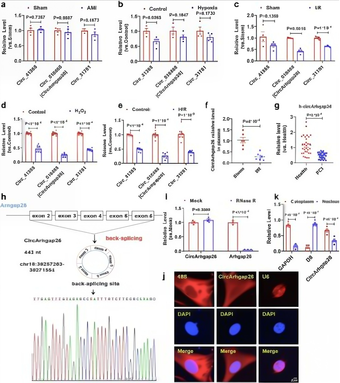
Fig.1Presents the Complete Characterization Data for CircArhgap26
Fig. 1a–e: qRT-PCR confirmed that circArhgap26 was significantly downregulated in I/R-injured hearts and in cardiomyocytes stimulated by H₂O₂ and H/R; Fig. 1f: Levels of circArhgap26 were reduced in the serum of I/R mice; Fig. 1g: h-circArhgap26 levels in the plasma of PCI patients were significantly lower than those in healthy controls (n = 30 vs. n = 30), highlighting its clinical translational value; Fig. 1h: Sanger sequencing validated the reverse splicing site; Fig. 1i: RNase R resistance assays confirmed the circular structure; Fig. 1j–k: FISH and nuclear-cytoplasmic separation experiments showed that circArhgap26 is primarily localized in the cytoplasm and lacks protein-coding potential.
II. Functional Validation Specific to Cardiomyocytes: circArhgap26 Is an Endogenous Protective Factor Against I/R Injury
To elucidate the biological function of circArhgap26, the research team used the AAV9-cTnT vector to achieve cardiomyocyte-specific overexpression. Following ischemia-reperfusion (I/R) modeling, the overexpression group showed significantly improved cardiac function (increased EF and FS, reduced ventricular diameter), decreased serum LDH and plasma cTnT levels, significantly reduced infarct size as demonstrated by Evans blue/TTC double staining, a decreased TUNEL-positive rate, and downregulation of the pro-apoptotic protein Bax coupled with upregulation of the anti-apoptotic protein Bcl-2. Conversely, myocardial-specific knockdown of circArhgap26 exacerbated cardiac dysfunction, enlarged the infarct area, and promoted apoptosis. In vitro H₂O₂ models and experiments using the human AC16 cell line yielded consistent results, and validation in female mice ruled out gender-specific bias.
This bidirectional “gain-of-function and loss-of-function” validation strategy established circArhgap26 as an endogenous protective factor against cardiac ischemia-reperfusion (I/R) injury.
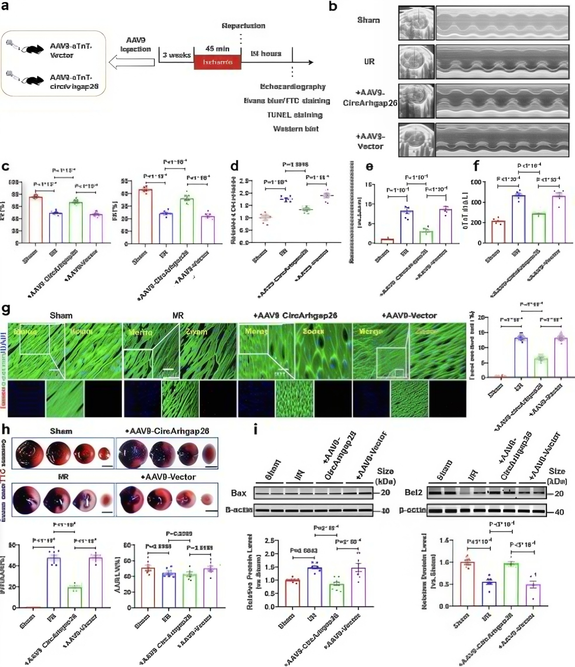
Fig. 2 Shows the Significant Protective Effect of CircArhgap26 Overexpression
Fig. 2b–c: Echocardiography showed increased EF and FS, indicating improved cardiac function; Fig. 2d–f: Decreased serum LDH and plasma cTnT levels indicated reduced myocardial injury; Fig. 2g: TUNEL staining showed reduced myocardial cell apoptosis; Fig. 2h: Evans blue/TTC double staining shows a significant reduction in the infarct area; Fig. 2i: Western blot analysis shows downregulation of Bax and upregulation of Bcl-2.
III. Capture of Downstream Target Proteins: From Mass Spectrometry Screening to PKP1 Domain Localization
The function of circRNA typically depends on its interaction with proteins. The research team used RNA pull-down combined with LC-MS/MS to identify proteins that bind to circArhgap26 in cardiac myocyte lysates. Metascape functional clustering analysis revealed that the pull-down proteins were significantly enriched in the cell adhesion protein apoptosis shear pathway, with PKP exhibiting the highest mass spectrometry score.
RIP experiments bidirectionally confirmed the direct interaction between circArhgap26 and PKP1; RNA pull-down combined with Western blot provided independent validation; molecular docking predicted their binding mode; and FISH/IF colocalization analysis demonstrated their colocalization in the cytoplasm. Most importantly, the team constructed full-length and truncated mutants of PKP1 (ARM1, ARM2–5, ARM6–9) and, through RIP experiments, precisely localized the binding site: circArhgap26 specifically binds to the ARM1 domain of PKP1. Additionally, the team found that PKP1 was significantly upregulated at the protein level in I/R-injured hearts and oxidatively stressed cardiomyocytes, suggesting that it may be a pro-pathogenic factor in I/R injury.
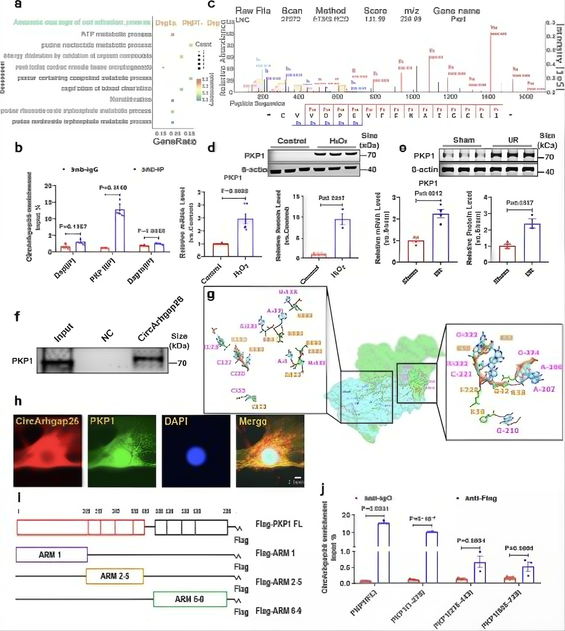
Fig. 3 Shows the Identification and Validation of PKP1
Fig. 3a: Metascape functional clustering shows that pull-down proteins are primarily enriched in the cell adhesion protein apoptosis shear pathway, which includes PKP1; Fig. 3b: RIP experiments confirm that circArhgap26 specifically binds to PKP1; Fig. 3c: LC-MS/MS mass spectrometry identified PKP1 peptides; Fig. 3d–e: PKP1 was significantly upregulated at the protein level in H₂O₂-stimulated cardiomyocytes and in ischemia-reperfusion (I/R)-induced hearts; Fig. 3f: RNA pull-down combined with Western blot validated the interaction; Fig. 3g: Molecular docking predicts the binding mode; Fig. 3h: FISH/IF colocalization analysis shows that the two proteins colocalize in the cytoplasm; Fig. 3i–j: Truncation mutation experiments confirm that the ARM1 domain of PKP1 is the key binding region for circArhgap26.
IV. Validation of PKP1’s Independent Function: Identification of Apoptosis-Inducing Factors
After identifying PKP1 as an interaction target of circArhgap26, the research team needed to determine whether PKP1 itself plays an independent role in I/R injury. The team generated myocardium-specific PKP1 knockdown mice and, following I/R modeling, observed protective effects nearly identical to those seen with circArhgap26 overexpression—improved cardiac function, reduced myocardial injury markers, decreased apoptosis, reduced infarct size, and reversal of the Bax/Bcl-2 ratio.
This result has dual significance: on the one hand, it confirms that PKP1 is a pro-apoptotic factor in I/R injury; on the other hand, it lays the logical foundation for subsequent “functional recovery” experiments—if circArhgap26 exerts its effects by regulating PKP1, then simultaneous overexpression of PKP1 should be able to reverse the protective effects of circArhgap26.
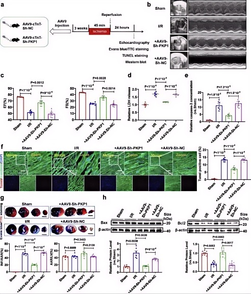
Fig. 4 Illustrates the Protective Effects of Myocardial-Specific PKP1 Knockdown
Fig. 4b–c: Increased EF and FS, decreased LVIDd/LVIDs; Fig. 4d–e: Decreased LDH and caspase-3 activity; Fig. 4f: Decreased TUNEL positivity rate; Fig. 4g: Reduced infarct size; Fig. 4h: Downregulation of Bax and upregulation of Bcl-2. Conclusion: PKP1 is an pro-apoptotic factor in I/R injury; PKP1 knockdown mimics the protective effects of circArhgap26 overexpression.
V. Functional Restoration Experiment: Establishing the circArhgap26→PKP1 Causal Regulatory Axis
The core of mechanistic research lies in establishing causal relationships. The research team designed rigorous functional rescue experiments: in mice with circArhgap26 overexpression, simultaneous overexpression of PKP1 was observed to significantly reverse the improvements in cardiac function, reduction in infarct size, and inhibition of apoptosis induced by circArhgap26; conversely, in mice with circArhgap26 knockdown, simultaneous knockdown of PKP1 partially rescued the deterioration in cardiac function. An in vitro H₂O₂ model similarly confirmed that PKP1 overexpression blocks the anti-apoptotic effects of circArhgap26.
This series of replicate experiments followed the logic of molecular biology research and clearly established that PKP1 is an essential mediator for the cardioprotective effects of circArhgap26.
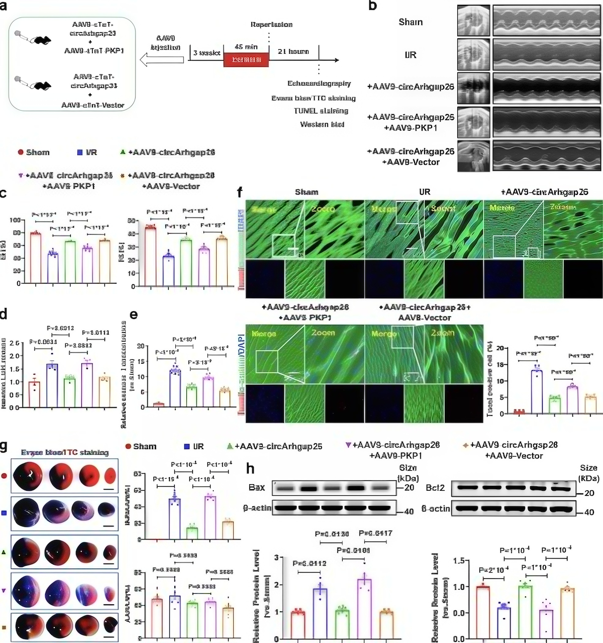
Fig. 5 PKP1 Mediates the Regulatory Role of CircArhgap26 in I/R Injury
In mice with circArhgap26 overexpression, concurrent overexpression of PKP1 significantly reversed the cardioprotective effect of circArhgap26; in mice with circArhgap26 knockdown, concurrent knockdown of PKP1 partially rescued the deterioration of cardiac function; consistent results were obtained in an in vitro H₂O₂ model.
VI. From Protein Stability to Palmitoylation: A Preliminary Exploration of the Molecular Mechanism by Which circArhgap26 Regulates PKP1
The next question to address is: How does circArhgap26 regulate PKP1? The research team first ruled out regulation at the transcriptional level—qRT-PCR showed that neither overexpression nor knockdown of circArhgap26 affected PKP1 mRNA levels, but significantly altered its protein levels, suggesting that regulation occurs at the post-transcriptional level.
CHX (streptomycin) tracing experiments revealed that circArhgap26 overexpression significantly accelerated PKP1 protein degradation and shortened its half-life. Further bioinformatics functional clustering suggested that circArhgap26 is closely associated with lipid modification. The team treated cardiomyocytes with the palmitoylation inhibitor 2-BP and found that PKP1 protein levels decreased in a dose-dependent manner; Subsequently, by directly measuring palmylation levels via an ABE (Acyl-Biotin Exchange) assay, the team ultimately confirmed that overexpression of circArhgap26 significantly reduced the level of palmylation modification of PKP1, thereby promoting its protein degradation.
This finding establishes, for the first time, a direct link between circRNA and protein palmylation modification in the field of cardiovascular research.
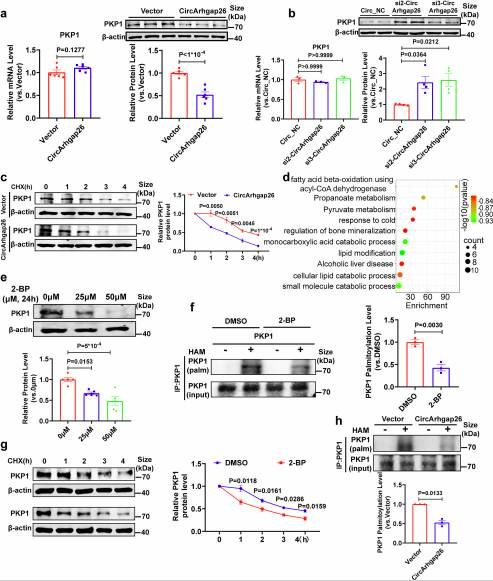
Fig. 6 Molecular Mechanism by Which circArhgap26 Regulates PKP1
circArhgap26 does not affect PKP1 mRNA levels but significantly reduces its protein levels (post-transcriptional regulation). Fig. 6c: CHX tracing experiments show that circArhgap26 overexpression accelerates PKP1 degradation and shortens its half-life; Fig. 6e: The palmitoylation inhibitor 2-BP dose-dependently reduced PKP1 protein levels; Fig. 6f–h: ABE (Acyl-Biotin Exchange) experiments confirmed that circArhgap26 overexpression significantly reduced PKP1 palmitoylation levels.
VII. Elucidation of the Competitive Inhibition Mechanism: Screening of ZDHHC1 and Competition for Binding Sites
Palmylation depends on ZDHHC family palmityltransferases. The research team systematically screened 23 ZDHHC family members and found that only ZDHHC1 and ZDHHC17 were upregulated during I/R; further functional validation showed that only ZDHHC1 knockdown could inhibit PKP1 palmitoylation, confirming that ZDHHC1 is the key enzyme mediating PKP1 palmitoylation.
Co-IP experiments confirmed a direct interaction between PKP1 and ZDHHC1, while overexpression of circArhgap26 competitively attenuated this interaction. Molecular docking and truncation experiments revealed a sophisticated competitive mechanism: ZDHHC1 also binds to the ARM1 domain of PKP1—a site that completely overlaps with the binding site of circArhgap26.
Using the CSS-Palm database to predict potential palmitoylation sites on PKP1 (C14, C136, C457, C672), the team constructed a series of point mutants. The results showed that the C14S and C136S double mutation almost completely eliminated PKP1 palmitoylation and accelerated its degradation; more importantly, the inhibitory effect of circArhgap26 on PKP1 palmitoylation was completely lost in the C14S/C136S mutant.
Thus, the core mechanism is clear: circArhgap26 competitively binds to the ARM1 domain of PKP1 with ZDHHC1, preventing palmitoylation at the C14/C136 sites, which leads to PKP1 protein instability and degradation.
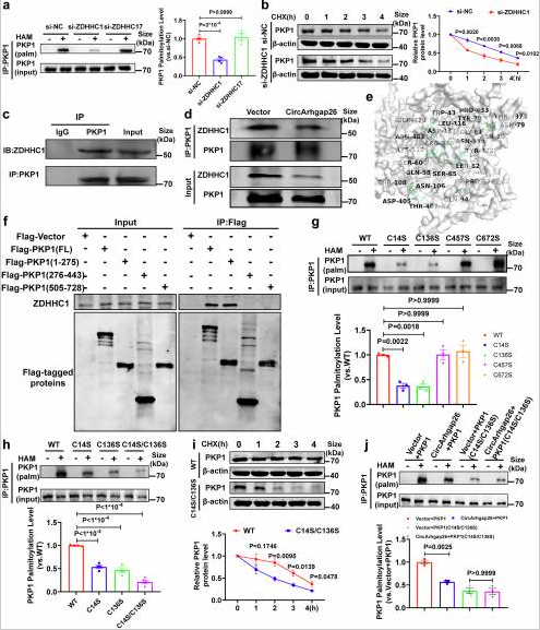
Fig. 7 ZDHHC1 Screening, Competitive Co-IP, Molecular Docking, and Site-Specific Mutation Validation
Among 23 ZDHHC family members screened, only ZDHHC1 and ZDHHC17 were upregulated during I/R; further validation confirmed that ZDHHC1 is the key enzyme mediating PKP1 palmitoylation; Fig. 7d: Co-IP confirmed that ZDHHC1 interacts with PKP1, and overexpression of circArhgap26 competitively attenuated this interaction; Fig. 7e: Molecular docking reveals that ZDHHC1 also binds to the ARM1 domain of PKP1; Fig. 7g–h: The C14S/C136S double mutation in PKP1 almost completely abolishes palmitoylation and accelerates degradation; Fig. 7i–j: The inhibitory effect of circArhgap26 on PKP1 palmitoylation is completely lost in the C14S/C136S mutant.
VIII. Analysis of the Downstream Apoptosis Pathway: The PKP1→APAF1→Caspase Axis
How does PKP1 promote apoptosis? Through predictions based on the STRING database and experimental validation, the research team discovered that APAF1 (apoptosis-promoting factor 1) is a key downstream target of PKP1. Mechanistic studies indicate that PKP1 does not affect APAF1 mRNA levels or protein degradation rates, but rather promotes translation initiation efficiency by directly binding to the 5' untranslated region (5'UTR) of APAF1 mRNA.
Both luciferase reporter assays and polysome profiling confirmed PKP1’s role in promoting APAF1 translation. Functional rescue experiments showed that APAF1 overexpression reverses the anti-apoptotic effect caused by PKP1 knockdown. Ultimately, the PKP1/APAF1 axis activates the Caspase-9/Caspase-3 apoptotic signaling pathway, while overexpression of circArhgap26 inhibits the activation of this pathway, forming a complete regulatory cascade: circArhgap26 → PKP1 → APAF1 → Caspase.
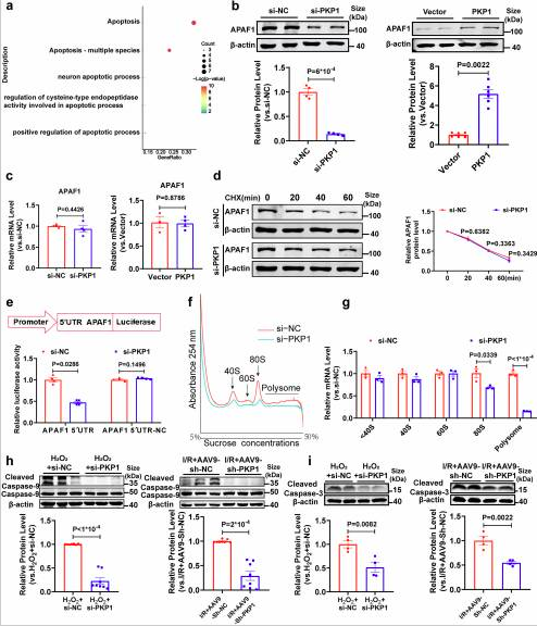
Fig. 8 APAF1 Protein Levels, Luciferase Reporter Assay, Polyribosome Analysis, and Caspase-9/3 Cleavage Validation
PKP1 does not affect APAF1 mRNA levels or protein degradation rates; Fig. 8e: A luciferase reporter assay confirmed that PKP1 binds to the 5' UTR of APAF1 mRNA and promotes its translation; Fig. 8g: Polysome profiling revealed that PKP1 knockdown reduces the distribution of APAF1 mRNA in the translationally active fraction; Fig. 8h–i: Overexpression of APAF1 reverses the anti-apoptotic effect of PKP1 knockdown, and overexpression of circArhgap26 inhibits Caspase-9/Caspase-3 activation.
IⅩ. Upstream m6A Modification Regulation: YTHDF2-Mediated Degradation of circArhgap26
The research team further investigated the upstream mechanisms underlying the downregulation of circArhgap26 during I/R. The bioinformatics software SRAMP predicted that circArhgap26 contains a high-confidence m6A modification site; MeRIP-qPCR confirmed that m6A levels in circArhgap26 were significantly elevated in I/R-treated hearts.
As a key m6A-reading protein, YTHDF2 was shown to bind to circArhgap26; knocking down YTHDF2 significantly upregulated the expression of circArhgap26. This suggests that during I/R injury, YTHDF2 promotes the degradation of circArhgap26 by recognizing the m6A modification, leading to its downregulation, which in turn releases the molecular constraint on PKP1 and ultimately exacerbates myocardial injury. This upstream mechanism, together with downstream palmitoylation regulation, forms a complete m6A–circRNA–palmitoylation dual-modification coupling network.
Ⅹ. Mechanism Integration: A Panoramic View of the circArhgap26/PKP1/APAF1 Signaling Axis
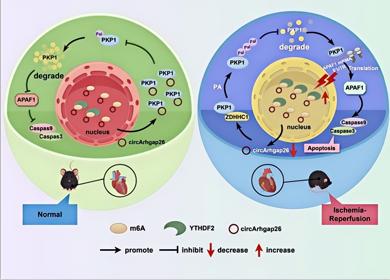
Fig. 9 Schematic Diagram of the Mechanism
Based on the above upstream and downstream studies, the team mapped out a complete regulatory pathway:
I/R injury → Upregulation of YTHDF2 + increased m6A modification → Degradation of circArhgap26 → Enhanced binding of PKP1 to ZDHHC1 → PKP1C14/ C136 palmitoylation → Stable accumulation of PKP1 protein → PKP1 binds to the 5' UTR of APAF1 mRNA → Upregulation of APAF1 translation → Activation of Caspase-9/Caspase-3 → Cardiomyocyte apoptosis → Deterioration of cardiac function
Conversely, overexpression of circArhgap26 can competitively occupy the ARM1 domain of PKP1, thereby blocking ZDHHC1-mediated palmitoylation, promoting PKP1 degradation, reducing APAF1 translation, and ultimately inhibiting apoptosis and protecting the heart. The significant downregulation of h-circArhgap26 in the plasma of PCI patients also provides clinical evidence for its use as a non-invasive prognostic biomarker.
The technical approach outlined in this STTT top-tier journal study is very comprehensive, ranging from circRNA screening, protein interaction identification, and post-translational modification validation to functional restoration experiments, covering the gold standard combination of techniques currently used in cardiovascular molecular biology research:
Technical Modules | Core Experiments | Key Reagent Requirements |
circRNA Characterization | qRT-PCR, RNase R resistance, FISH, nuclear-cytoplasmic separation | Highly Specific circRNA Probes, Internal Control Antibodies |
Functional Validation | AAV9 Myocardial-Specific Overexpression/Knockdown, Echocardiography, TTC Staining | Assay Kits for Detection of Cardiac Injury Markers |
Protein-Protein Interaction Identification | RNA pull-down、LC-MS/MS、RIP、Co-IP | High-Affinity Antibodies Against PKP1, ZDHHC1, and YTHDF2 |
Verification of Palmitoylation | ABE Assay, 2-BP Inhibitor Treatment, Site-Specific Mutations | PKP1, ZDHHC1 Antibodies (Co-IP Grade) |
Apoptosis Pathway Assay | Western blot(Bax、Bcl2、Caspase-3/9、APAF1)、TUNEL | Complete Set of Antibodies for Apoptosis Signaling Pathways |
m6A Modification Validation | MeRIP-qPCR、RIP | YTHDF2、METTL3 and m6A Antibodies |
KMD Bioscience, as a professional provider of antibody R&D and manufacturing services, also offers a complete set of core target antibodies relevant to this study, all of which have undergone rigorous validation across multiple platforms, including WB, IHC, IF, and IP:
Product Name | Target | Use Cases | KMD Bioscience Product Advantages | CAS |
| ZDHHC17 | WB, IHC | Customizable, highly specific antibodies suitable for multiple species, including humans, mice, and rats | PA15150 |
Q1. What is the core mechanism of action of circArhgap26?
circArhgap26 is a circular RNA that exerts a protective effect against myocardial ischemia-reperfusion injury; its core mechanism involves alleviating myocardial injury by competitively inhibiting the palmitoylation of PKP1. Specifically, circArhgap26 competitively binds to the ARM1 domain of PKP1 with the palmitoyltransferase ZDHHC1, preventing palmitoylation at positions C14 and C136. This leads to PKP1 protein instability and degradation, thereby reducing the translation of downstream APAF1, inhibiting the activation of the Caspase-9/Caspase-3 apoptotic pathway, and ultimately protecting cardiomyocytes from I/R injury.
Q2. What was the key experiment in this study that established the circArhgap26→PKP1 causal regulatory axis?
Functional rescue experiments were key to establishing this causal relationship. The research team simultaneously overexpressed PKP1 in mice with circArhgap26 overexpression and found that this significantly reversed the cardioprotective effects induced by circArhgap26; conversely, simultaneous PKP1 knockdown in mice with circArhgap26 knockdown partially rescued the deterioration of cardiac function. An in vitro H₂O₂ model similarly confirmed that PKP1 overexpression blocks the anti-apoptotic effects of circArhgap26. This series of rigorous rescue experiments clearly demonstrates that PKP1 is an essential intermediary molecule for circArhgap26 to exert its cardioprotective effects.
Q3. What is the role of palmitoylation in I/R injury, and what are the intervention strategies for circArhgap26?
Palmylation is an important post-translational lipid modification catalyzed by ZDHHC family transferases. During I/R injury, ZDHHC1 expression is upregulated, catalyzing the palmylation of PKP1 at positions C14 and C136, leading to the stable accumulation of the PKP1 protein and subsequently promoting apoptosis. The intervention strategy for circArhgap26 lies in its ability to competitively bind to the ARM1 domain of PKP1—a site that completely overlaps with the ZDHHC1 binding site—thereby blocking palmitoylation and promoting PKP1 degradation. This competitive inhibition mechanism provides a theoretical basis for developing small-molecule inhibitors targeting the PKP1-ARM1 domain or the active site of ZDHHC1.
Q4. How does the m6A-YTHDF2 axis regulate the expression of circArhgap26?
During ischemia-reperfusion (I/R) injury, the level of m6A modification on circArhgap26 is significantly elevated. As a key m6A reader protein, YTHDF2 recognizes and binds to m6A modification sites on circArhgap26, promoting its degradation. Experiments show that knocking down YTHDF2 significantly upregulates the expression of circArhgap26. This upstream regulatory mechanism suggests that I/R injury promotes the degradation of circArhgap26 through YTHDF2-mediated recognition of m6A modifications, thereby releasing the molecular constraint on PKP1 and exacerbating myocardial injury. Together with downstream palmitoylation regulation, this mechanism forms a dual-modification coupling network involving m6A, circRNA, and palmitoylation.
Q5. What are the core advantages of KMD Bioscience’s antibody products in cardiovascular research?
A comprehensive validation system ensures that all antibodies undergo rigorous validation across multiple platforms—including WB, IHC, IF, IP, and FCM—to guarantee reliable performance in various experimental scenarios of cardiovascular disease research and meet comprehensive needs ranging from protein expression detection to interaction analysis.
Comprehensive target coverage: We offer a complete suite of antibody products for core targets in cardiovascular research, including PKP1, ZDHHC1, YTHDF2, Bax, Bcl-2, Caspase-3/9, and APAF1, covering cutting-edge research areas such as the apoptotic pathway, palmitoylation, and m6A modification.
Strong customization capabilities: We offer custom antibody services for multi-species reactivity based on research needs, supporting various sample types including human, mouse, and rat samples. These services are particularly well-suited for research on animal models of cardiovascular disease, facilitating the translation of scientific findings into clinical applications.
References
Zhang MY, Ji DN, Qi WY, Wang S, Dai CY, Xu FY, Dong Z, Xiong C, Zhang BW, Wang Y, Guo XF, Zhang B, Cheng WZ, Zhang XY, Jin SD, Guan XX, Hong H, Zhou T, Li SF, Zhang R, Sun LH, Xuan LN, Jiang Y, Yang BF, Xu CQ. M6A-modified circArhgap26 attenuates cardiac ischemia‒reperfusion injury by suppressing plakophilin-1 palmitoylation. Signal Transduct Target Ther. 2026 Mar 17;11(1):99. doi: 10.1038/s41392-026-02609-4. PMID: 41844577; PMCID: PMC12996625.

- Protein Platform
- Protein Expression Services
- Protein Modification & Detection Services
- Protein Interaction Services
- Protein Purification Service
- Antibody Discovery Platform
- VHH Antibody Platform
- Phage Display Platform
- Antibody Humanization Platform
- Single B Cell Screening Platform
- Antibody Expression & Validation Platform
- Peptide Library Platform
- Custom Antibody Platform
- Innovative Drug Discovery Platform
- Product Center
- Proteins
- Antibodies
- Raw Materials Reagents
- Small Molecule Reagents
- Peptide Products
- Nanoparticle Products
- Contact Us
- Phone:400-621-6806
- Mobile:13110936064(WeC
- E-mail:[email protected]
- Address:Room 108, Building 10, No. 6 Hechang Road, Wuqing Development Zone, Tianjin City
-

WeChat
-

Service Account
-
 400-621-6806
-
 3599163699
-
 Monday to Friday 9:00-18:00
-

-
 0
-
