


The Definitive Overview of Phage Display Technology in Microbiology
- 2026-07-29
- 136
In the rapidly evolving landscape of biotherapeutics and diagnostic reagent development, relying strictly on traditional animal immunization protocols is no longer a viable commercial strategy. The inherent limitations of the mammalian immune system—specifically its inability to generate robust responses against highly conserved or toxic antigens—create massive bottlenecks in the drug discovery pipeline. If you are attempting to isolate highly specific recombinant antibodies for challenging targets, hybridoma technology will frequently fail you.
This is where an operational understanding of Phage Display Technology in Microbiology becomes a critical asset. Awarded the Nobel Prize in Chemistry in 2018, this high-throughput screening methodology fundamentally altered the biotechnology sector by completely bypassing in vivo immunization. By genetically fusing a protein or peptide sequence to the coat protein of a bacteriophage, scientists created a direct, physical linkage between genotype and phenotype. In this comprehensive, opinionated guide, we will dissect the mechanics of phage display, evaluate its commercial viability against competing technologies, and provide actionable intelligence on whether your laboratory should internalize this platform or outsource it to specialized contract research organizations.
Quick Answer: Is Phage Display Worth the Investment?
Yes, adopting Phage Display Technology in Microbiology is absolutely essential for modern biologic development. We recommend this technology unequivocally for discovering recombinant antibodies (such as scFv, Fab, and VHH nanobodies) against non-immunogenic, toxic, or highly conserved targets where traditional animal models fail. It condenses the discovery timeline from several months to just a few weeks. However, building a diverse, high-capacity phage library (exceeding 10^10 variants) requires massive capital and specialized molecular biology infrastructure. For most commercial entities, outsourcing the screening process to an established Phage Display Platform provides a superior return on investment compared to attempting to build and validate a naive library from scratch in-house.
What is Phage Display Technology in Microbiology?
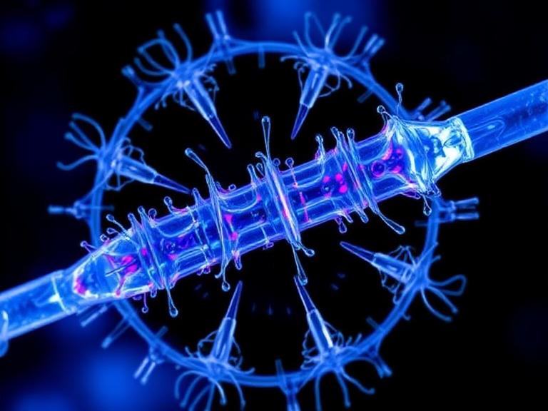
At its core, Phage Display Technology in Microbiology is a sophisticated selection technique used to study protein-protein, protein-peptide, and protein-DNA interactions. A bacteriophage is a virus that specifically infects bacteria. In commercial applications, the M13 filamentous phage is the most commonly utilized vector. Scientists genetically engineer the phage genome by inserting a foreign DNA sequence directly into the gene that encodes one of the phage's surface coat proteins (typically pIII or pVIII).
As the phage replicates inside its bacterial host (E. coli), it expresses this foreign sequence on its outer surface, "displaying" the resulting peptide or antibody fragment to the outside world, while the DNA blueprint for that protein remains safely encapsulated inside the phage particle. This physical connection is the genius of the system. You can create a "library" containing billions of unique phages, wash them over a target molecule, and physically capture the specific phages that bind. Because the DNA is inside the captured phage, you instantly know the exact genetic sequence required to manufacture that specific binder.
Quick Summary Table: Phage Display Specifications
| Parameter | Industry Standard Specifications |
|---|---|
| Typical Host Vector | M13 Filamentous Bacteriophage, T7 Phage |
| Target Coat Proteins | pIII (low valency/high affinity), pVIII (high valency/low affinity) |
| Standard Library Size | 10^9 to 10^11 unique variants |
| Discovery Timeline | 3 to 5 weeks (Significantly faster than in vivo methods) |
| Common Output Formats | scFv, Fab fragments, VHH (Nanobodies), Custom Peptides |
How It Works: The Mechanics of Biopanning
The actual execution of Phage Display Technology in Microbiology is conducted through an iterative screening process known as "biopanning." In our testing and daily laboratory operations, the biopanning protocol dictates the ultimate success or failure of the discovery campaign. The process involves four meticulous steps:
Incubation (Binding): The vast library of displaying phages is exposed to an immobilized target antigen. This antigen is typically coated onto a microtiter plate or conjugated to magnetic beads. The phages that possess a complementary shape will physically bind to the antigen.
Washing: This is where precision is paramount. The immobilized target is aggressively washed with buffer solutions. Unbound or weakly bound phages are flushed away. In most professional situations, the stringency of the wash buffer is increased in subsequent rounds to isolate only the most aggressive, high-affinity binders.
Elution: The strong binders that survived the washing phase are chemically or enzymatically detached (eluted) from the target antigen.
Amplification: These elite phages are then used to infect fresh E. coli cultures. The bacteria act as factories, multiplying the winning phages by the millions. This enriched pool is collected, and the entire panning cycle is repeated 3 to 5 times.
By the end of the process, what started as a highly diverse library of billions is narrowed down to a handful of hyper-specific binders. Their DNA is sequenced, and the genetic code is transferred into an Antibody Expression & Validation Platform for large-scale production.
The Unmatched Benefits of Phage Display
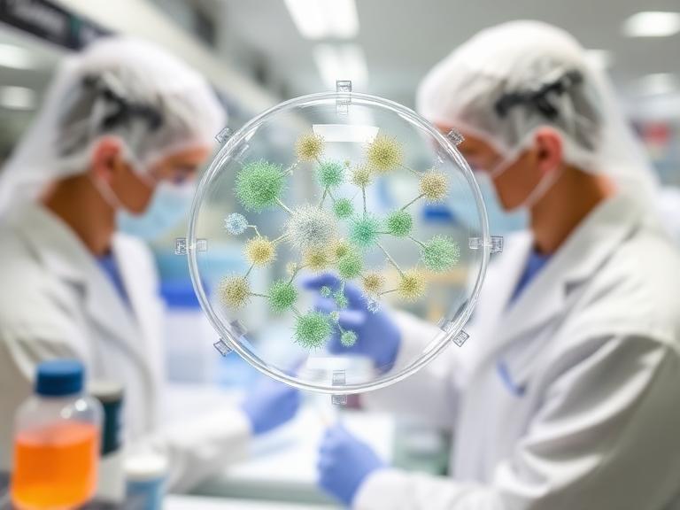
The commercial and scientific advantages of phasing out animal models in favor of in vitro screening are overwhelming. For heavy-duty applications in therapeutics, Phage Display Technology in Microbiology offers distinct operational leverage.
First, it completely circumvents the phenomenon of immunological tolerance. Mammalian immune systems are programmed not to attack their own proteins (self-antigens). Because phage display occurs entirely in a test tube, you can easily generate antibodies against highly conserved human targets. Furthermore, you can target lethal molecules. You cannot inject a mouse with a potent venom or deadly pathogen and expect it to survive long enough to mount an immune response. Phage display handles toxic antigens effortlessly.
Finally, the speed is unparalleled. Traditional hybridoma generation can take upwards of four to six months. A highly optimized Peptide Library Platform or antibody library can yield validated sequences in under four weeks, drastically accelerating your time-to-market.
Limitations and Drawbacks
We are industry practitioners, and we must emphasize that no technology is flawless. Phage Display Technology in Microbiology has distinct limitations that must be factored into your cost-benefit analysis.
The most glaring limitation is the lack of eukaryotic post-translational modifications. Because the proteins are expressed by bacterial hosts (E. coli), they do not undergo glycosylation. If the functional binding of your target relies heavily on specific sugar chains, phage-derived antibodies may struggle to recognize the native mammalian protein. Additionally, during the creation of synthetic or naive libraries, the natural pairing of heavy and light chains is randomized. This loss of natural in vivo pairing can occasionally result in antibodies with lower thermodynamic stability, requiring extensive downstream engineering via an Antibody Humanization Platform.
Pros and Cons Table: Phage Display Technology
| The Pros (Advantages) | The Cons (Limitations) |
|---|---|
| Requires zero animal immunization (ethically superior and faster). | Proteins expressed in E. coli lack post-translational modifications. |
| Capable of screening massive libraries (up to 10^11 variants) simultaneously. | Building a high-quality, diverse naive library is incredibly expensive. |
| Successfully targets non-immunogenic, conserved, or highly toxic antigens. | Random VH/VL pairing can lead to suboptimal stability in some binders. |
| Provides immediate access to the genetic sequence for rapid recombinant production. | Requires highly specialized molecular biology infrastructure to execute properly. |
Who Should Use It
For commercial users: Biotechnology firms developing novel oncology therapeutics, diagnostic manufacturers seeking robust VHH nanobodies for assay kits, and academic institutions pushing the boundaries of an Innovative Drug Discovery Platform must utilize phage display. If your competitive edge relies on securing intellectual property for unique, fully humanized recombinant antibodies rapidly, this technology is mandatory.
Who Does Not Need It
For beginners and routine diagnostic labs: If you are running standard western blots, ELISAs, or immunohistochemistry on well-documented pathways, do not attempt to commission a custom phage display project. The cost will obliterate your budget. For routine laboratory work, we strongly recommend purchasing validated antibody products or sourcing reliable antibody reagents from an established primary antibody supplier.
Comparison Table: Phage Display vs. Traditional Methods
| Feature / Technology | Phage Display Screening | Traditional Hybridoma (In Vivo) | Single B Cell Screening |
|---|---|---|---|
| Discovery Timeline | 3 - 5 Weeks | 4 - 6 Months | 2 - 4 Weeks |
| Animal Dependency | None (In Vitro) | Required (Mice/Rabbits) | Required (Alpaca/Rabbit/Human) |
| Target Restrictions | None (Can target toxins/self-antigens) | Fails on toxic or conserved antigens | Requires an immunogenic response |
| Natural Chain Pairing | Randomized (Loss of natural pairing) | Preserved | Preserved |
| Best Use Case | Fully human libraries, toxic targets | Standard monoclonal reagent production | Rapid isolation of naturally affinity-matured binders |
Common Mistakes in Phage Display Screening
From our experience auditing failed screening campaigns, the most catastrophic mistake researchers make is working with an underpowered library. If your library only contains 10^6 variants, your chances of pulling a sub-nanomolar affinity binder are practically zero. A commercial-grade library must exceed 10^9 variants.
Another frequent error is poor panning stringency. If your washing steps are too gentle, you will elute thousands of false positives (background binders). Conversely, if you are too aggressive in round one, you will wash away the rare, high-affinity clones. Mastering the titration of detergents (like Tween-20) during the washing phase is what separates amateur laboratories from industry leaders.
Buying Considerations: Outsourcing vs. In-House
When deciding how to implement Phage Display Technology in Microbiology, you face a critical build-versus-buy scenario. Establishing a 10^10 capacity naive library in-house requires years of labor, sequencing validation, and millions of dollars in infrastructure. For 95% of biotech firms, this is not a practical use of capital.
When selecting a Contract Research Organization (CRO) to handle your project, you must verify their library metrics. Ask to see their next-generation sequencing (NGS) data to prove library diversity. Ensure they offer a seamless pipeline that transitions from discovery directly into a Custom Antibody Platform for expression. If a CRO hands you a DNA sequence but cannot physically manufacture the recombinant protein for you, you will lose months transferring the project to a new vendor.
Expert Recommendation from KMD Bioscience
Why Partner with KMD Bioscience?
From our extensive industry experience, piecing together a discovery pipeline from multiple disjointed vendors leads to catastrophic delays. You need a partner who controls the entire vertical architecture of recombinant development.
KMD Bioscience has extensive experience in Recombinant Antibody development and research, supported by a robust technical system that provides a distinct edge within the industry. We deliver premium scientific services to our clients. We have achieved outstanding results in preparing scFv, Fab, and VHH Antibodies, earning the trust of clients globally. Utilizing Phage Display Antibody Library Construction followed by multiple rounds of ELISA Screening, we efficiently identify high-affinity antibodies for clients. Furthermore, we conduct diverse antibody screening assays tailored to specific client requirements to generate highly specific and stable antibodies.
Our Verdict: Do not waste your budget attempting to build naive libraries in-house. Leverage our pre-validated infrastructure. If you require research antibodies for labs or are pursuing complex therapeutic targets, integrating your project into our proven phage display and Single B Cell Screening Platform is the most commercially sound decision you can make in 2026.
Frequently Asked Questions (FAQ)
What is the primary advantage of Phage Display Technology in Microbiology over hybridoma?
The primary advantage is that phage display is an entirely in vitro process, eliminating the need for animal immunization. This allows researchers to discover antibodies against highly toxic, lethal, or non-immunogenic self-antigens that mammalian immune systems cannot process. It also reduces discovery timelines from several months to just a few weeks.
What types of antibodies can be produced using phage display?
Phage display is highly versatile and is typically used to discover smaller recombinant antibody fragments such as scFv (single-chain variable fragments), Fab fragments, and VHH nanobodies. Because you instantly obtain the DNA sequence, these fragments can easily be engineered into full-length monoclonal antibodies (IgG) in downstream expression platforms.
Is phage display suitable for discovering therapeutic drugs?
Absolutely. Phage display is a foundational technology in modern drug discovery. Several blockbuster therapeutic antibodies currently on the market, including Adalimumab (Humira), were discovered and fully humanized using phage display libraries, making it a gold standard for commercial pharmaceutical development.
Authoritative References & Industry Standards
To ensure your discovery pipeline aligns with global scientific standards and regulatory compliance, we recommend reviewing the foundational literature and guidelines provided by the following authoritative organizations:
The Nobel Prize Organization: The Nobel Prize in Chemistry 2018 – Awarded jointly to George P. Smith and Sir Gregory P. Winter for the pioneering development of the phage display of peptides and antibodies, validating its global scientific impact.
National Institutes of Health (NIH) / PubMed: Phage Display Technology: Clinical Applications and Recent Innovations – Peer-reviewed comprehensive literature detailing the biochemical mechanics and therapeutic successes of biopanning in modern medicine.
U.S. Food and Drug Administration (FDA): Drug Development and Approval Process – Regulatory frameworks outlining the necessary characterization, humanization, and clinical validation required for biologics and recombinant antibodies derived from in vitro display platforms.
- Phage Display Technology in Microbiology

- Protein Platform
- Protein Expression Services
- Protein Modification & Detection Services
- Protein Interaction Services
- Protein Purification Service
- Antibody Discovery Platform
- VHH Antibody Platform
- Phage Display Platform
- Antibody Humanization Platform
- Single B Cell Screening Platform
- Antibody Expression & Validation Platform
- Peptide Library Platform
- Custom Antibody Platform
- Innovative Drug Discovery Platform
- Product Center
- Proteins
- Antibodies
- Raw Materials Reagents
- Small Molecule Reagents
- Peptide Products
- Nanoparticle Products
- Contact Us
- Phone:400-621-6806
- Mobile:13110936064(WeC
- E-mail:[email protected]
- Address:Room 108, Building 10, No. 6 Hechang Road, Wuqing Development Zone, Tianjin City
-

WeChat
-

Service Account
-
 400-621-6806
 -
 3599163699
 -
 Monday to Friday 9:00-18:00
 -

-
 0
0
-
