


Vaccine Efficacy Repeatedly Compromised? Possibly Due to the ‘Immunogenicity Engine’at the E484 Site Not Being Tuned Correctly
- 2026-07-11
- 115
The continuous mutation of SARS-CoV-2 has led to a high frequency of mutations in the receptor-binding domain (RBD) of the spike protein, which not only enhances viral transmission and immune evasion but also reduces the protective efficacy of existing vaccines and the reliability of antibody detection assays. As the core region mediating viral entry by binding to the host ACE2 receptor, RBD serves as a critical antigenic target for the development of recombinant protein vaccines and COVID-19 antibody detection kits. Therefore, screening for RBD variants with high immunogenicity and broad-spectrum recognition is of central importance for developing epidemic-fighting tools tailored to emerging variants.
In September 2022, a research team from Southern Medical University published a study in Viruses, demonstrating that the expression of various SARS-CoV-2 RBD mutants using an E. coli expression system confirmed that amino acid mutations at position E484 significantly enhance the immunogenicity of the RBD protein. This finding provides a key candidate target and technical support for the design of broadly protective COVID-19 vaccines and the development of multi-variant-compatible antibody detection assays.
I. Overview
SARS-CoV-2 infection relies on the binding of the spike protein RBD to the host cell ACE2 receptor. K417, L452, E484, and N501 are high-frequency mutation sites on RBD. Among these, the E484 site exhibits multiple mutations such as E484K, E484Q, and E484A, which are widely distributed in major circulating variants including Beta, Gamma, Kappa, and Omicron, directly affecting viral infectivity and immune evasion characteristics.
In this study, using the wild-type RBD as a template, nine RBD mutants (six single-point mutants, one double-point mutant, and two triple-point mutants) were constructed. The proteins were prepared using a prokaryotic expression system in E. coli, and active proteins were obtained through inclusion body denaturation and renaturation followed by affinity purification. The antigenicity in vitro was validated by Western blot, and the immunogenicity in vivo was systematically evaluated through mouse immunization experiments. Ultimately, highly immunogenic mutants such as E484K and E484Q were identified, confirming the key regulatory role of the E484 site in RBD function and immunogenicity. This study also validates that E. coli-expressed RBD can serve as an efficient platform for vaccine development and antibody detection tool development.
II. Prokaryotic Expression and Purification of RBD Mutants
In this study, nine target mutants (six single-point mutants, one double-point mutant, and two triple-point mutants) were constructed using inverse PCR. After prokaryotic expression and protein purification, active recombinant RBD proteins with a molecular weight of approximately 30 kDa were obtained. Affinity chromatography combined with ultrafiltration was used to remove endotoxins, and the endotoxin levels in all mutants were below 2.0 EU/mL, meeting the requirements for animal experiments and subsequent applications.
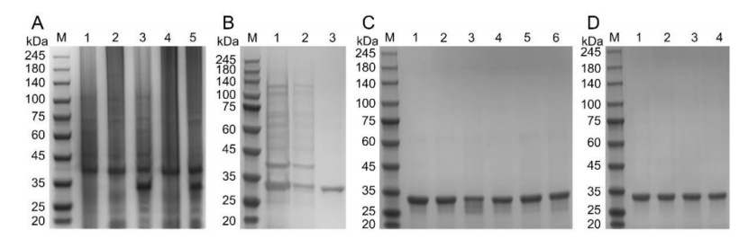
Fig 1. Expression and purification of RBD protein mutants
III. RBD Mutants Exhibit Strong In Vitro Antigenicity
The biological activity of purified wild-type and mutant RBD proteins was assessed by Western blot. Both wild-type and all nine mutant proteins displayed clear specific bands at approximately 30 kDa, confirming that all RBD mutants possess good in vitro antigenicity with stable and reliable antigenic activity, rendering them suitable for subsequent key experiments such as in vivo immunogenicity evaluation in mice.
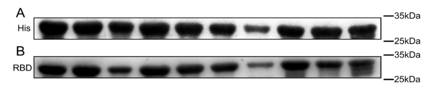
Fig 2. Western blot analysis of RBD protein mutants
IV. Mutation at the E484 Site Is the Core Key to Enhancing RBD Immunogenicity
BALB/c mice were immunized with RBD proteins, and ELISA results showed that no anti-RBD antibodies were detected in pre-immune sera from any mouse. On day 13 post-immunization, both wild-type and all mutants successfully induced specific RBD antibodies. By day 25, antibody levels had increased substantially, with significant differences in antibody titers induced by different mutants. Specifically, the antibody titers induced by E484K, E484Q, K417T‑E484K‑N501Y, and K417N‑E484K‑N501Y were 4.7‑fold, 4.8‑fold, 6.8‑fold, and 5.0‑fold higher than that of wild-type RBD, respectively, demonstrating significantly superior immunogenicity. The K417N, L452R, and L452R‑E484Q groups showed titers approximately 2‑fold higher than wild‑type. The single-point mutants K417T and N501Y showed no significant difference from wild‑type. These results directly demonstrate that mutation at the E484 site is the core key to enhancing RBD immunogenicity.
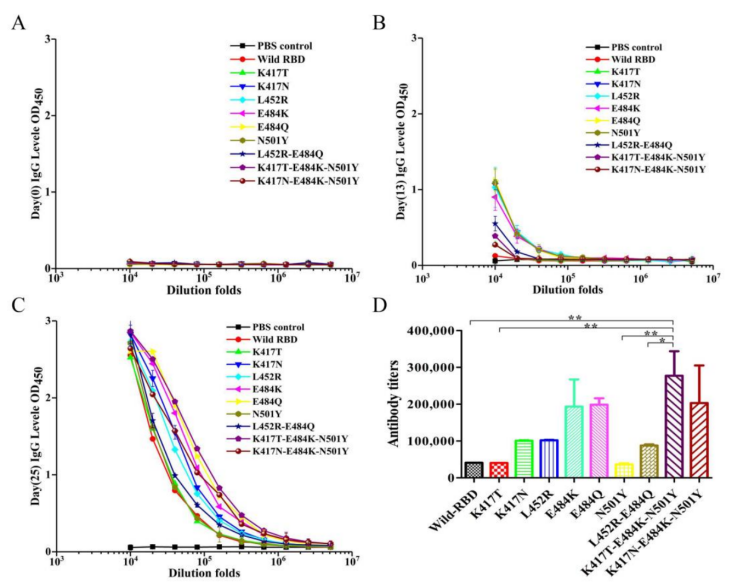
Fig 3. Detection of antibody levels against SARS-CoV-2 RBD in mice by enzyme-linked immunosorbent assay (ELISA)
V. Key Regulatory Role of the E484 Site in Broad-Spectrum RBD Antigenicity
In this study, E484K, E484Q, K417T‑E484K‑N501Y, and K417N‑E484K‑N501Y were used as coating antigens to assess the cross-binding titers of sera from immunized mice. The results showed that the E484K antigen efficiently recognized antibodies from all immunization groups, exhibiting excellent binding ability with antibodies induced by wild‑type and various mutants, demonstrating outstanding broad‑spectrum reactivity. The E484Q antigen induced antibodies with high binding titers against a broad range of RBD mutants, showing strong cross‑recognition capacity. In contrast, the two triple‑point mutant antigens only showed good recognition of antibodies from the E484K and E484Q immunization groups, with no significant advantage for other groups, making them unsuitable as broad‑spectrum detection antigens. This study also confirmed that the binding affinity between RBD and ACE2 does not directly determine its immunogenicity, a key finding that provides a novel theoretical basis for the design and optimization of COVID‑19 vaccine antigens.
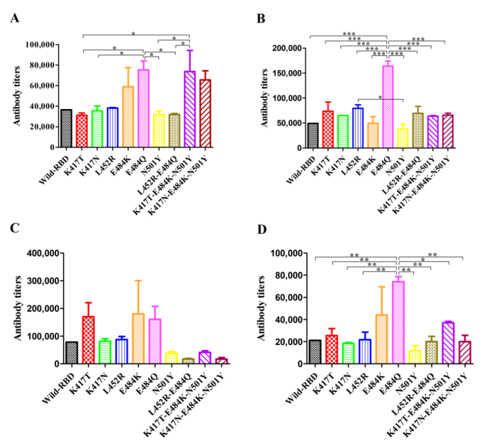
Fig 4. Comprehensive analysis of the antigenicity of RBD variant proteins
This study successfully constructed and validated the in vitro antigenicity and in vivo immunogenicity of nine SARS‑CoV‑2 RBD mutants, and demonstrated that amino acid changes at the E484 site can significantly enhance the immunogenicity of the RBD protein. The research not only improves the understanding of the functional regulatory mechanisms of SARS‑CoV‑2 RBD mutants but also provides critical experimental data on key antigenic targets for the development of broad‑spectrum recombinant protein vaccines and multi‑variant antibody detection reagents. It holds significant theoretical value and application prospects for enhancing the ability to respond to viral variants and strengthening the COVID‑19 detection system.
The recombinant human anti-SARS-CoV-2 S1 RBD monoclonal antibodies used in this study were purchased from KMD Bioscience (Tianjin, China), including three different types of core reagents with catalog numbers MAV101, MAV102, and MAV103. These were primarily used for Western blot antigenicity identification and ELISA antibody titer analysis, enabling accurate validation of the in vitro antigenic activity and in vivo immunogenicity of RBD mutants, thereby providing critical experimental support for SARS-CoV-2 RBD functional studies and vaccine antigen screening.
KMD Bioscience has established a mature antibody research and development technology platform, encompassing a multi-species antibody discovery platform covering hybridoma, phage display, and Single B cell screening. This platform enables the development of various antibody types, including mouse, rabbit, human, and alpaca VHH nanobodies, and provides integrated solutions ranging from monoclonal/polyclonal antibody preparation to antibody humanization, affinity maturation, and bispecific antibody construction. Furthermore, by integrating BLI and SPR affinity analysis as well as flow cytometry and ELISA-based specificity validation technologies, the platform can screen antibodies with affinity down to the pM level, providing robust technical support for the translation of research outcomes into industrial applications.
[1] Zhang Z, Wan X, Li X, Cai S, Wan C. Enhancing the Immunogenicity of RBD Protein Variants through Amino Acid E484 Mutation in SARS-CoV-2. Viruses. 2022;14(9):2020.
——FAQs——
Q1: Why is the RBD region of SARS-CoV-2 a key target for vaccine and diagnostic reagent development?
A: The receptor-binding domain (RBD) of the SARS-CoV-2 spike (S) protein is the core functional region through which the virus recognizes and binds to the host cell ACE2 receptor. The RBD binds to ACE2 with high affinity through conformational changes, mediating the fusion of the viral envelope with the cell membrane and thereby completing the invasion process. Therefore, blocking the RBD-ACE2 interaction is the primary mechanism by which neutralizing antibodies exert their protective effects, and it also serves as the logical starting point for vaccine design and antibody detection reagent development.
The RBD offers multiple advantages as an antigenic target. First, it contains several major neutralizing epitopes, and antibodies generated after immunization can effectively block viral binding. Second, compared to the full-length S protein, the RBD is relatively conserved in structure and smaller in molecular weight, making it easier to produce via prokaryotic or eukaryotic recombinant expression systems, thereby reducing production costs. However, with the continuous emergence of variants such as Omicron, high-frequency mutations at sites including K417, L452, E484, and N501 on the RBD have led to enhanced viral immune evasion, reducing the efficacy of existing vaccines and detection reagents.
Q2: What are the advantages and challenges of using the E. coli expression system to produce RBD proteins?
A: The E. coli expression system is the most commonly used and lowest-cost prokaryotic system for recombinant protein production. Its advantages include a well-understood genetic background, ease of manipulation, short culture cycles, straightforward scale-up, and high target protein yield, making it suitable for industrial preparation. For functional domains such as RBD where glycosylation is not critical, the E. coli system can produce milligram-to-gram quantities of recombinant protein in a short time, greatly reducing R&D costs.
However, the E. coli expression system also presents significant challenges. Due to the lack of eukaryotic post-translational modification machinery, complex proteins often form inclusion bodies (inactive insoluble aggregates). Additionally, endotoxins (lipopolysaccharides, LPS) in the cell wall of Gram-negative bacteria can strongly activate immune cells, affecting both in vitro and in vivo experimental results. For RBD proteins, the technical difficulties lie in how to renature inclusion bodies into correctly folded, active proteins and how to reduce endotoxin levels to a safe threshold.
Q3: Why does mutation at the E484 site significantly enhance the immunogenicity of the RBD protein? What is the mechanism?
A: The E484 site is located at the edge of the RBD-ACE2 binding interface and is a highly exposed flexible loop region. Mutations such as E484K, E484Q, and E484A frequently occur at this site in multiple variants (Beta, Gamma, Kappa, Omicron). The ability of amino acid changes at this site to significantly enhance RBD immunogenicity involves three mechanistic levels: antigen structure, epitope exposure, and immune recognition.
First, from a structural perspective, the wild-type RBD carries negatively charged glutamic acid at position 484. Mutations to positively charged lysine (E484K) or neutral glutamine (E484Q) alter local charge distribution and side‑chain conformation, potentially inducing subtle adjustments in the overall RBD folding pattern. Such conformational changes may render previously hidden or partially hidden immunodominant epitopes (e.g., neutralizing antibody binding epitopes) more exposed on the protein surface, thereby enhancing recognition efficiency by B‑cell receptors.
Second, E484 mutations may alter the aggregation state or degradation kinetics of RBD. In this study, the antibody titers induced by E484K and E484Q mutants in mice reached 4.7‑fold and 4.8‑fold those of wild‑type RBD, respectively, whereas single‑point mutations K417T or N501Y showed no significant improvement. This suggests that the E484 site acts as a unique “immunogenicity‑enhancing switch.” This is likely because E484 mutations increase the efficiency of protein uptake, processing, and presentation by antigen‑presenting cells, thereby more effectively activating Tfh cells and germinal center reactions.
Finally, the study also found that when used as a coating antigen, the E484K mutant efficiently recognized antibodies from all immunization group sera (including those induced by wild‑type and various mutants), demonstrating excellent broad‑spectrum reactivity. This indicates that E484 mutation not only enhances its own immunogenicity but may also induce broad‑spectrum antibodies targeting conserved regions of RBD. The underlying mechanisms warrant further investigation through crystal structure determination and B‑cell epitope mapping.
Q4: Does the binding affinity between RBD protein and ACE2 determine its immunogenicity?
A: Conventional thinking often suggests that a higher binding affinity between a viral antigen and its host receptor may imply that the antigenic epitope is more "critical," thereby inducing a stronger immune response. However, through systematic comparison of ACE2 binding capacity and in vivo immunogenicity among different RBD mutants, this study indicates that RBD‑ACE2 binding affinity does not directly determine immunogenicity.
In contrast, the E484K and E484Q mutations have a relatively minor impact on ACE2 affinity (in some contexts even slightly reducing binding) yet significantly enhance immunogenicity. This suggests that immunogenicity is primarily determined by antigen conformational stability, epitope exposure, proteasomal degradation patterns, and interactions with innate immune receptors, rather than solely by receptor binding capacity.
Q5: What is the impact of endotoxin levels on in vitro and in vivo experiments with recombinant proteins?
A: Endotoxin, also known as lipopolysaccharide (LPS), is a major component of the cell wall of Gram‑negative bacteria such as E. coli. During recombinant protein production, residual endotoxin, even after purification, can severely interfere with subsequent experiments.
In vitro, endotoxin activates macrophages, dendritic cells, and other cell types, inducing the release of inflammatory cytokines such as TNF‑α, IL‑1β, and IL‑6, leading to increased background in Western blots and reduced cell viability (a common cause of cell death).
In vivo, endotoxin directly triggers innate immune responses, causing fever, inflammation, or even death in mice, while also non‑specifically enhancing or distorting adaptive immune responses, thereby compromising the reliability of vaccine immunogenicity evaluation. Therefore, for recombinant proteins used in animal immunization, endotoxin levels are generally required to be below 5 EU/mL.
- ADCs
- scFv antibodies
- targeted therapy
- immunogenicity

- Protein Platform
- Protein Expression Services
- Protein Modification & Detection Services
- Protein Interaction Services
- Protein Purification Service
- Antibody Discovery Platform
- VHH Antibody Platform
- Phage Display Platform
- Antibody Humanization Platform
- Single B Cell Screening Platform
- Antibody Expression & Validation Platform
- Peptide Library Platform
- Custom Antibody Platform
- Innovative Drug Discovery Platform
- Product Center
- Proteins
- Antibodies
- Raw Materials Reagents
- Small Molecule Reagents
- Peptide Products
- Nanoparticle Products
- Contact Us
- Phone:400-621-6806
- Mobile:13110936064(WeC
- E-mail:[email protected]
- Address:Room 108, Building 10, No. 6 Hechang Road, Wuqing Development Zone, Tianjin City
-

WeChat
-

Service Account
-
 400-621-6806
 -
 3599163699
 -
 Monday to Friday 9:00-18:00
 -

-
 0
0
-
