


What is the protocol for phage display library screening?
- 2026-05-25
- 153
In contemporary drug discovery and monoclonal antibody engineering, the identification of high-affinity binding leads depends heavily on functional display platforms. Phage display technology allows researchers to link phenotype directly to genotype by displaying variable peptide or antibody fragments on the coat proteins of filamentous bacteriophages. Extracting specific binder candidates from a vast complex mixture requires a series of selection steps known as biopanning. Establishing a precise protocol for phage display library screening dictates your target specificity, candidate pool diversity, and overall lead discovery timelines.
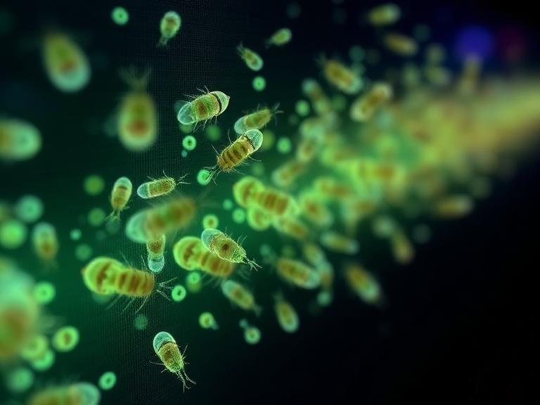
From our experience at KMD Bioscience, baseline screening failures or the selection of low-specificity binders are rarely caused by poor library diversity. Instead, they typically trace back to unbalanced target immobilization parameters, inadequate blocking steps, or uncalibrated elution stresses that release non-specific background binders. When a screen lacks carefully calculated selection dynamics, target-unrelated phages outgrow authentic binders during expansion phases, which ruins the discovery cycle. This technical brief details the core steps, validation methodologies, and biochemical controls needed to optimize your biopanning workflows.
Table of Contents
2. Detailed Execution: Protocol for Phage Display Library Screening
3. Biopanning Phase and Thermodynamic Parameter Summary Matrix
4. Critical Adjustments to Increase Affinity and Avoid Common Pitfalls
5. Post-Screening Validation: Characterization and Sequencing Pipelines
6. Sourcing Full Turnkey Biologics Support: The KMD Bioscience Edge
1. Biochemical Foundations of Linear Biopanning Cascades
To accurately configure a protocol for phage display library screening, discovery teams must analyze the physical binding mechanics taking place inside the selection well or liquid matrix. The entire process balances kinetic association and dissociation constraints. When a diverse, variable display population encounters an immobilized antigen target, binder candidates form non-covalent structural links with specific target epitopes, while weak variants remain free in the surrounding liquid.
We recommend establishing strict baseline negative control screens alongside your main experimental pipelines. If your protocol overlooks a pre-clearing phase, variable clones can bind to plastic polystyrene container walls or chemical biotin-binding streptavidin matrices instead of your actual target. Performing an early negative selection step removes these background binding elements, ensuring that your remaining population targets the true antigen structures during subsequent wash steps.
2. Detailed Execution: Protocol for Phage Display Library Screening
The following detailed directives map out the linear execution phases required to complete a comprehensive round of biopanning, tracking the process from early target preparation to viral amplification.
Phase 1: Target Immobilization and Surface Blocking
The selection loop begins by attaching the purified antigen target to a solid substrate, such as a high-binding polystyrene ELISA plate well or magnetic micro-beads. For structural targets, we recommend applying an antigen concentration between 10 to 50 micrograms per milliliter in a basic carbonate coating fluid, allowing the mixture to settle overnight at 4 degrees Celsius to ensure dense coverage.
Following target attachment, the remaining open plastic areas must be blocked completely using an alternative protein solution, such as 3% bovine serum albumin or 5% skim milk liquid. This blocking step prevents non-specific virion attachment to raw plastic surfaces. It is vital to alternate your blocking reagents across subsequent selection cycles to prevent selecting antibodies or peptides that inadvertently target the blocking proteins themselves.
Phase 2: Library Incubation and Negative Pre-Clearing
Before introducing the main virion collection to the coated target wells, the library must be run through an uncoated, blocked container. This pre-clearing phase allows any background binders to attach to the raw blocking components, effectively filtering them out of the main stream. Once pre-cleared, the remaining solution is transferred into the antigen-coated wells.
Incubation should be managed for 1 to 2 hours at room temperature under soft orbital shaking. This quiet phase allows the variable coat proteins to interact with the target surfaces, establishing steady kinetic links. For target antigens that are sensitive to conformation changes, this step can be conducted at 4 degrees Celsius to preserve fragile structural epitopes.
Phase 3: High-Stringency Washing and Contaminant Clearance
Once incubation concludes, unbound or loosely attached viral particles must be removed using a buffered wash fluid, typically Phosphate-Buffered Saline mixed with varying percentages of Tween-20 detergent (PBS-T). The number and duration of these wash steps dictate the selection pressure of your protocol for phage display library screening.
During early rounds, keeping the Tween-20 concentration around 0.1% removes heavy background contaminants while safeguarding weak, authentic binders. As you move into later rounds, increasing the detergent concentration to 0.5% and performing up to 15 intense manual wash cycles raises selection stringency. This high pressure removes fast-dissociating clones, ensuring only your highest-affinity candidates remain bound.
Phase 4: Elution Modes and Complete Particle Recovery
Recovering the highly specific, bound particles requires breaking the non-covalent structural links without harming the underlying viral infectivity. This elution step can be achieved using chemical adjustments or competitive displacement methods.
The standard method uses an acidic solution, typically 0.1 M Glycine-HCl adjusted to a low pH of 2.2. The acid disrupts structural links quickly, releasing the bound particles into the solution. Once collected, the fluid must be neutralized instantly with 1 M Tris-HCl at pH 9.0 to bring the environment back to physiological levels, safeguarding the viral coat structure and preserving its capacity to infect subsequent bacterial hosts.
Phase 5: Bacterial Infection and Viral Amplification
Because the volume of eluted virions from early screening rounds is too small for immediate characterization, the collected population must undergo an intermediate expansion cycle. The neutralized solution is mixed with an active culture of exponential-phase Escherichia coli cells, such as ER2738 or TG1 strain variations.
These specialized bacterial hosts allow the viral DNA to replicate rapidly. Following an overnight incubation period, the amplified particles are purified from the bacterial debris using a standard Polyethylene Glycol (PEG-NaCl) precipitation protocol. This step concentrates the viral pool, providing a rich population ready for subsequent screening cycles.
3. Biopanning Phase and Thermodynamic Parameter Summary Matrix
To help discovery directors and laboratory technicians manage their selection parameters, the following responsive matrix table aggregates the core operational targets across a typical three-round screening sequence.
| Screening Cycle Round | Antigen Target Concentration | Wash Fluid Formulation (PBS-T) | Elution Chemistry Chosen | Primary Operational Selection Objective |
|---|---|---|---|---|
| Round 1: Initial Capture | High (10 - 50 µg/mL) | Low Stringency; 5x cycles with 0.1% Tween-20 | 0.1 M Glycine-HCl (pH 2.2) basic asset | Maximize clone recovery; capture all authentic binder lineages |
| Round 2: Mid-Tier Push | Medium (5 - 10 µg/mL) | Moderate; 10x cycles with 0.1% Tween-20 | 0.1 M Glycine-HCl (pH 2.2) or Triethylamine | Filter out moderate background binders; select solid affinity leads |
| Round 3: Final Selection | Low (1 - 2 µg/mL) | High Stringency; 15x cycles with 0.5% Tween-20 | Competitive Antigen Displacement Option | Isolate highest-affinity binders; clear fast-dissociating clones |
4. Critical Adjustments to Increase Affinity and Avoid Common Pitfalls
Achieving an exceptional affinity rating on a newly discovered monoclonal sequence requires strategic adjustments to your screening parameters across consecutive selection cycles. Simply repeating identical steps will cause low-affinity clones to outgrow specific binders due to minor advantages in replication speed. We recommend lowering the antigen concentration by half in each consecutive round to keep selection pressure high.
Additionally, introducing competitive displacement elution provides an elegant way to target specific functional sites. By adding free, unattached antigen or a known competing ligand straight to the well during your final round, you can displace clones that bind specifically to your target's active sites. This competitive approach isolates binders that target precise, therapeutically relevant epitopes while leaving non-functional wall binders behind.
5. Post-Screening Validation: Characterization and Sequencing Pipelines
Once you complete your final high-stringency screening round, the isolated clone pool must undergo characterization to verify binding performance. Individual colonies are picked from agar plates and evaluated using a monoclonal phage ELISA setup. This test screens individual clones against your target antigen and negative control elements, identifying the top performers.
The confirmed positive binders are then analyzed using Sanger sequencing or Next-Generation Sequencing (NGS) paths to identify their variable region sequences. This data allows you to group candidates by sequence homology and CDR3 loop lengths, mapping out distinct binder families. Clones with promising sequences are then advanced into expression systems to produce soluble antibody fragments for detailed binding and functional testing.
6. Sourcing Full Turnkey Biologics Support: The KMD Bioscience Edge
While mastering a protocol for phage display library screening is essential for small-scale discovery projects, advancing a lead into therapeutic validation requires an enterprise-level contract research partner. KMD Bioscience has been committed to becoming a leading provider of therapeutic antibody discovery and related support services since its establishment in 2022. Focusing on technological research and development, the company provides high-quality Contract Research Organization (CRO) services to scientists and research institutions worldwide, aiming to promote the development and innovation of medical science and technology. As a high-tech enterprise, it has been recognized as a National Patent Pilot Unit and obtained ISO9001:2015 Quality Management System certification for its laboratories, ensuring your discovery programs meet strict international data traceability standards.
Operating our own advanced discovery facilities allows us to support clients from early immunogen design to final hit characterization. For organizations launching comprehensive antibody discovery campaigns, we recommend utilizing our fully integrated Phage Display Platform to access specialized single-chain variable fragment (scFv) or single-domain VHH library arrays. Sourcing teams can also explore our cross-functional therapeutic discovery pipelines by checking our Innovative Drug Discovery Platform.
Furthermore, evaluating new candidate leads requires manufacturing stable, high-purity protein targets. To optimize your early validation steps, your design teams can integrate our custom Protein Expression Services or leverage our scalable, high-throughput Protein Expression Platform to obtain high-purity antigens. For structural characterization projects that require precise post-translational changes or custom label attachments, engineers can review our specialized Protein Modification & Detection Services portfolio.
To measure the binding kinetics and verify the precise interaction paths of your newly discovered leads, we recommend using advanced real-time metrology tools. Discoverers can access our analytical pipelines by exploring our Protein Interaction Services core, or run final quality-assurance audits by leveraging our centralized, ISO-certified Detection Platform to secure your company's therapeutic discovery investments.
7. Frequently Asked Questions (FAQs)
What are the absolute single best adjustments to include in a protocol for phage display library screening?
The single most effective approach is combining a pre-clearing negative selection phase with step-wise reductions in target antigen concentrations across consecutive rounds, while increasing Tween-20 wash stringency to filter out low-affinity background binders.
Why must the acidic elution fluid be neutralized instantly with Tris-HCl during particle collection?
Filamentous bacteriophages are sensitive to prolonged extreme pH exposure. Leaving eluted virions in an acidic glycine environment for extended periods can denature their coat proteins and destroy their capacity to infect E. coli hosts, disrupting bacterial amplification steps.
What is the functional difference between an experimental well and a negative control well during library validation?
An experimental well is coated with your targeted antigen to capture specific binders, whereas a negative control well contains only the blocking protein matrix. Comparing clone binding scores across both wells via ELISA allows you to filter out non-specific plastic or blocking-buffer binders.
How does a platform like KMD Bioscience lower operational data risks for therapeutic discovery labs?
KMD Bioscience operates laboratories certified under ISO9001:2015 standards, ensuring that all sample tracking, buffer manufacturing, and automated biopanning processes match strict quality metrics. This rigorous framework guarantees highly reproducible data and traceable hit sequences for international patent filings.
8. Peer-Reviewed Academic and Metrology References
1. International Organization for Standardization. (2021). Quality Management Systems — Requirements for Biotechnology Research and Development Laboratories (ISO Standard No. 9001:2015). Available via (https://www.iso.org/)
2. Journal of Molecular Biology. (2024). Thermodynamic Stabilization, Selection Stringency, and Kinetic Dissociation Trajectories across Filamentous Phage Display Screening Cascades. Biologics Reference Manual Portfolio.
3. National Institutes of Health. (2025). Evaluation of Monoclonal Sequence Enrichment, Next-Generation Sequencing Audits, and Epitope Targeting Validation within Synthetic Single-Chain Fragment Libraries. Government Health Reference Database.
- protocol for phage display library screening

- Protein Platform
- Protein Expression Services
- Protein Modification & Detection Services
- Protein Interaction Services
- Protein Purification Service
- Antibody Discovery Platform
- VHH Antibody Platform
- Phage Display Platform
- Antibody Humanization Platform
- Single B Cell Screening Platform
- Antibody Expression & Validation Platform
- Peptide Library Platform
- Custom Antibody Platform
- Innovative Drug Discovery Platform
- Product Center
- Proteins
- Antibodies
- Raw Materials Reagents
- Small Molecule Reagents
- Peptide Products
- Nanoparticle Products
- Contact Us
- Phone:400-621-6806
- Mobile:13110936064(WeC
- E-mail:[email protected]
- Address:Room 108, Building 10, No. 6 Hechang Road, Wuqing Development Zone, Tianjin City
-

WeChat
-

Service Account
-
 400-621-6806
 -
 3599163699
 -
 Monday to Friday 9:00-18:00
 -

-
 0
0
-
