


Analyzing the difference between phage display and yeast display in Antibody Discovery
- 2026-04-01
- 367
In the rapidly advancing field of biopharmaceuticals, the ability to isolate high-affinity, highly specific therapeutic molecules from massive combinatorial libraries is the cornerstone of modern drug development. Among the various in vitro selection technologies available to researchers today, two platforms consistently dominate the landscape: phage display and yeast surface display. Understanding the technical nuances, molecular limitations, and specific advantages of each system is critical for structuring an efficient discovery pipeline. For pharmaceutical scientists and bioengineers, clearly defining the difference between phage display and yeast display often dictates the ultimate success and manufacturability of a clinical candidate.
At KMD Bioscience Co., Ltd., we have been committed to becoming a leading provider of therapeutic antibody discovery and related support services since our establishment in 2022. As an ISO9001:2015 Quality Management System certified high-tech enterprise, and a recognized National Patent Pilot Unit with over 30 granted patents and 4 registered trademarks, our laboratories are deeply entrenched in these exact molecular evaluations. Over 90% of our technical service team members hold a master degree or higher, providing expert-level Contract Research Organization (CRO) services to research institutions and biotechnology firms globally. Each year, we deliver nearly a thousand technical service consultations and solution designs. From our experience managing complex screening campaigns within our Antibody Drug Discovery Platform, we recommend selecting a display technology not based on convenience, but on the specific biochemical requirements of your target antigen. In this authoritative guide, we will dissect the definitive difference between phage display and yeast display, exploring library capacity, post-translational modifications, and sorting methodologies.
Table of Contents
4. The Core difference between phage display and yeast display
5. Strategic Application in Drug Discovery at KMD Bioscience
1. The Fundamentals of In Vitro Display Technologies
Before examining the difference between phage display and yeast display, it is essential to understand the shared principle that makes both technologies so powerful: the physical linkage of genotype and phenotype. In any display system, the genetic material encoding a specific protein variant is encapsulated within or carried by a biological host, while the protein itself is expressed and presented on the exterior surface of that host.
This physical linkage allows researchers to expose a massive library of variants (the phenotype) to an immobilized target antigen. The variants that bind to the target are captured, while non-binders are washed away. Because the binding proteins are physically attached to their genetic blueprints, researchers can then recover the bound hosts, extract the DNA, and sequence it to identify the exact genetic code of the successful binder. Both our Phage Display Platform and other eukaryotic expression platforms rely on this fundamental mechanism to bypass the slow, tedious process of traditional hybridoma immunization.
2. The Mechanics of Phage Display
Phage display typically utilizes filamentous bacteriophages (such as M13) that infect Escherichia coli (E. coli). In this system, the DNA sequence of the library—which could be a Peptide Library Platform or a highly diverse scFv/Fab library—is fused to the gene encoding a phage coat protein, most commonly pIII or pVIII. When the E. coli host is infected, it acts as a factory, assembling new phage particles that display the foreign protein on their outer coat.
The primary advantage of phage display is its unparalleled scale. Because E. coli transformation is highly efficient, researchers can construct libraries containing between 10 billion and 1 trillion distinct variants. This massive diversity is crucial when screening naive or synthetic libraries where the probability of finding a high-affinity binder is statistically low. From our experience executing thousands of projects through our VHH Antibody Platform, phage display remains the gold standard for initial high-throughput screening campaigns, quickly narrowing down a massive pool of candidates to a manageable number of viable leads.
3. The Mechanics of Yeast Surface Display
Yeast surface display utilizes the eukaryotic organism Saccharomyces cerevisiae. In this architecture, the protein of interest is typically fused to the Aga2p subunit of the yeast a-agglutinin receptor complex, which is anchored to the yeast cell wall via disulfide bonds to the Aga1p subunit. As a eukaryotic host, yeast possesses the intricate cellular machinery required to properly fold complex mammalian proteins, form correct disulfide bonds, and execute necessary post-translational modifications (PTMs) such as glycosylation.
Unlike phage, which rely on solid-phase biopanning (washing away unbound phage from a coated plate), yeast display seamlessly integrates with Fluorescence-Activated Cell Sorting (FACS). By labeling the target antigen with a fluorescent dye, researchers can pass the yeast library through a flow cytometer. The machine reads the fluorescence of every individual yeast cell, allowing for the precise, quantitative isolation of variants with the highest binding affinities. This capability makes yeast display exceptionally powerful during the affinity maturation phase of antibody engineering.
4. The Core difference between phage display and yeast display
For scientists looking to optimize their CRO outsourcing strategies, recognizing the specific difference between phage display and yeast display ensures that resources are allocated correctly across the discovery timeline. We recommend evaluating these four critical parameters.
4.1 Transformation Efficiency and Library Size
The most glaring difference between phage display and yeast display is the absolute library capacity. E. coli takes up foreign DNA via electroporation with extreme efficiency, allowing phage libraries to routinely reach sizes of 10^10 to 10^11 variants. Yeast transformation is biologically more cumbersome and less efficient. Consequently, yeast display libraries typically max out between 10^7 and 10^9 variants. If your project requires screening a completely naive, unimmunized library where maximum diversity is required to find a rare binder, phage display is the undisputed choice.
4.2 Post-Translational Modifications and Protein Folding
Because bacteriophages rely on a prokaryotic bacterial host (E. coli), they lack the endoplasmic reticulum and Golgi apparatus required for complex eukaryotic folding and glycosylation. A significant difference between phage display and yeast display is that phage may successfully display a protein that binds the target in vitro, but that same protein might aggregate, misfold, or fail to express when transitioned to a mammalian production cell line. Yeast, being eukaryotic, possesses a quality control mechanism in its secretory pathway. Proteins displayed on yeast are far more likely to fold correctly and express efficiently when moving downstream to our Recombinant Antibody and Protein Expression and Purification Platform.
4.3 Biopanning vs. Flow Cytometry (FACS)
The isolation methodology heavily defines the difference between phage display and yeast display. Phage display relies on biopanning—a process where phages are incubated with immobilized antigens, washed, and eluted. While this allows for processing massive volumes of phage quickly, it is a largely qualitative "blind" process. You know a binder was captured, but you cannot easily determine its exact affinity during the wash step. Yeast display utilizes FACS. This enables researchers to visually plot the binding affinity and expression levels of thousands of cells per second in real-time. You can set highly specific sorting gates to capture only the top 0.1% of high-affinity binders, making yeast display vastly superior for fine-tuning and affinity maturation.
4.4 Valency and Avidity Effects
Valency refers to the number of target-binding molecules presented on the surface of the host. In traditional pIII phage display, valency is usually restricted to 1 to 5 copies per phage particle. In contrast, a single yeast cell can display between 10,000 and 100,000 copies of the fusion protein. This high density on the yeast surface can lead to avidity effects—where multiple weak interactions combine to create a strong overall binding strength, potentially masking true, single-molecule high-affinity interactions. Experienced researchers must carefully control antigen concentration in FACS to mitigate this difference between phage display and yeast display.
5. Strategic Application in Drug Discovery at KMD Bioscience
At KMD Bioscience, our comprehensive suite of services ensures that we do not force a project into a single methodology. By deeply understanding the difference between phage display and yeast display, we frequently employ a tandem approach within our Innovative Drug Discovery Platform.
From our experience, we recommend initiating de novo discovery projects using our Phage Display Platform. Whether utilizing our Custom Antibody Platform or our VHH Antibody Platform, the massive 10^11 diversity of phage libraries ensures that we capture a broad spectrum of unique functional binders. Once a panel of lead candidates is identified, we transition these sequences into our Antibody Engineering and Optimization Platform. Here, we may utilize yeast display to perform precise affinity maturation, utilizing FACS to isolate variants that offer picomolar affinities. Finally, the optimized sequences are transitioned to our Stable Cell Line Development and Microbial Strain Engineering Platform, or directly into our Antibody Expression & Validation Platform for comprehensive biophysical characterization. This integrated, multi-platform approach mitigates the individual weaknesses of any single technology.
6. Summary Table: Display Technologies Compared
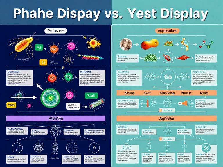
To assist our global partners in visualizing the operational difference between phage display and yeast display, we have compiled the following technical matrix.
| Technical Parameter | Phage Display (M13) | Yeast Surface Display |
|---|---|---|
| Host Organism | Escherichia coli (Prokaryotic) | Saccharomyces cerevisiae (Eukaryotic) |
| Max Library Size | 10^10 to 10^11 variants | 10^7 to 10^9 variants |
| Protein Folding & PTMs | Basic; lacks mammalian glycosylation | Advanced; features eukaryotic secretory quality control |
| Screening Methodology | Solid-phase Biopanning | Fluorescence-Activated Cell Sorting (FACS) |
| Real-Time Affinity Sorting | No (Qualitative washing) | Yes (Quantitative gating via flow cytometry) |
| Valency | Low (1 to 5 copies via pIII) | High (10,000 to 100,000 copies) |
| Primary Application Stage | De novo discovery, naive library screening | Affinity maturation, developability profiling |
7. Frequently Asked Questions (FAQs)
Why is the library size difference between phage display and yeast display so important?
When screening a naive library (derived from non-immunized sources), the statistical probability of finding a high-affinity antibody against a specific antigen is incredibly low. You need a massive pool of variants to find a match. The 100-to-1000 fold larger capacity of phage display makes it mathematically superior for this initial "needle in a haystack" search compared to yeast display.
Can KMD Bioscience perform Single B Cell Screening instead of display technologies?
Yes. While understanding the difference between phage display and yeast display is critical for in vitro synthetic libraries, our Single B Cell Screening Platform offers an alternative in vivo approach. We can directly isolate antigen-specific B cells from immunized animals or human donors, preserving the natural pairing of heavy and light chains, which is highly advantageous for certain therapeutic targets.
Does yeast display guarantee successful mammalian expression later?
While yeast is a eukaryote and folds proteins much more similarly to human cells than bacteria do, it is not identical. Yeast glycosylation patterns differ from mammalian patterns (often being high-mannose). However, from our experience, if a complex antibody format folds well on yeast, it has a significantly higher probability of successful expression in CHO or HEK293 cells during our Antibody Expression & Validation Platform phases compared to a sequence pulled directly from phage.
What is KMD Bioscience's Antibody Humanization Platform?
When an antibody is discovered using animal immunization or an animal-derived phage library, the sequence must be humanized to prevent immune rejection in patients. Our Antibody Humanization Platform uses advanced structural modeling and CDR-grafting techniques to replace animal framework regions with human sequences while retaining original binding affinity, often confirmed through rigorous downstream screening.
8. Industry References
To further explore the molecular biology, library construction protocols, and screening methodologies governing therapeutic antibody discovery, we recommend consulting the following authoritative academic resources:
- difference between phage display and yeast display

- Protein Platform
- Protein Expression Services
- Protein Modification & Detection Services
- Protein Interaction Services
- Protein Purification Service
- Antibody Discovery Platform
- VHH Antibody Platform
- Phage Display Platform
- Antibody Humanization Platform
- Single B Cell Screening Platform
- Antibody Expression & Validation Platform
- Peptide Library Platform
- Custom Antibody Platform
- Innovative Drug Discovery Platform
- Product Center
- Proteins
- Antibodies
- Raw Materials Reagents
- Small Molecule Reagents
- Peptide Products
- Nanoparticle Products
- Contact Us
- Phone:400-621-6806
- Mobile:13110936064(WeC
- E-mail:[email protected]
- Address:Room 108, Building 10, No. 6 Hechang Road, Wuqing Development Zone, Tianjin City
-

WeChat
-

Service Account
-
 400-621-6806
 -
 3599163699
 -
 Monday to Friday 9:00-18:00
 -

-
 0
0
-
